Mostrando entradas con la etiqueta células. Mostrar todas las entradas

lunes, 25 de agosto de 2014

Sorting cells with sound waves

Acoustic device that separates tumor cells from blood cells could help assess cancer’s spread.
Illustration: Christine Daniloff/MIT
Researchers from MIT, Pennsylvania State University, and Carnegie Mellon University have devised a new way to separate cells by exposing them to sound waves as they flow through a tiny channel. Their device, about the size of a dime, could be used to detect the extremely rare tumor cells that circulate in cancer patients’ blood, helping doctors predict whether a tumor is going to spread.
This microfluidic device uses sound waves to sorts cells as they flow through the channel, from left to right. Image courtesy of the researchers
Separating cells with sound offers a gentler alternative to existing cell-sorting technologies, which require tagging the cells with chemicals or exposing them to stronger mechanical forces that may damage them.

“Acoustic pressure is very mild and much smaller in terms of forces and disturbance to the cell. This is a most gentle way to separate cells, and there’s no artificial labeling necessary,” says Ming Dao, a principal research scientist in MIT’s Department of Materials Science and Engineering and one of the senior authors of the paper, which appears this week in the Proceedings of the National Academy of Sciences.

Subra Suresh, president of Carnegie Mellon, the Vannevar Bush Professor of Engineering Emeritus, and a former dean of engineering at MIT, and Tony Jun Huang, a professor of engineering science and mechanics at Penn State, are also senior authors of the paper. Lead authors are MIT postdoc Xiaoyun Ding and Zhangli Peng, a former MIT postdoc who is now an assistant professor at the University of Notre Dame.

The researchers have filed for a patent on the device, the technology of which they have demonstrated can be used to separate rare circulating cancer cells from white blood cells.

To sort cells using sound waves, scientists have previously built microfluidic devices with two acoustic transducers, which produce sound waves on either side of a microchannel. When the two waves meet, they combine to form a standing wave (a wave that remains in constant position). This wave produces a pressure node, or line of low pressure, running parallel to the direction of cell flow. Cells that encounter this node are pushed to the side of the channel; the distance of cell movement depends on their size and other properties such as compressibility.

However, these existing devices are inefficient: Because there is only one pressure node, cells can be pushed aside only short distances.

The new device overcomes that obstacle by tilting the sound waves so they run across the microchannel at an angle — meaning that each cell encounters several pressure nodes as it flows through the channel. Each time it encounters a node, the pressure guides the cell a little further off center, making it easier to capture cells of different sizes by the time they reach the end of the channel.


ORIGINAL: MIT
Anne Trafton | MIT News Office 
August 25, 2014

domingo, 27 de abril de 2014

Coming Soon: New Smart Biosensor That Directs Cells To Kill Cancer. Surgery Glasses

These biosensors can further be customised to recognise factors of relevance to various patients' needs. 

Monday, April 21, 2014: Biologists at the Northwestern University's McCormick school of engineering and applied science have developed a ground breaking technology that could modify human cells to create therapeutics used in turn to selectively target and destroy tumour cells in the human body without disrupting healthy cells. The unique protein biosensor engineers cells to kill cancer by helping them effectively distinguish between healthy and cancerous cells.

Add caption
While sitting on the surface of a cell, the biosensor can be programmed to sense its immediate environment for specific factors following which it sends a signal to the engineered cell's nucleus. This triggers a gene expression programme within the cell. "Till date, there was no way to engineer cells in a manner that allowed them to sense key pieces of information about their environment, which could indicate whether the engineered cell is in healthy tissue or sitting next to a tumour," Joshua Leonard, an assistant professor at Northwestern University's McCormick school of engineering and applied science was quoted as saying. 

Moreover, the programme is activated only in the vicinity of tumour cells, thereby minimising any side effects. These biosensors can further be customised to recognise factors of relevance to various patients' needs. "In that way, you could programme a cell-based therapy to specify which cells it should kill," Leonard added.

Meanmwhile, a team of scientists at Washington University School of Medicine in St. Louis (WUSTL) and the University of Arizona (UA) have developed a new pair of hi-tech glasses that can help surgeons to detect cancer cells. These glasses will help surgeons to visualise cancer cells which will glow blue when viewed through these glasses during surgeries. Cancer cells are invisible in normal optics even if you are viewing through a high-powered magnifying device. This innovative technology incorporates a custom video, a head mounted display and then inject a blue dye into a patient. This will specifically bind to cancer cells and makes them glow. Doctors can then easily differentiate cancer cells from healthy cells and can make sure that no tumour cells are left over during surgery. It can detect and remove tumours as small as 1mm. 

Saurabh Singh, EFYTIMES News Network 

ORIGINAL: EFY Times

jueves, 25 de julio de 2013

Scientists transplant photoreceptors from retina grown ‘in a dish’

ORIGINAL: UCL
21 July 2013
Professor Robin Ali, UCL Institute of Ophthalmology (Credit: UCL)
UCL scientists have carried out the first successful transplant of light-sensitive photoreceptor cells extracted from a synthetic retina, grown ‘in a dish’ from embryonic stem cells.
When transplanted into night-blind mice these cells appeared to develop normally, integrating into the existing retina and forming the nerve connections needed to transmit visual information to the brain.

The study, funded by the Medical Research Council (MRC) and published today in Nature Biotechnology, suggests that embryonic stem cells could in future provide a potentially unlimited supply of healthy photoreceptors for retinal cell transplantations to treat blindness in humans.

The loss of photoreceptors – light sensitive nerve cells that line the back of the eye – is a leading cause of sight loss in degenerative eye diseases such as age-related macular degeneration, retinitis pigmentosa and diabetes-related blindness.

There are two types of photoreceptor in the eye – rods and cones. Rod cells are especially important for seeing in the dark as they are extremely sensitive to even low levels of light.

The new 3D technique more closely mimics normal development, which means we are able to pick out and purify the cells at precisely the right stage to ensure successful transplantation. The next step will be to refine this technique using human cells to enable us to start clinical trials.

Professor Robin Ali, UCL Institute of Ophthalmology
Previous work by Professor Robin Ali and his team at the UCL Institute of Ophthalmology and Moorfields Eye Hospital has shown that transplanting immature rod cells from the retinas of healthy mice into blind mice can restore their sight. However, in humans this type of therapy would not be practical for the thousands of patients in need of treatment.

Using a new laboratory technique involving 3D culture and differentiation of mouse embryonic stem cells, which was developed recently in Japan, Prof Ali’s team were able to grow retinas containing all the different nerve cells needed for sight.

Professor Ali, UCL Institute of Ophthalmology and Moorfields Eye Hospital, who led the research, said: “Over recent years scientists have become pretty good at working with stem cells and coaxing them to develop into different types of adult cells and tissues. But until recently the complex structure of the retina has proved difficult to reproduce in the lab. This is probably because the type of cell culture we were using was not able to recreate the developmental process that would happen in a normal embryo.

“The new 3D technique more closely mimics normal development, which means we are able to pick out and purify the cells at precisely the right stage to ensure successful transplantation. The next step will be to refine this technique using human cells to enable us to start clinical trials.”

The researchers grew retinal precursor cells using the new 3D culture method and compared them closely with cells developed normally, looking for different markers at different stages of development. They also carried out tests to look at the genes being expressed by the two types of cells to make sure they were biologically equivalent.

They then transplanted around 200,000 of the artificially grown cells by injecting them into the retina of night blind mice. Three weeks after transplantation the cells had moved and integrated into the recipient mouse retina and were beginning to look like normal mature rod cells. These cells were still present six weeks after transplantation. The researchers also saw nerve connections (synapses), suggesting that the transplanted cells were able to connect with the existing retinal circuitry.

Dr Rob Buckle, Head of Regenerative Medicine at the MRC, said: “Regenerative medicine holds a great deal of promise for treating degenerative diseases and the eye is one area in particular where scientists are making very rapid progress. This study is an important milestone on the road to developing a widely available cell therapy for blindness as it proves unequivocally that embryonic stem cells can provide a renewable source of photoreceptors that could be used in treatments.”

-Ends-

Media contact: David Weston


Links:
Full paper in Nature Biotechnology (£)
Related images on the UCL Flickr stream
A nascent retina, generated from a 3D embryonic stem cell culture
A nascent retina, generated from a 3D embryonic stem cell culture, containing photoreceptor precursors expressing normal photoreceptor proteins, including the visual pigment, Rhodopsin (green) and the transcription factor, Crx (red). The precursors from such retinae can be isolated and transplanted into adult mice.
Image by Anai Gonzalez-Cordero. Read the press release at: bit.ly/1dPdfrp

miércoles, 10 de octubre de 2012

Nobel de Química 2012: Robert J. Lefkowitz y Brian K. Kobilka

ORIGINAL: La Vanguardia
Josep Corbella. Barcelona
10/10/2012

La Real Academia de Ciencias de Suecia ha concedido el galardón a los científicos estadounidenses Robert Lefkowitz y Brian Kobilka por sus investigaciones sobre un tipo de receptores de la membrana de las células que regulan múltiples funciones biológicas

Los científicos estadounidenses Robert J. Lefkowitz y Brian K. Kobilka han sido galardonados con el Premio Nobel de Química 2012 AP / EFE / Archivo
Los científicos estadounidenses Brian Kobilka y Robert Lefkowitz han ganado el premio Nobel de Química 2012 por sus investigaciones sobre un tipo de receptores de la membrana de las células que regulan múltiples funciones biológicas.

De los receptores acoplados a proteínas G, como se denominan, depende la actividad de hormonas como la adrenalina o la leptina, así como de neurotransmisores como la serotonina o la dopamina. Regulan, por lo tanto, desde el apetito al estado de ánimo, pasando por la tensión arterial, el tono muscular o las reacciones ante situaciones de estrés.

Aproximadamente la mitad de los fármacos existentes actualmente basan su eficacia en la acción de estos receptores, ha destacado la Real Academia de Ciencias de Suecia al anunciar el galardón. Su conocimiento detallado, gracias a las investigaciones de Kobilka y Lefkowitz ayudará a desarrollar nuevos fármacos más eficaces y con menos efectos secundarios.

Kobilka (Little Falls, Minnesota,1955) es profesor de la Universidad de Stanford en California. Lefkowitz (Nueva Yordk, 1943) es profesor de la Universidad Duke en Durham (Carolina del Norte). Ambos compartirán los 8 millones de coronas suecas (unos 900.000 euros) del premio.

La presente edición de los Premios Nobel arrancó el lunes con la concesión al británico John B. Gurdon y al nipón Shinya Yamanaka del de Medicina, y prosiguió ayer con el anuncio de que el Nobel de Física recayó en el francés Serge Haroche y el estadounidense David J. Wineland.

En los próximos días, la Academia Sueca seguirá dando a conocer el nombre de los ganadores de los Premios Nobel por este orden: mañana jueves 11 será el turno del Nobel de Literatura, seguido por el Nobel de la paz, el viernes día 12.

Por último, el próximo 15 se conocerá el ganador del premio Nobel de Economía. El de Economía, por cierto, es el único de los galardones que no quedó estableció por Alfred Nobel en su testamento, sino que se incorporó a la lista en 1969.

jueves, 13 de septiembre de 2012

Keep your distance! Why cells and organelles don't get stuck

ORIGINAL: eScience News

August 30, 2012 - 10:10 in Biology & Nature
Repulsion by water molecules is the dominant mechanism when the separation between two biomembranes is larger than around one nanometer. The molecules are oriented toward the lipids in the membrane and lose their preferred spatial arrangement –repulsion arises, keeping the membranes apart. Copyright Dr. Emanuel Schneck

If the distance between membranes is less than around one nanometer, the influence of lipid molecules prevails: At small distances they block each others’ mobility (image below).

Biomembranes enclose biological cells like a skin. They also surround organelles that carry out important functions in metabolism and cell division. Scientists have long known in principle how biomembranes are built up, and also that water molecules play a role in maintaining the optimal distance between neighboring membranes-otherwise they could not fulfill their vital functions. Now, with the help of computer simulations, scientists of the Technische Universität München (TUM) and the Freie Universität Berlin have discovered two different mechanisms that prevent neighboring membrane surfaces from sticking together. Their results appear in the Proceedings of the National Academy of Sciences.

Biomembranes consist of lipids, chain-like fat molecules stacked side by side. In the aqueous environment of cells, the lipids organize themselves into a so-called bilayer with fat-soluble "hydrophobic" ends of the molecular chains facing inward and water-soluble "hydrophilic" ends facing outward. If the water-soluble surfaces of two membranes come too close to each other, a pressure is generated-hydration repulsion-that prevents the membrane surfaces from touching. Between two intact biomembranes there is always a film of water just a few nanometers thick. Until now, however, it was unclear how hydration repulsion works on the molecular level.

By means of complex simulations, the scientists discovered two different mechanisms whose contribution depends on the distance between the membranes.
If the membranes are separated by around one nanometer or more, the water molecules play the decisive role in holding them apart. Since they have to orient themselves simultaneously to the lipids of both membrane surfaces, they give up their preferred spatial arrangement. Then they function like "bumpers," pushing the membranes apart.
When the separation is smaller, the lipids in the opposing membrane surfaces mutually inhibit their own mobility, and the repulsive force is increased.

For some time, these two mechanisms had been discussed as possible explanations for hydration repulsion. Now, with their computer simulations, the scientists from TUM and FU Berlin have for the first time correctly predicted the strength of the pressure, that is, in agreement with experimental results. In doing so, they have elucidated in detail the significance of the different mechanisms. "We were able to predict the water pressure so accurately because we determined the chemical potential of the water precisely in our computations," explains Dr. Emanuel Schneck, formerly a member of the work group of FU Berlin Professor Roland Netz (then at TUM). Schneck is currently a researcher at the Institute Laue Langevin (ILL). "The chemical potential indicates how 'willing' the water molecules are to stay in a particular spot. In order for us to obtain correct results, the potential at the membrane surfaces and the potential in the surrounding water must have the same value in our simulations."

The researchers now want to translate their results to many more biological surfaces and, in the process, initiate considerably more complex computer models.

This research was supported by the German Research Foundation (DFG SFB 765), and by the Ministry for Economy and Technology (BMWi) in an Allianz Industrie Forschung (AiF) project framework.

lunes, 2 de julio de 2012

IBM researchers unleash plastic 'ninjas' to fight deadly bacteria

ORIGINAL: IBMLabs

Having discovered that materials could be manipulated at the atomic level to control their movement, IBM researchers used this knowledge to create staph-killing 'ninja polymers' that leave healthy cells alone. They move quickly to target infected cells in the body, destroy the harmful content inside, and then disappear from the body by biodegrading without damaging side effects or accumulating in the organs.

viernes, 1 de junio de 2012

Discovery Uses 'Fracture Putty' to Repair Broken Bone in Days

ORIGINAL: ScienceDaily

ScienceDaily (Feb. 7, 2012) — Broken bones in humans and animals are painful and often take months to heal. Studies conducted in part by University of Georgia Regenerative Bioscience Center researchers show promise to significantly shorten the healing time and revolutionize the course of fracture treatment.

A team of University of Georgia researchers created a new "fracture putty" to speed healing of bone fractures. From left to right are Steve Stice, Jennifer Mumaw, Erin Jordan and John Peroni. (Credit: Image courtesy of University of Georgia)
"Complex fractures are a major cause of amputation of limbs for U.S. military men and women," said Steve Stice, a Georgia Research Alliance Eminent Scholar, animal and dairy scientist in the UGA College of Agricultural and Environmental Sciences and director of the UGA Regenerative Bioscience Center.

"For many young soldiers, their mental health becomes a real issue when they are confined to a bed for three to six months after an injury," he said. "This discovery may allow them to be up and moving as fast as days afterward."

Stice is working with Dr. John Peroni to develop a fast bone healing process. "This process addresses both human and veterinary orthopedic needs," said Peroni, an associate professor of large animal surgery in the UGA College of Veterinary Medicine and a member of the RBC.

Peroni and Stice are leading a large animal research project funded by the U.S. Department of Defense. The project includes scientists and surgeons from the Baylor University College of Medicine, Rice University and the University of Texas, who conducted the early studies.

Engineering new bone

"Healing of critical-size defects is a major challenge to the orthopedic research community," Peroni said. "Large-bone defects must be stabilized and necessitate technologies that induce rapid bone formation in order to replace the missing tissue and allow the individual to return to rapid function. To date, no single material can suffice."

The group they lead is a multidiscipline and multi-institutional group actively working on bone tissue engineering.

"Our group has been working productively together on numerous projects through the last several years," Stice said, "So, a collegial relationship and successful collaborative working relationship is already established."

Between 2009 and 2011, the collaborations received a $1.4 million grant from the DOD for the use of stem cells in fracture healing to be tested in sheep.

"In our experiences with large animal models, following the guidelines established by our animal care and use committee," Stice said, "we have been successful in formulating a product that contains mesenchymal stem cells and allows them to survive in the environment of the fracture long enough to elicit the rapid formation of new bone."

This year, the group showed bone can be generated in sheep in less than four weeks. The speed in which bone is formed is one of the truly unique features of this study.

Fracture putty

To start the bone regeneration process, the RBC used adult stem cells that produce a protein involved in bone healing and generation. They then incorporated them into a gel, combining the healing properties into something Stice calls "fracture putty."

With Peroni's assistance, the Houston-based team used a stabilizing device and inserted putty into fractures in rats. Video of the healed animals at two weeks shows the rats running around and standing on their hind legs with no evidence of injury. The RBC researchers are testing the material in pigs and sheep, too.

"The small-animal work has progressed, and we are making good progress in large animals," he said.

More work is needed to get to human medical trials, but the threat of losing federal funding for biomedical work through the DOD means they will have to find new ways to fund the project.

Next steps

"The next step is to show that we can rapidly and consistently heal fractures in a large animal," Peroni said, "then to convert it to clinical cases in the UGA [College of Veterinary Medicine] clinics where clinicians treat animals with complex fractures all the time."

Once they have something that works for animals, it will be passed over to the DOD for human use.

Peroni, who is chairman of the North American Veterinary Regenerative Medicine Association, is hopeful this material will be promoted to the veterinary and human medical fields through the educational efforts of NAVRMA and the RBC.

However, the RBC isn't the only group working on a faster fix for broken bones.

"Our approach is biological with the putty," Stice said. "Other groups are looking at polymers and engineering approaches like implants and replacements which may eventually be combined with our approach. We are looking at other applications, too, using this gel, or putty, to improve spinal fusion outcomes."

One of the best hopes for the fracture putty is in possible facial cranial replacements, an injury often seen on the battlefield.


The project ends in mid-2012. "By then we are to deliver the system to the DOD," Stice said.

lunes, 7 de mayo de 2012

Solar-Powered Sea Slugs

ORIGINAL: U Maine | Nature
Imagine being able to turn on solar-powered cells whenever food became limiting in your environment to exploit the sun’s energy to produce chemical energy. Further imagine the advantage of being mobile and camouflaged with a green, rippling leaf appearance in a sea filled with predators in search of soft-bodied creatures. The sacoglossan mollusc Elysia chlorotica (Gould) possesses all of these traits. It is a shell-less, green “walking leaf” that will feed on algae when they are available, stealing the chloroplasts, and using them for solar-power when food is scarce.


ORIGINAL: Nature
The slug pictured to the right, Elysia chlorotica, is a symbiont thief.


Elysia chlorotica eats the alga Vaucheria litorea but does not digest it. The slug cuts open algal filaments and sucks out the contents, transferring the living chloroplasts to its own tissue. Chloroplasts are organisms that have lived symbiotically within plant cells for many millions of years. They harness energy from the sun, which they give to the plant or alga cell they live within. Most animals digest the chloroplasts entirely when they eat plants, but not Elysia. By keeping the chloroplasts intact and transferring them to its own tissue, Elysia allows them to continue photosynthesizing, producing energy for the slug. The slug can then live for months without eating as long as sunlight is available, and can maintain the same chloroplasts for its entire adult life. This is an extremely unique relationship between an animal and plant symbionts.

Many other animals form associations with photosynthetic organisms. Corals such as the one depicted below have a symbiosis with multiple single-celled organisms called zooxanthellae. This is a multiple-level symbiosis because corals house the entire chloroplast-containing zooxanthellae cells within their tissue. This is different from Elysia chlorotica, who has cut out the middleman — instead of incorporating entire cells, it only retains the chloroplasts.
The photograph of Elysia chowing down was taken by Nicholas E. Curtis and Ray Martinez. The second photograph of Elysia is courtesy of Mary S. Tyler, and was the cover of PNAS when this paper was published. The lower picture is the coral Porites as photographed by Casey Dunn.

You can watch two amazing videos of the slugs in action, here and here, both of which were included in the PNAS paper.
--Freya Goetz


These sea slugs feed by slicing or puncturing siphonaceous algal cells and sucking out the cell contents. All of the contents, including the algal nucleus, are discarded except the chloroplasts which are engulfed phagocytotically into the digestive cells. (The figure to the right shows the "stolen" chloroplasts, or kleptoplasts, within the cells of the slug's digestive tract.)

By distributing the “photosynthetic factories“ throughout their extensively branched digestive system just one cell layer beneath the epidermis, the sea slugs not only blend into the green algal bed (the figure to the right shows the sea slug on strands of the alga Vaucheria litorea ), they also capture light energy to fuel photoautotrophic CO2 fixation. In some cases, the resulting carbon products can totally sustain the sea slugs for several months in the absence of an algal food source and serve as precursors for synthesis of chemical defense compounds and the copious mucus which bathes and protects the sea slugs.


Symbiosis
Symbiotic associations between organisms, even of different kingdoms, is not that unusual. However, in almost all cases they represent an association between two intact, free-living organisms, both of which have retained their complete cellular genetic make-up. Such associations are typically intercellular and, if intracellular, the symbiont is frequently isolated from the host’s cytosol by sequestration in a vacuole or host-provided membrane. What makes the sea slug/algal chloroplast symbiosis so remarkable is that the symbiont is a “naked,” foreign organelle sustained intracellularly in direct contact with the host sea slug cytosol and the symbiont remains functional for several months despite the absence of any algal nucleo-cytosolic influence.

There is some disagreement on whether an association between an organism and an isolated organelle such as a chloroplast constitutes symbiosis since the symbiont (chloroplast) is not a free-living organism. The term symbiosis was first defined as, “unlike organisms” living together. “Unlike organisms” came to mean different species and symbiosis changed to reflect, “prolonged physical associations without respect to outcome.” In the early 1900's, the Russian scientist K.S. Mereschkovsky proposed that chloroplasts originated from blue-green algae (cyanobacteria), a process he named symbiogenesis or “the origin of evolutionary novelty via symbiosis.”

In 1975, Robert Trench defined intracellular symbiosis as, “the coexistence of at least two genomes of divergent evolutionary origins occupying the same cytoplasmic environment.”

In her book on “Symbiotic Interactions,” Angela Douglas (1994) emphasizes that symbiosis is not dependent on mutual benefit to the partners, rather that at least one of the partners acquires a new metabolic property. Considering both Trench’s and Douglas’ definitions, we conclude that the intracellular association of algal chloroplasts with molluscan cells can be considered a unique symbiotic association. The chloroplast represents a symbiont genome and the host mollusc acquires a new metabolic capability, photosynthesis. Still, others prefer to use the term kleptoplasty or “something borrowed” to describe the chloroplast symbiosis. Regardless of definition or term used, today it is universally recognized that great biological novelty and diversity come from symbiotic associations and symbiosis is a widespread biological phenomenon.

PRIMARY, SECONDARY AND TERTIARY ENDOSYMBIOSIS
The endosymbiotic events leading to a solar-powered sea slug.

The endosymbiont theory traces the origin of the chloroplast to a free-living cyanobacterium that was engulfed by a eukaryote giving rise to the primary lineages of glaucophytes, red algae (rhodophytes), and the green plants and algae (the viridiplantae or streptophytes and chlorophytes, respectively).

Subsequently, secondary endosymbiosis, the uptake of a eukaryotic alga (green or red lineage) by another heterotrophic eukaryotic host, gave rise to a diverse group of secondary or complex algae, including the heterokont Vaucheria litorea.
Tertiary symbiosis is most commonly associated with dinoflagellates and the replacement of their endosymbiont with a new secondary endosymbiont. Here, we propose that the engulfment of secondary chloroplasts by a sea slug also represents a tertiary endosymbiotic association.

Kleptoplasty by Elysia chlorotica.
History
The first reports of "green bodies" within molluscs were made in 1904 by Brüel. However, it was not until the mid 1960's that these "green bodies" were studied and seen to be functional chloroplasts. From this time on, there has been much interest in the phenomenon by which sea slugs find, acquire, and maintain chloroplasts as an essential component of their life-cycle.

Kleptoplasty (“stealing” of chloroplasts) by Elysia chlorotica is remarkable for at least three reasons.
  1. First, the “symbiont” in this case is not another autonomous organism with an intact genome, but rather a “naked,” foreign organelle (chloroplast).
  2. Second, the symbiont is housed intracellularly and not sequestered between cells or within a vacuolar membrane.
  3. Third, and perhaps most remarkable, the semi-autonomous kleptoplasts remain functional for as long as ten months within the foreign host cytosol despite the absence of any of its own algal nucleo-cytosolic components.
This type of long-term activity by isolated plastids is unprecedented and astounding considering that chloroplasts are derived from once free-living cyanobacteria and have lost the majority of their genes. Hence, they are dependent on their own nucleo-cytosol for protein synthesis, targeting, and regulation of just about every function of the organelle. In turn, chloroplasts are fragile organelles and very sensitive to physical and chemical changes in their environment, including osmotic stress. Evidently, the advantages of photoautotrophy have provided a strong selective pressure for the evolution of this association.
Whether the establishment of this symbiotic association is fueled by photoautotrophy pressure to sustain energy production for the sea slug when food is scarce or the need for camouflage protection when facing life without a protective shell in a predatory environment, or both, the association has not progressed to a hereditary one where the plastids are passed from one generation of slugs to the next. Instead, the association must be established anew each year and is required for the slug to develop into a mature adult sea slug, at least in laboratory experiments.

Establishment of the Kleptoplastic Association
We have now succeeded in culturing Elysia chlorotica in the laboratory including establishing the symbiotic or kleptoplastic association with Vaucheria litorea chloroplasts. Adult E. chloroticaproduce eggs, devoid of plastids, typically in late spring and planktonic veligers hatch within 4 to 5 d and then spend about 5 d feeding on unicellular algae (Rhodomonas or Isochrysis in the lab). When subsequently provided with filaments of V. litorea, metamorphosis of the veligers into juvenile sea slugs occurs within 1 to 2 d.
The endosymbiosis is established when young juvenile sea slugs grasp and then puncture the siphonaceous algal cells and suck out the cell contents. All of the algal contents are “discarded” except the chloroplasts, which are engulfed phagocytotically into the digestive cells. The captured chloroplasts fill the growing, extensively branched digestive tubules that lie just one cell layer beneath the epidermis.

Over the next several months the sea slugs may continue to feed onVaucheria if it is available and/or sustain themselves by photoautotrophic CO2 fixation using their newly acquired chloroplasts.


In laboratory culture, the sea slugs are kept apart from the algae, thus sustaining themselves totally by photosynthesis for up to ten months. Interestingly, death of the adults occurs almost synchronously late each spring in the lab and field (Pierce et al. 1999 Biol. Bulletin on virus).

The alga vaucheria
Vaucheria: Synonyms = water felt
Reproduction = asexual by fragmentation of filaments or zoospores and sexual by oogamy.

Vaucheria is a salt-water, yellow-green alga (Xanthophyte), in the Heterokont Kingdom.
Xanthophytes (yellow-green algae): >600 species, PS, fresh and marine waters, Chl a and c1 and c2 and NO fucoxanthin or Chl b. Cell wall, but not of cellulose or chitin. Food reserve = oil or fat, not starch typically. Haploid nucleus with outer membrane continuous with ctER. Sessile or free-living; motile flatellated unicells to colonies to siphonaceous (coenocytic) multinucleated filaments, to multicellular filaments.


Heterokont (=stramenopile = chromista)
description: Members of the Kingdom Chromista or Heterokontae exhibit the following characteristics: tubular mitochondria, motile cell with two different flagella. They vary from unicellular flagellates to large, siphonaceous filaments. They include diatoms, raphidiophytes, chrysophytes (golden algae), oomycota (water molds), phaeophytes (brown algae and kelps), haptophytes, silicoflagellata, and the xanthophyta (yellow-green algae including Vaucheria).
Vaucheria exhibits a siphonaceous morphology with a large vacuole and a thin layer of multinucleate cytoplasm containing numerous chloroplasts.

Chloroplasts
The chloroplasts ofVaucheria litorea have the standard four membranes surrounding them when seen within the algal cytoplasm. When isolated, they have only the standard double envelope.

Vaucheria litorea.
Taxonomy:

Kingdom Chromista (=Heterokontae)

Division (Phylum): Ochrophyta
Class: Xanthophyceae
Order: Vaucheriales
Family: Vaucheriaceae
Genus: Vaucheria (De Candolle, 1801)
Species: Vaucheria litorea Hofman ex C. Agardh, 1823

Culturing the alga vaucheria
The heterokont alga (= chromophytes [chl a and c], autotrophic stramenopiles), Vaucheria litorea, is maintained in culture in enriched quarter-strength (tolerates ¼ to full-strength sea water) Instant Ocean and a modified f/2 medium with natural lighting and daily manual swirling.

about elysia chlorotica
Found in saltwater marshes along the east coast of the US as far north as Nova Scotia to NC in the south and some times down to FL; waters of widely varying salinity, but usually brackish salt marshes. Our specimens are routinely collected from an intertidal marsh on Martha’s Vineyard Island, MA.

Synonyms = “leaves that crawl,” “solar-powered sea slugs,” “walking leaf,” “eastern emerald elysia,”
Reproduction = hermaphroditic, but self-fertilization is not common, shed fertilized eggs
Size = 2 to 3 cm common; some up to 6cm

jueves, 19 de abril de 2012

Pionero en células madre e ingeniero en código abierto son galardonados con el "Premio Millennium de Tecnología 2012"

ORIGINAL: TechnologyAcademyFinnland
19 de abril 2012


La Academia de Tecnología de Finlandia ha declarado hoy que dos destacados innovadores, Linus Torvalds y el Dr. Shinya Yamanaka, ganadores del importante premio a la innovación tecnológica Premio Millenium de Tecnología de 2012, . Los galardonados, siguen los pasos de los vencedores anteriores, como el creador de la World Wide Web, Sir Tim Berners-Lee, en una ceremonia que se celebrará en Helsinki, Finlandia, el miércoles 13 de junio de 2012, cuando el ganador del Gran Premio será anunciado. La bolsa de premios es superior a 1 millón de euros.

Linus Torvalds, Finlandia / EE.UU.
Linus Torvalds. ZDnet
En reconocimiento a su creación de un nuevo sistema operativo de código abierto utilizado en computadores que llevan a que el núcleo de Linux. La libre disponibilidad de Linux en la Web rápidamente causó una reacción en cadena que conduce a un cuyo gran desarrollo y puesta a punto equivalen a 73.000 años-hombre. Hoy, millones de computadoras, teléfonos inteligentes y las grabadoras de video digital como TiVo  se ejecutan en Linux. El logro de Linus Torvalds logros han tenido un gran impacto en el desarrollo de software compartido, trabajo en red y la apertura de la web, haciéndola accesible a millones, si no miles de millones.

El Dr. Shinya Yamanaka, Japón
Dr. Shinya Yamanaka. Wikipedia
En reconocimiento a su descubrimiento de un nuevo método para desarrollar células madre pluripotentes inducidas para la investigación médica que no se basan en el uso de células madre embrionarias. Usando su método para crear células madre, los científicos de todo el mundo están haciendo grandes avances en la investigación en las pruebas de drogas y la biotecnología médica que deberían conducir, algún día a el crecimiento exitoso de los tejidos de implantes para la cirugía clínica y la lucha contra enfermedades incurables como el cáncer, la diabetes y de Alzheimer. El Dr. Yamanaka está específicamente nombrado por su destacado trabajo en la metodología éticamente sostenible.


Premio Millennium de Tecnología
El Premio Millennium de Tecnología es un homenaje de Finlandia a la vida, la mejora de la innovación tecnológica. El premio se concede cada dos años por una innovación tecnológica que mejora significativamente la calidad de vida humana, actual y en el futuro. Es otorgado por la Technology Academy Finland, una fundación independiente constituida por la industria finlandesa en asociación con el Estado finlandés. Los galardonados fueron seleccionados por el Patronato de la Fundación sobre la base de las recomendaciones formuladas por el Comité de Selección Internacional.

Linus Torvalds
El Dr. Shinya Yamanaka

Comité de Selección Internacional tiene amplia experiencia tecnológica
Las candidaturas elegibles han sido examinados por el Comité de Selección Internacional, una red distinguido de los principales científicos finlandeses e internacionales y tecnólogos. La decisión final sobre todos los laureados se realiza por el Consejo de la Technology Academy Finland sobre la base de una propuesta de la CAI. El ganador del Gran Premio serán anunciados en una ceremonia festiva en Helsinki el 13 de junio de 2012.

Linus Torvalds dijo:

"El software es muy importante en el mundo moderno para no ser desarrollado a través de fuentes abiertas. El impacto real de Linux es semejante a una forma de permitir a las personas y empresas construir en la parte superior del mismo para hacer su propia cosa. Finalmente estamos llegando al punto en que "los datos son sólo de datos", y no tenemos todos estos canales especiales absurdos de comunicación para las diferentes formas de datos ".

El Dr. Yamanaka dijo:

"En el siglo 21, la biología médica avanza a un ritmo más rápido que antes y la medicina personalizada estará fácilmente disponible en un futuro no lejano. Las iPS derivadas de células células diferenciadas podría tratar la anemia de células falciformes y lesiones de la médula espinal. Ya hay planes para llevar a cabo un ensayo clínico en algunos pacientes con enfermedad macular relacionada con la edad en los próximos años."

"Mis objetivos en la década incluyen el desarrollo de nuevos fármacos para enfermedades intratables mediante el uso de tecnología de células iPS y llevar a cabo ensayos clínicos psra usarlos en algunos pacientes con enfermedad de Parkinson, diabetes o enfermedades de la sangre."

El Dr. Ainomaija Haarla, el presidente de la Academia de Tecnología de Finlandia, dijo:

"Hemos tenido muchas nominaciones dignas sobre las cuales deliberar, pero al final las hemos reducido a estos dos candidatos que han hecho un impacto tan significativo en el campo de la informática y la investigación con células madre. Espero que este anuncio conduzca a un mayor reconocimiento para estos científicos extraordinarios y las tecnologías que han desarrollado. De estos dos hombres además se hablará durante los siglos por venir. "

Notas para los editores:
1. Para preguntas de los medios internacionales, por favor póngase en contacto con:
Rob Blackhurst Apollo Relaciones Públicas rob@apollopublicrelations.com 00 44 787 9423 341

2. Para preguntas de los medios finlandeses, por favor póngase en contacto con:
Niina Suhonen, Jefe de Comunicaciones y Marketing, Technology Academy Finland
nombre.apellido (a) technologyacademy.fi, tel. +358 40 8439 438

3. Contactos en la Academia de Tecnología de Finlandia
El Dr. Tech. Ainomaija Haarla, Presidente y CEO
nombre.apellido (a) technologyacademy.fi, tel: +358 40 716 0703
Canciller Jarl-Thure Eriksson, Abo Akademi University, Presidente del Comité Internacional de Selección, nombre.apellido (a) abo.fi, tel. +358 40 5012 570

Más información sobre el Premio y los miembros del Comité Internacional de Selección en: www.millenniumprize.fi

Información adicional acerca de Technology Academy Finland en:
www.technologyacademy.fi

Más información sobre los premiados y sus innovaciones:
Historias de caso, entrevistas, fotografías y vídeos sobre los premiados se encuentran disponibles en www.millenniumprize.fi

Academia de Tecnología de Finlandia (TAF) es una fundación independiente con la misión de apoyar la investigación científica y las nuevas tecnologías que beneficien a la humanidad y mejorar la calidad de vida de las personas. TAF otorga el bienal Premio del Milenio Tecnología y ejecuta eventos asociados, tales como el Campamento Anual de la Juventud del Milenio. También promueve la TAF Finlandia como un país de alta tecnología, participando activamente en las redes mundiales en la comunidad científica, las empresas y las organizaciones no gubernamentales.

Entre los ganadores anteriores
El Premio Millennium de Tecnología se ha adjudicado en cuatro ocasiones. El Premio inaugural fue otorgado en 2004 a Sir Tim Berners-Lee, inventor de la World Wide Web. En 2006, el Premio se otorgó al profesor Shuji Nakamura, inventor del revolucionario nuevas fuentes de luz - de color azul brillante, verde y blanco LED y un láser azul. En 2008, el profesor Robert Langer ganó el premio por su trabajo innovador en la liberación controlada de fármacos y para el desarrollo de biomateriales innovadores para su uso en la regeneración de tejidos. El cuarto premio fue otorgado al profesor Michael Grätzel en 2010 por sus desarrollos innovadores en tinte de células solares sensibilizadas. La nueva tecnología tendrá un impacto significativo en el desarrollo de soluciones energéticas del futuro, y las células de Grätzel se espera que desempeñen un papel importante y extensa en aplicaciones de energía renovable.

Lo que el comité de selección de dicho acerca de laureados de este año

Linus Torvalds
"En reconocimiento de la creación libre de prejuicios de un nuevo sistema operativo de código abierto que lleva a gran parte del núcleo de Linux explotación. La disponibilidad gratuita en la Web rápidamente causó una reacción en cadena que conduce a un mayor desarrollo y puesta a punto vale la pena el equivalente de 73.000 años-hombre. Hoy en día el número estimado de usuarios es de 30 millones de dólares. El logro de Linus Torvalds ha tenido un gran impacto en el desarrollo de software y en cuestiones culturales y éticas de la red y la apertura de la web. "

El Dr. Shinya Yamanka
"En reconocimiento del descubrimiento de un nuevo método y el desarrollo de los procedimientos técnicos necesarios para producir células madre pluripotentes inducidas a partir de tejido celular normal. El logro tiene un gran impacto en la investigación en la medicina y la biotecnología, las células madre pluripotentes ya se utilizan para las pruebas de drogas médicas y el crecimiento de los tejidos de implantes. El descubrimiento del Dr. Yamanaka también tiene una influencia ética fundamental, ya que elimina la necesidad de que las células madre embrionarias. Él es, sin duda, identificado como el padre de la innovación ".

Socios del Premio de Tecnología del Milenio
Kemira, Neste Oil, Nokia, Outotec, SEB,
Cargotec, la FIM, Fortum, Gasum, Halton, Lönnberg, Metso, Metsägroup, Ruukki, Vaisala, Wärtsilä

sábado, 3 de marzo de 2012

Immortal worms defy aging

ORIGINAL: KurzweilAI
February 29, 2012

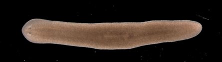
Planarian flatworm (credit: The University of Nottingham)
Researchers from The University of Nottingham have discovered how planarian flatworms overcome the aging process to be potentially immortal: they can rejuvenate their telomeres.

The discovery, funded by the Biotechnology and Biological Sciences Research Council (BBSRC) and Medical Research Council (MRC), may eventually lead to alleviating aging and age-related characteristics in human cells.

Planarian worms have amazed scientists with their apparently limitless ability to regenerate. Researchers have been studying their ability to replace aged or damaged tissues and cells in a bid to understand the mechanisms underlying their longevity.

“We’ve been studying two types of planarian worms; those that reproduce sexually, like us, and those that reproduce asexually, simply dividing in two,” said Dr. Aziz Aboobaker from the University’s School of Biology.

“Both appear to regenerate indefinitely by growing new muscles, skin, guts and even entire brains over and over again.

“Usually when stem cells divide — to heal wounds, or during reproduction or for growth — they start to show signs of aging. This means that the stem cells are no longer able to divide and so become less able to replace exhausted specialized cells in the tissues of our bodies.

“Our aging skin is perhaps the most visible example of this effect. Planarian worms and their stem cells are somehow able to avoid the aging process and to keep their cells dividing.”

Each time an animal cell divides, the protective telomere “cap” gets shorter. When they get too short, the cell loses its ability to renew and divide. In an immortal animal, we would therefore expect cells to be able to maintain telomere length indefinitely so that they can continue to replicate. Aboobaker predicted that planarian worms actively maintain the ends of their chromosomes in adult stem cells, leading to theoretical immortality.

Previous work, leading to the award of the 2009 Nobel Prize for Physiology or Medicine, had shown that telomeres could be maintained by the activity of an enzyme called telomerase. In most sexually reproducing organisms, the enzyme is most active only during early development. So as we age, telomeres start to reduce in length.

This project identified a possible planarian version of the gene coding for this enzyme and turned down its activity. This resulted in reduced telomere length and proved it was the right gene. They were then able to confidently measure its activity and resulting telomere length and found that asexual worms dramatically increase the activity of this gene when they regenerate, allowing stem cells to maintain their telomeres as they divide to replace missing tissues.

“The next goals for us are to understand the mechanisms in more detail and to understand more about how you evolve an immortal animal,” said Aboobaker.

“The worms are a model system in which we can ask questions, like is it possible for a multicellular animals to be immortal and avoid the effects of aging?,” he told KurzweilAI.

“If so, how does this animal do this in comparison to animals that don’t? Of course we hope that this impacts humans, that’s why we do it. But we aren’t planning on making any drugs or medicines… other people are, I’m sure.”

Ref.: Thomas C. J. Tan et al., , Telomere maintenance and telomerase activity are differentially regulated in asexual and sexual worms, Proceedings of the National Academy of Sciences, 2012 [DOI: 10.1073/pnas.1118885109] (open access)

miércoles, 1 de febrero de 2012

"Captura del Cloroplasto", cómo la información genética migra de una planta a otra.

ORIGINAL: ScienceDaily

ScienceDaily (01 de febrero 2012) - Para generar árboles filogenéticos e investigar las relaciones entre los organismos, los científicos suelen buscar similitudes y diferencias en el ADN. Los científicos de plantas fueron confundidos por el hecho de que el ADN extraído de los cloroplastos de las plantas verdes a veces mostraron mayor similitud con especies relacionadas que crecieron en la misma zona. 

Trataron de explicar el fenómeno con la suposición en el que de vez en cuando especies sexualmente incompatibles para ser normalmente cruzadas produjeran descendencia con una nueva combinación de los genomas nuclear y del cloroplasto.

Un injerto natural entre un abedul (izquierda) y el roble (derecha). Especies sexualmente incompatibles pueden intercambiar genomas de cloroplastos en las zonas de injertos
Se acuñó el término "captura del cloroplasto" para ilustrar lo que ellos pensaban que estaba sucediendo.

Ahora, científicos allegados a Ralph Bock, del Instituto Max Planck de Fisiología Molecular de Plantas en Potsdam descubrieron que la transferencia de los cloroplastos en un todo, o al menos sus genomas, puede ocurrir en zonas de contacto entre las plantas. El cruce entre especies no es necesario. El genoma del cloroplasto si lo hace, será transmitida a la siguiente generación y, por tanto, dará origen a una planta con rasgos nuevos. Estos hallazgos son de gran importancia para la comprensión de la evolución, así como la crianza de nuevas variedades vegetales.

Muchas plantas de madera, especialmente de árboles frutales y rosa, son deliberadamente dañadas por los jardineros. Les cortan ramas o cortar abolladuras en la corteza con el fin de poner las partes de otra planta en las ranuras. La planta cuyas raíces tocan el suelo se llama "stock", mientras que heredero es el término técnico para la rama que se coloca sobre él. La razón detrás de las "atrocidades" del jardinero es la de reproducir las variedades con un rendimiento especialmente alto, sin que las leyes de Mendel entren en juego. Según Mendel, sólo algunas partes de los descendientes muestran los mismos rasgos que sus padres. El resto de la descendencia será más probable es que sea menos valiosa. Al poner una rama de una variedad de manzana exitosa en una población nueva, la manzana del árbol deseado es fácilmente clonada. Sin embargo, las uniones del injerto no siempre tiene que ser hechas por el hombre. Las plantas que crecen sólo en las cercanías de los demás pueden fusionarse.

En las zonas de contacto mencionados arriba, llamadas de transferencia horizontal de genes (HGT Horizontal Gene Transfer), la transferencia de genes sin reproducción sexual, puede ocurrir. Durante mucho tiempo, los científicos creían que HGT se limitaba a los procariotas, los organismos sin núcleo.