Mostrando entradas con la etiqueta Señalización celular. Mostrar todas las entradas

domingo, 27 de abril de 2014

Coming Soon: New Smart Biosensor That Directs Cells To Kill Cancer. Surgery Glasses

These biosensors can further be customised to recognise factors of relevance to various patients' needs. 

Monday, April 21, 2014: Biologists at the Northwestern University's McCormick school of engineering and applied science have developed a ground breaking technology that could modify human cells to create therapeutics used in turn to selectively target and destroy tumour cells in the human body without disrupting healthy cells. The unique protein biosensor engineers cells to kill cancer by helping them effectively distinguish between healthy and cancerous cells.

Add caption
While sitting on the surface of a cell, the biosensor can be programmed to sense its immediate environment for specific factors following which it sends a signal to the engineered cell's nucleus. This triggers a gene expression programme within the cell. "Till date, there was no way to engineer cells in a manner that allowed them to sense key pieces of information about their environment, which could indicate whether the engineered cell is in healthy tissue or sitting next to a tumour," Joshua Leonard, an assistant professor at Northwestern University's McCormick school of engineering and applied science was quoted as saying. 

Moreover, the programme is activated only in the vicinity of tumour cells, thereby minimising any side effects. These biosensors can further be customised to recognise factors of relevance to various patients' needs. "In that way, you could programme a cell-based therapy to specify which cells it should kill," Leonard added.

Meanmwhile, a team of scientists at Washington University School of Medicine in St. Louis (WUSTL) and the University of Arizona (UA) have developed a new pair of hi-tech glasses that can help surgeons to detect cancer cells. These glasses will help surgeons to visualise cancer cells which will glow blue when viewed through these glasses during surgeries. Cancer cells are invisible in normal optics even if you are viewing through a high-powered magnifying device. This innovative technology incorporates a custom video, a head mounted display and then inject a blue dye into a patient. This will specifically bind to cancer cells and makes them glow. Doctors can then easily differentiate cancer cells from healthy cells and can make sure that no tumour cells are left over during surgery. It can detect and remove tumours as small as 1mm. 

Saurabh Singh, EFYTIMES News Network 

ORIGINAL: EFY Times

sábado, 22 de febrero de 2014

The Discovery and Potential of Nerve Growth Factor (NGF) by Rita Levi-Montalcini

Rita Levi-Montalcini was working with chick embryos in 1938, investigating how neurons find their way to the limbs they are to innervate, when she was barred from the University of Turin, a Jew in Mussolini’s Italy. She continued work in a laboratory she set up in her bedroom in Turin, then in the countryside.

“At the end of this difficult period, there was a seed for what has been one of most fantastic developments in one field of neuroscience”—the identification of nerve growth factor (NGF), said Piergiorgio Strata, president of Italy’s National Institute of Neuroscience, and a member of the European Dana Alliance for the Brain (EDAB).

The occasion was a memorial symposium for Levi-Montalcini, a founding member of EDAB who died in 2012 at the age of 103. Neuroscientists who knew, worked or studied with Levi-Montalcini honored her life by elaborating her legacy—a morning's tour through research that followed the groundbreaking discoveries for which she received The Nobel Prize in Physiology or Medicine in 1986.

Working at Washington University in St. Louis after the war, she and Stanley Cohen (with whom she shared the Nobel) identified a compound, expressed by peripheral cells, that attracted spinal neurons and induced neurite formation, then isolated this substance—NGF—from tumors, snake venom, and mouse salivary glands—all in the face of relentless skepticism from the scientific community.

Speakers at the symposium, presented by the Italian Cultural Institute and Centro Primo Levi in NYC, stressed the characteristics that enabled her to flourish intellectually and prevail in adversity: 
  • a powerful, charismatic personality, 
  • enormous drive and passion for her work, and 
  • an approach that combined intuition with analysis. 
"She often noted that she viewed herself as an artist more than a scientist," said a Neuron obituary.

The importance of this work, speakers said, could hardly be overestimated. "If we look at the history of 20th century neuroscience, Rita ranks with the giants... she was the first major molecular neurobiologist," wrote fellow Nobelist (and member of the Dana Alliance for Brain Initiatives) Eric Kandel, in a tribute read at the meeting. "Her extraordinary discovery of NGF affected all aspects of our field."

As the first identified growth factor, NGF introduced a radically new concept, said Lloyd Greene of Columbia University. "We knew from insulin that organs could communicate via substances that went into the bloodstream. A major implication of Rita's findings was that there was another means of communication between cells, at short range.” Her inquiry into embryonic development illuminated key processes in mature neurons—survival, plasticity, neuroprotection—and beyond, “an explosion of findings within and outside the nervous system."

Ralph Bradshaw of University of California, San Francisco, called NGF a "Rosetta Stone" that helped decode key aspects of nervous system function, proteins, receptors, and cancer biology.

He reviewed some of his involvement in elaborations of the NGF discovery—beginning with the sequencing, in collaboration with Ruth Angeletti (who had been Levi-Montalcini's only PhD student, now at Albert Einstein College of Medicine), of the NGF molecule. The structure, he said, suggested a compound that acted like insulin on target cells. "It turned out we were right, but not for all the right reasons."
“The idea that NGF was an endocrine-like substance led to pursuit of the receptor,” Bradshaw said, summarizing research that eventually characterized not one but two receptors (a fact that “befuddled the field for 15-20 years”) and then to elucidation of the molecular signaling pathways by which NGF and related compounds modulate cellular function.

“The picture started to evolve that these factors were not only involved in growth and development, but also as regulators in growth disorders, namely cancer… that these were very important discoveries,” Bradshaw said.

Greene’s research exemplified this importance. "In science, you start working on one thing and end up with something far different," he said. “NGF led us, in ways we never would have anticipated, to a potential treatment for brain tumors.”

It began with studies in the 1990s to explore how NGF regulates genes. Using serial analysis of gene expression, Greene’s research team identified hundreds of genes that became more or less active after exposure to the compound. The researchers then focused on transcription factors—proteins that determine whether genes are turned on or off. They found that one of these compounds, ATF5, was particularly abundant in neural progenitor cells, but not in mature neurons or astrocytes, and that NGF shut down production of ATF5.

“This led us to the idea that ATF5 is important for proliferation of [stem] cells that eventually give rise to the brain. When they encounter growth factor, they turn into differentiated cells and stop proliferating,” Greene said. Neural progenitor cells that were experimentally deprived of ATF5 differentiated prematurely and failed to migrate. Cells infected with a retrovirus to keep on producing the transcription factor never differentiated and continued to divide—much like a tumor.

“We wondered: is ATF5 present in glioblastomas?” he said.

It was; cells from 29 of these highly virulent, virtually incurable tumors all expressed the transcription factor. When the researchers silenced ATF5 in cultured glioblastoma cells, the cells died.

In subsequent in vivo studies, the researchers gave mice with experimentally induced glioblastomas subcutaneous injections of a molecule that hybridized dominant-negative ATF5 protein, which neutralizes ATF5, with penetratin, a peptide that crosses the blood-brain barrier.

Within days of treatment, tumor cells began to die; 19 days and 6 months later, the tumors had disappeared on MRI. Treated animals all survived for 6 months, while 60% of the others died. There was no apparent kidney, brain, liver, or blood toxicity.

As work proceeds with other animals, “we’re collecting data to go to the FDA for possible clinical trials,” Greene said. The approach “could work for other tumors as well.”

Antonio Cattaneo of the European Brain Research Institute in Rome (which Levi-Montalcini helped establish in 2002), described research linking the NGF system to Alzheimer’s disease pathology, and suggesting a novel treatment strategy.

Using antibodies that target NGF, he showed that neutralizing the growth factor in the brains of adult mice initiated a process of neuroinflammation and neurodegeneration. While the effect on cholinergic neurons—a key population in Alzheimer’s disease (AD)—was first implicated, it became clear that astrocytes and glia were compromised as well.

Further studies characterized this neurodegeneration process as an imbalance between NGF and a precursor protein, proNGF, and showed that the same result could be achieved by modifying mouse brain cells to overexpress proNGF.

Cattaneo has been exploring ways to “strengthen the balance by increasing NGF.” When mice, genetically modified to express AD-like pathology, were given a modified form of NGF intranasally, amyloid plaques regressed, and learning and memory deficits improved.

“This may be a viable candidate for a non-invasive therapeutic approach to AD,” he said. “We’re collaborating with the pharmaceutical industry to get clinical trials.”

Looking toward the future of NGF-related research, Cattaneo cited an "agenda" that Levi-Montalcini proposed in 2009, at the age of 100. In addition to work (like the studies described above) aiming to develop its therapeutic potential, she urged investigations of the NGF system’s role earlier in embryonic development than the nervous system, and in more primitive species.

Her agenda called for studies of NGF in other tissues, particularly the reproductive system. “Rita predicted it would be found to participate in processes like activation of sperm or implantation of ova,” Cattaneo said.

Her scientific intuitions were still reliable, he said. In a paper published three years later, researchers described their work identifying a substance in the semen of diverse mammals that induces ovulation. It was NGF. 

ORIGINAL: DANA Foundation
by Carl Sherman

February 20, 2014

viernes, 17 de enero de 2014

World-first working eukaryotic cell made from plastic


Researchers at the Institute for Molecules and Materials at Radboud University Nijmegen used a water droplet as the structure upon which they built the first polymer cell

Previously, chemists have managed to create artificial cell walls and developed synthetic DNA to produce self-replicating, synthetic bacterial cells. Now, for the first time, researchers have used polymers to produce an artificial eukaryotic cell capable of undertaking multiple chemical reactions through working organelles.

Eukaryotic cells are the building blocks for complex life-forms like plants and animals. The main distinction between the simpler and more ancient prokaryotic cells and eukaryotes is the presence of organelles in the latter. Organelles are specialized subunits within a cell that have a specific function, and which allow cells to undertake multiple chemical processes in an extremely small space.

This compartmentalization was one of the key features developed by nature during the early evolution of early life on Earth. It is also of interest to chemists as eukaryotic cells are capable of efficient chemistry at a very small scale, something which is difficult to replicate in the lab. That might be all about to change now chemists at Radboud University Nijmegen in The Netherlands have built the world’s first eukaryotic cell using plastic.

“Competing groups are working closer to biology; making cells from fatty acids, for example. We would like to do the same in the future," says Professor Jan van Hest who created the organelles with his PhD candidate Ruud Peters. "Another step would be to make cells that produce their own energy supply."

The researchers used a water droplet as the structure upon which they built the polymer cell. To create the organelles, they produced tiny polystyrene-b-poly spheres filled with enzymes designed to undertake set chemical processes. These sub-micrometric nanoreactors were then encapsulated in a coating of a polymer called polybutadiene-b-poly polymersome using emulsion-centrifugation to form a cell wall.

This formed a compartmentalized structure resembling nature’s eukaryotic cell. Within this, a multistep chemical reaction was undertaken which resembled a natural enzyme pathway. Using fluorescence, van Hest and Peters were able to show a chain of chemical reactions within the cell, proof they had created a polymer cell with working organelles.

"We are also working on ways of controlling the movement of chemicals within the cell, towards organelles," says van Hest. "By simulating these things, we are able to better understand living cells. One day we will even be able to make something that looks very much like the real thing."

Their work was published in the journals Angewandte Chemie and highlighted in Nature Chemistry.


ORIGINAL: GizMag
January 15, 2014

martes, 20 de agosto de 2013

Being Printed, Living Tissue

ORIGINAL: NYTimes
By HENRY FOUNTAIN
August 18, 2013

Being Printed, Living Tissue: At labs around the world, researchers have been experimenting with bioprinting, but there are many formidable obstacles to overcome.


SAN DIEGO — Someday, perhaps, printers will revolutionize the world of medicine, churning out hearts, livers and other organs to ease transplantation shortages. For now, though, Darryl D’Lima would settle for a little bit of knee cartilage.

Printing Out a Biological Machine (August 20, 2013)


Darryl D'Lima, an orthopedic specialist, worked with a bioprinter in his research on cartilage at Scripps Clinic in San Diego. . Sandy Huffaker for The New York Times

Dr. D’Lima, who heads an orthopedic research lab at the Scripps Clinic here, has already made bioartificial cartilage in cow tissue, modifying an old inkjet printer to put down layer after layer of a gel containing living cells. He has also printed cartilage in tissue removed from patients who have undergone knee replacement surgery.

There is much work to do to perfect the process, get regulatory approvals and conduct clinical trials, but his eventual goal sounds like something from science fiction: to have a printer in the operating room that could custom-print new cartilage directly in the body to repair or replace tissue that is missing because of injury or arthritis.

Just as 3-D printers have gained in popularity among hobbyists and companies who use them to create everyday objects, prototypes and spare parts (and even a crude gun), there has been a rise in interest in using similar technology in medicine. Instead of the plastics or powders used in conventional 3-D printers to build an object layer by layer, so-called bioprinters print cells, usually in a liquid or gel. The goal isn’t to create a widget or a toy, but to assemble living tissue.

At labs around the world, researchers have been experimenting with bioprinting, first just to see whether it was possible to push cells through a printhead without killing them (in most cases it is), and then trying to make cartilage, bone, skin, blood vessels, small bits of liver and other tissues. There are other ways to try to “engineer” tissue — one involves creating a scaffold out of plastics or other materials and adding cells to it. In theory, at least, a bioprinter has advantages in that it can control the placement of cells and other components to mimic natural structures.

But just as the claims made for 3-D printing technology sometimes exceed the reality, the field of bioprinting has seen its share of hype. News releases, TED talks and news reports often imply that the age of print-on-demand organs is just around the corner. (Accompanying illustrations can be fanciful as well — one shows a complete heart, seemingly filled with blood, as the end product in a printer).

The reality is that, although bioprinting researchers have made great strides, there are many formidable obstacles to overcome.

“Nobody who has any credibility claims they can print organs, or believes in their heart of hearts that that will happen in the next 20 years,” said Brian Derby, a researcher at the University of Manchester in Britain who reviewed the field last year in an article in the journal Science.

For now, researchers have set their sights lower. Organovo, for instance, a San Diego company that has developed a bioprinter, is making strips of liver tissue, about 20 cells thick, that it says could be used to test drugs under development.

A lab at the Hannover Medical School in Germany is one of several experimenting with 3-D printing of skin cells; another German lab has printed sheets of heart cells that might some day be used as patches to help repair damage from heart attacks. A researcher at the University of Texas at El Paso, Thomas Boland, has developed a method to print fat tissue that may someday be used to create small implants for women who have had breast lumpectomies. Dr. Boland has also done much of the basic research on bioprinting technologies. “I think it is the future for regenerative medicine,” he said.

Dr. D’Lima acknowledges that his dream of a cartilage printer — perhaps a printhead attached to a robotic arm for precise positioning — is years away. But he thinks the project has more chance of becoming reality than some others.

“Printing a whole heart or a whole bladder is glamorous and exciting,” he said. “But cartilage might be the low-hanging fruit to get 3-D printing into the clinic.”

One reason, he said, is that cartilage is in some ways simpler than other tissues. Cells called chondrocytes sit in a matrix of fibrous collagens and other compounds secreted by the cells. As cells go, chondrocytes are relatively low maintenance — they do not need much nourishment, which simplifies the printing process.

Keeping printed tissue nourished, and thus alive, is one of the most difficult challenges facing researchers. Most cells need to be within a short distance — usually a couple of cell widths — of a source of nutrients. Nature accomplishes this through a network of microscopic blood vessels, or capillaries.

But trying to emulate capillaries in bioprinted tissue is difficult. With his fat tissue, Dr. Boland’s approach is to build channels into the degradable gel containing the fat cells, and line the channels with the kind of cells found in blood vessels. When the printed fat is implanted, the tubes “start to behave as micro blood vessels,” he said.

The body naturally produces chemical signals that would cause it to start growing small blood vessels into the implant, Dr. Boland said, but the process is slow. With his approach, he said, “we expect this will be sped up, and hopefully keep the cells alive.”

With cartilage, Dr. D’Lima does not need to worry about blood vessels — the chondrocytes get the little nourishment they need through diffusion of nutrients from the joint lining and bone, which is aided by compression of the cartilage as the joints move. Nor does he need to be concerned with nerves, as cartilage lacks them.

But there is still plenty to worry about. Although it is less than a quarter of an inch thick, cartilage of the type found in the knee or hip has a complex structure, with several layers in which collagen and other fibrous materials are oriented differently.

“The printing demands change with every layer,” Dr. D’Lima said. “Most 3-D printers just change the shape. We are changing the shape, the composition, the type of cells, even the orientation of the cells.”

Dr. D’Lima has been involved in orthopedic research for years; one of his earlier projects, a sensor-laden knee-replacement prosthesis called the electronic knee, has provided invaluable data about the forces that act on the joint. So he was aware of other efforts to make and repair cartilage. “But we didn’t want to grow tissue in the lab and then figure how to transplant it into the body,” he said. “We wanted to print it directly in the body itself.”

He and his colleagues began thinking about using a thermal inkjet printer, in which tiny channels containing the ink are heated, producing a vapor bubble that forces out a drop. The technology is very reliable and is used in most consumer printers, but the researchers were wary because of the heat produced. “We thought it would kill the cells,” Dr. D’Lima said.

But Dr. Boland, then at Clemson University, and others had already done the basic research that showed that the heat pulse was so rapid that most cells survived the process.

Dr. D’Lima’s group soon discovered another problem: the newest thermal inkjets were too sophisticated for their work. “They print at such high resolution that the print nozzles are too fine for cells to squeeze through,” he said.

They found a 1990s-era Hewlett-Packard printer, a Deskjet 500, with bigger nozzles. But that printer was so old that it was difficult finding ink cartridges; the researchers finally located a supplier in China who had some.

Their idea was to replace the ink in the cartridges with their cartilage-making mixture, which consisted of a liquid called PEG-DMA and the chondrocytes. But even that created a problem — the cells would settle out of the liquid and clog the printhead. So the researchers had to devise a way to keep the mixture stirred up.

The mixture also has to be liquid to be printed, but once printed it must become a gel — otherwise the end product would just be a watery mess. PEG-DMA becomes a gel under ultraviolet light, so the solution was to keep the print area constantly exposed to UV light to harden each drop as it was printed. “So now you’re printing tissue,” Dr. D’Lima said.

But Dr. D’Lima and his group are investigating other materials for their gel. While PEG-DMA is biocompatible (and approved for use by the Food and Drug Administration), it would remain in the body and might eventually cause inflammation. So they are looking for substances that could degrade over time, to be replaced by the matrix produced by the chondrocytes. The printed material could be formulated to degrade at the same rate as the natural matrix is produced.

There are plenty of other challenges as well, Dr. D’Lima said, including a basic one — how to get the right kinds of cells, and enough of them, for the printer. It would not make much sense to use a patient’s own limited number of cartilage cells from elsewhere in the body. So his lab is investigating the use of stem cells, precursor cells that can become chondrocytes. “The advantage of stem cells is that it would mean a virtually unlimited supply,” Dr. D’Lima said.

Dr. D’Lima’s team is investigating other technologies that might be used in combination with bioprinting, including electrospinning, a method of creating the fibers in the matrix, and nanomagnetism, a way to orient the fibers. His lab takes a multidisciplinary approach — he even attends Siggraph, the large annual computer graphics convention, to get ideas. “They’re like 10 years ahead of medical technology,” he said.

Meanwhile, the lab has upgraded its printing technology. The Deskjet is still around, but it has not been used in more than a year. It has been supplanted by a much more sophisticated device from Hewlett-Packard — essentially a programmable printhead that allows the researchers to adjust drop size and other characteristics to optimize the printing process.

Dr. D’Lima said the biggest remaining hurdles were probably regulatory ones — including proving to the F.D.A. that printed cartilage can be safe — and that most of the scientific challenges had been met. “I think in terms of getting it to work, we are cautiously optimistic,” he said.

lunes, 8 de julio de 2013

Tissue engineering: How to build a heart

ORIGINAL: Nature
03 July 2013

With thousands of people in need of heart transplants, researchers are trying to grow new organs.


The heart makers
Harald Ott and his collaborators test the durability of bioengineered heart parts.

Doris Taylor doesn't take it as an insult when people call her Dr Frankenstein. “It was actually one of the bigger compliments I've gotten,” she says — an affirmation that her research is pushing the boundaries of the possible. Given the nature of her work as director of regenerative medicine research at the Texas Heart Institute in Houston, Taylor has to admit that the comparison is apt. She regularly harvests organs such as hearts and lungs from the newly dead, re-engineers them starting from the cells and attempts to bring them back to life in the hope that they might beat or breathe again in the living.

Taylor is in the vanguard of researchers looking to engineer entire new organs, to enable transplants without the risk of rejection by the recipient's immune system. The strategy is simple enough in principle. First remove all the cells from a dead organ — it does not even have to be from a human — then take the protein scaffold left behind and repopulate it with stem cells immunologically matched to the patient in need. Voilà! The crippling shortage of transplantable organs around the world is solved.

Free podcast
Brendan Maher and Takanori Takebe discuss the techniques being used to create hearts and livers for transplant.

In practice, however, the process is beset with tremendous challenges. Researchers have had some success with growing and transplanting hollow, relatively simple organs such as tracheas and bladders (see go.nature.com/zvuxed).

But growing solid organs such as kidneys or lungs means getting dozens of cell types into exactly the right positions, and simultaneously growing complete networks of blood vessels to keep them alive. The new organs must be sterile, able to grow if the patient is young, and at least nominally able to repair themselves. Most importantly, they have to work — ideally, for a lifetime. The heart is the third most needed organ after the kidney and the liver, with a waiting list of about 3,500 in the United States alone, but it poses extra challenges for transplantation and bioengineering. The heart must beat constantly to pump some 7,000 litres of blood per day without a back-up. It has chambers and valves constructed from several different types of specialized muscle cells called cardiomyocytes. And donor hearts are rare, because they are often damaged by disease or resuscitation efforts, so a steady supply of bioengineered organs would be welcome.

Taylor, who led some of the first successful experiments to build rat hearts1, is optimistic about this ultimate challenge in tissue engineering. “I think it's eminently doable,” she says, quickly adding, “I don't think it's simple.” Some colleagues are less optimistic. Paolo Macchiarini, a thoracic surgeon and scientist at the Karolinska Institute in Stockholm, who has transplanted bioengineered tracheas into several patients, says that although tissue engineering could become routine for replacing tubular structures such as windpipes, arteries and oesophagi, he is “not confident that this will happen with more complex organs”.

Yet the effort may be worthwhile even if it fails, says Alejandro Soto-Gutiérrez, a researcher and surgeon at the University of Pittsburgh in Pennsylvania. “Besides the dream of making organs for transplantation, there are a lot of things we can learn from these systems,” he says — including a better basic understanding of cell organization in the heart and new ideas about how to fix one.

NIK SPENCER/NATURE
The scaffold
For more than a decade, biologists have been able to turn embryonic stem cells into beating heart-muscle cells in a dish. With a little electrical pacemaking from the outside, these engineered heart cells even fall into step and maintain synchronous beating for hours.

But getting from twitching blobs in a Petri dish to a working heart calls for a scaffold to organize the cells in three dimensions. Researchers may ultimately be able to create such structures with three-dimensional printing — as was demonstrated earlier this year with an artificial trachea2 (see Nature http://doi.org/m2q; 2013). For the foreseeable future, however, the complex structure of the human heart is beyond the reach of even the most sophisticated machines. This is particularly true for the intricate networks of capillaries that must supply the heart with oxygen and nutrients and remove waste products from deep within its tissues. “Vascularity is the major challenge,” says Anthony Atala, a urologist at Wake Forest University in Winston-Salem, North Carolina, who has implanted bioengineered bladders into patients3 and is working on building kidneys (see Nature http://doi.org/dw856h; 2006).

The leading techniques for would-be heart builders generally involve reusing what biology has already created. One good place to see how this is done is Massachusetts General Hospital in Boston, where Harald Ott, a surgeon and regenerative-medicine researcher, demonstrates a method that he developed while training under Taylor in the mid 2000s.

Suspended by plastic tubes in a drum-shaped chamber made of glass and plastic is a fresh human heart. Nearby is a pump that is quietly pushing detergent through a tube running into the heart's aorta. The flow forces the aortic valve closed and sends the detergent through the network of blood vessels that fed the muscle until its owner died a few days before. Over the course of about a week, explains Ott, this flow of detergent will strip away lipids, DNA, soluble proteins, sugars and almost all the other cellular material from the heart, leaving only a pale mesh of collagen, laminins and other structural proteins: the 'extracellular matrix' that once held the organ together.

Related stories

The scaffold heart does not have to be human. Pigs are promising: they bear all the crucial components of the extracellular matrix, but are unlikely to carry human diseases. And their hearts are rarely weakened by illness or resuscitation efforts. “Pig tissues are much safer than humans and there's an unlimited supply,” says Stephen Badylak, a regenerative-medicine researcher at the University of Pittsburgh.

The tricky part, Ott says, is to make sure that the detergent dissolves just the right amount of material. Strip away too little, and the matrix might retain some of the cell-surface molecules that can lead to rejection by the recipient's immune system. Strip away too much, and it could lose vital proteins and growth factors that tell newly introduced cells where to adhere and how to behave. “If you can use a milder agent and a shorter time frame, you get more of a remodelling response,” says Thomas Gilbert, who studies decellularization at ACell, a company in Columbia, Maryland, that produces extracellular-matrix products for regenerative medicine.

Through trial and error, scaling up the concentration, timing and pressure of the detergents, researchers have refined the decellularization process on hundreds of hearts and other organs. It is probably the best-developed stage of the organ-generating enterprise, but it is only the first step. Next, the scaffold needs to be repopulated with human cells.

The cells
'Recellularization' introduces another slew of challenges, says Jason Wertheim, a surgeon at Northwestern University's Feinberg School of Medicine in Chicago, Illinois. “One, what cells do we use? Two, how many cells do we use? And three, should they be mature cells, embryonic stem cells, iPS [induced pluripotent stem] cells? What is the optimum cell source?”

Using mature cells is tricky to say the least, says Taylor. “You can't get adult cardiocytes to proliferate,” she says. “If you could, we wouldn't be having this conversation at all” — because damaged hearts could repair themselves and there would be no need for transplants.

Most researchers in the field use a mixture of two or more cell types, such as endothelial precursor cells to line blood vessels and muscle progenitors to seed the walls of the chambers. Ott has been deriving these from iPS cells — adult cells reprogrammed to an embryonic-stem-cell-like state using growth factors — because these can be taken from a patient in need and used to make immunologically matched tissues.

In principle, the iPS-cell approach could provide the new heart with its full suite of cell types, including vascular cells and several varieties of heart-muscle cell. But in practice, it runs into its own problems. One is the sheer size of a human heart. The numbers are seriously under-appreciated, says Ott. “It's one thing to make a million cells; another to make 100 million or 50 billion cells.” And researchers do not know whether the right cell types will grow when iPS cells are used to recapitulate embryonic development in an adult heart scaffold.

A decellularized human heart awaits rebuilding with an injection of precursor cells.
OTT LAB/MASSACHUSETTS GENERAL HOSPITAL
As they colonize the scaffold, some of the immature cells will take root and begin to grow. But urging them to become functional, beating cardiomyocytes requires more than just oxygenated media and growth factors. “Cells sense their environment,” says Angela Panoskaltsis-Mortari, who has been trying to build lungs for transplant at the University of Minnesota in Minneapolis. “They don't just sense the factors. They sense the stiffness and the mechanical stress,” which in turn pushes the cells down their proper developmental path.
So researchers must put the heart into a bioreactor that mimics the sensation of beating. Ott's bioreactors use a combination of electrical signals — akin to a pacemaker — to help to synchronize the beating cardiomyocytes seeded on the scaffold, combined with physical beating motions induced by a pump (see 'Customized organs'). But researchers face a constant battle in trying to ape the conditions present in the human body, such as changes in heart rate and blood pressure, or the presence of drugs. “The body reacts to things and changes the conditions so quickly it's probably impossible to mimic that in a bioreactor,” says Badylak.

When Taylor and Ott were first developing bioreactors, for decellullarized and repopulated rat hearts, they had to learn as they went along. “There was a lot of duct tape in the lab,” Ott says. But eventually the hearts were able to beat on their own after eight to ten days in the bioreactor, producing roughly 2% of the pumping capacity of a normal adult rat heart1. Taylor says that she has since got hearts from rats and larger mammals to pump with as much as 25% of normal capacity, although she has not yet published the data. She and Ott are confident that they are on the right path.

The beat
The final challenge is one of the hardest: placing a newly grown, engineered heart into a living animal, and keeping it beating for a long time.

The integrity of the vasculature is the first barrier. Any naked bit of matrix serves as a breeding ground for clots that could be fatal to the organ or the animal. “You're going to need a pretty intact endothelium lining every vessel or you're going to have clotting or leakage,” says Gilbert.

Ott has demonstrated that engineered organs can survive for a time. His group has transplanted a single bioengineered lung into a rat, showing that it could support gas exchange for the animal, but the airspace fairly quickly filled with fluids4. And an engineered rat-kidney transplant that Ott's group reported early this year survived without clotting, but had only minimal ability to filter urine, probably because the process had not produced enough of the cell types needed by the kidney5 (see Nature http://doi.org/m2r; 2013). Ott's team and others have implanted reconstructed hearts into rats, generally in the neck, in the abdomen or alongside the animal's own heart. But although the researchers can feed the organs with blood and get them to beat for a while, none of the hearts has been able to support the blood-pumping function. The researchers need to show that a heart has much higher ability to function before they can transplant it into an animal bigger than a rat.

With the heart, says Badylak, “you have to start with something that can function pretty well” from the moment the transplant is in place. “You can't have something pumping just 1 or 2 or 5% of the ejection fraction of the normal heart and expect to make a difference,” he says, referring to a common measure of pumping efficiency. There is little room for error. “We're just taking baby steps,” says Panoskaltsis-Mortari. “We're where people were with heart transplant decades ago.”

The decellularization process being cultivated by Ott and others is already informing the development of improved tissue-based valves and other parts of the heart and other organs. A bioengineered valve, for example, may last longer than mechanical or dead-tissue valves because they have the potential to grow with a patient and repair themselves. And other organs may not need to be replaced entirely. “I'd be surprised if within the next 5–7 years you don't see the patient implanted with at least part of an artery, lobes of a lung, lobes of a liver,” says Badylak.

Taylor suspects that partial approaches could aid patients with severe heart defects such as hypoplastic left heart syndrome, in which half the heart is severely underdeveloped. Restoring the other half, “essentially forces you to build the majority of the things you need”, she says.

And these efforts could hold lessons for the development of cell therapies delivered to the heart. Researchers are learning, for example, how heart cells develop and function in three dimensions. In the future, partial scaffolds, either synthetic or from cadavers, could allow new cells to populate damaged areas of hearts and repair them like patches.

The jars of ghostly floating organs might seem like a gruesome echo of the Frankenstein story, but Taylor says her work is a labour of love. “There are some days that I go, 'Oh my god, what have I gotten into?' On the other hand, all it takes is a kid calling you, saying 'Can you help my mother?' and it makes it all worthwhile.” Nature499,20–22(04 July 2013)
doi:10.1038/499020a

See Editorial page 6

References
Ott, H. C. et al. Nature Med. 14, 213–221 (2008).


Zopf, D. A., Hollister, S. J., Nelson, M. E., Ohye, R. G. & Green, G. E. N. Engl. J. Med. 368, 2043–2045 (2013).

Atala, A., Bauer, S. B., Soker, S., Yoo, J. J. & Retik, A. B. Lancet 367, 1241–1246 (2006).

Ott, H. C. et al. Nature Med. 16, 927–933 (2010).


Song, J. J. et al. Nature Med. 19, 646–651 (2013).

jueves, 4 de julio de 2013

Vascularized and functional human liver from an iPSC-derived organ bud transplant.

ORIGINAL: Yokohama City University

Critical shortage of donor organs for treating end-stage organ failure highlights the urgent need for generating organs from pluripotent stem cells. Despite many reports describing functional “cell” differentiation, no studies have succeeded in generating a three-dimensional vascularized “organ” such as liver so far.

Takanori Takebe and Hideki Taniguchi at Yokohama City University showed the generation of vascularized and functional human liver from human induced pluripotent stem cells (hiPSCs) by transplantation of in vitro grown liver buds (rudimentary liver). This study demonstrates a proof-of-concept that organ bud transplantation offers an alternative approach for treating organ failure by generating a 3D and vascularized organ.

This study will be published in the journal Nature (6 pm London time Wednesday 3rd July ) .

This work was supported by grants to H. Taniguchi from Strategic Promotion of Innovative Research and Development (S-innovation, 09158881) from Japan Science and Technology Agency (JST). This work was also supported by grants to T. Takebe from Grant-in-Aid for Scientific Research on Innovative Areas (No. 24106510) and Grant-in-Aid for Young Scientists (A) (24689052) by the Ministry of Education, Culture, Sports, Science and Technology of Japan.

Background and results
Since the discovery of embryonic stem cells in1981, decades of laboratory studies have failed to generate a complex vascularized organ such as liver from pluripotent stem cells (PSCs), giving rise to the  prevailing belief that in vitro recapitulation of the complex interactions among cells and tissues during organogenesis is considered to be essentially impractical. Here, we challenge this notion by focusing on the earliest process of organogenesis, i.e. cellular interactions during “organ bud” development.

Figure 1. Generation of Liver Bud from pluripotent stem cells by mimicking early organogenesis.

During the early liver organogenesis, liver progenitor cells delaminate from the foregut endodermal sheet and form a three-dimensional liver bud (LB) (Figure1), a condensed tissue mass that is soon vascularized. Such large-scale morphogenetic changes depend on the orchestration of signals between 
  • liver, 
  • mesenchymal and 
  • endothelial 
progenitors prior to blood perfusion. These observations led us to hypothesise that three-dimensional (3D) liver bud formation can be mimicked in vitro by culturing hepatic endoderm cells with endothelial and mesenchymal lineages.

Figure 2. Self-formation of three-dimensional liver bud from human iPSC in vitro.

Here, we found that, although cells were plated on 2D conditions, hiPSC-derived liver progenitos organised into macroscopically visible 3D liver bud (hiPSC-LBs, or “rudimentary liver”) by cultivating with human endothelial cells and human mesenchymal cells, presumably mimicking the above stated early developmental interactions (Figure2, upper). Surprisingly, we observed a formation of developing endothelial networks along with homogenously distributed hiPSC-liver progenitors even in vitro (Figure2, lower). Immunostaining and gene expression analyses revealed resemblance between in vitro grown hiPSC-LBs and in vivo liver buds.

Figure 3. Therapeutic potential of in vitro grown hiPSC-LB by mesenteric transplantation model.


Transplantation of hiPSC-LBs resulted in a formation of functional human vasculatures in l by connecting to the host vessels just within 48 hours (Figure3, left). The formation of functional vasculatures stimulated the maturation of hiPSC-LBs into tissue resembling the adult liver with multiple liver-specific functions such as protein production and human-specific drug metabolism. Furthermore, mesenteric transplantation of hiPSC-LBs rescued the drug-induced lethal liver failure model (Figure3, right). Thus, to our knowledge, we firstly demonstrated that the generation of functional human organ from pluripotent stem cells.

Future perspectives

Manipulation of iPSCs holds great promise for regenerative medicine. However, clinical trials of “cell transplantation”, that is a current main target of stem cell-based approach, have presented unsatisfactory results. Our study demonstrates a proof-of-concept that “organ bud transplantation” offers an alternative approach resulting in the generation of a 3D, vascularized organ. These results highlight the enormous therapeutic potential using in vitro grown organ bud transplantation for treating organ failure.

The liver is significantly active in metabolizing compounds and the major target for toxicity caused by drugs. Liver toxicity and alterations of hepatic physiology are frequently occurring reasons for preclinical failure during drug development. Prioritization of compounds based on human hepatotoxicity potential is currently a key unmet need in drug discovery. Current hepatocyte model systems include primary human hepatocytes harvested from cadavers, immortalized cell lines, hiPSC-derived hepatocyte-like cells and animal models. However, each of these model systems presents limitations in functionality, reproducibility, and/or availability. Our results represent the potential for predicting humanized profile of drug metabolism mimicking in vivo human physiology. Fully functioning hepatocytes differentiated from hiPSCs in liver bud will aid to assess the drug safety and efficacy for preclinical drug discovery, hepatotoxicity testing, and disease research.

For inquiries regarding this press release
Yokohama City University Department of Regenerative Medicine
Hideki Taniguchi, Takanori Takebe
Phone: +81-45-787-2621; FAX: +81-45-787-8963
E-mail:rtanigu@med.yokohama-cu.ac.jp (Taniguchi)
    ttakebe@med.yokohama-cu.ac.jp (Takebe)
URL:http://www-user.yokohama-cu.ac.jp/~saisei/

miércoles, 20 de marzo de 2013

Nanotechnology delivers potent anti-cancer agent where it needs to go

ORIGINAL: Foresight

(Credit: Courtesy of UCLA Engineering) 
One of the most promising near-term applications of current nanotechnology is in targeted drug delivery to treat cancer. Despite the fact that a number of approaches based on very different areas of nanoscience have shown promise in delivering a wide variety of agents in different animal models of cancer, a number of challenges remain, principally involving the stability of the nanoparticles in the circulatory system, getting them into cancer cells, releasing the cargo to kill the cells, and the fact that cancer cells often have defenses against anti-cancer drugs. A core-shell nanoparticle has been cleverly adapted to deliver a particularly effective agent to where it is needed. A hat tip to ScienceDaily for reprinting this UCLA news release “Tiny capsule effectively kills cancer cells“: 

Devising a method for more precise and less invasive treatment of cancer tumors, a team led by researchers from the UCLA Henry Samueli School of Engineering and Applied Science has developed a degradable nanoscale shell to carry proteins to cancer cells and stunt the growth of tumors without damaging healthy cells. 

In a new study, published online Feb. 1 in the peer-reviewed journal Nano Today [abstract], a group led by Yi Tang, a professor of chemical and biomolecular engineering and a member of the California NanoSystems Institute at UCLA, reports developing tiny shells composed of a water-soluble polymer that safely deliver a protein complex to the nucleus of cancer cells to induce their death. The shells, which at about 100 nanometers are roughly half the size of the smallest bacterium, degrade harmlessly in non-cancerous cells. 

The process does not present the risk of genetic mutation posed by gene therapies for cancer, or the risk to healthy cells caused by chemotherapy, which does not effectively discriminate between healthy and cancerous cells, Tang said. 

“This approach is potentially a new way to treat cancer,” said Tang. “It is a difficult problem to deliver the protein if we don’t use this vehicle. This is a unique way to treat cancer cells and leave healthy cells untouched.” 

The cell-destroying material, apoptin, is a protein complex derived from an anemia virus in birds. This protein cargo accumulates in the nucleus of cancer cells and signals to the cell to undergo programmed self-destruction. 

The polymer shells are developed under mild physiological conditions so as not to alter the chemical structure of the proteins or cause them to clump, preserving their effectiveness on the cancer cells. 

Tests done on human breast cancer cell lines in laboratory mice showed significant reduction in tumor growth. 

“Delivering a large protein complex such as apoptin to the innermost compartment of tumor cells was a challenge, but the reversible polymer encapsulation strategy was very effective in protecting and escorting the cargo in its functional form,” said Muxun Zhao, lead author of the research and a graduate student in chemical and biomolecular engineering at UCLA. 

Tang’s group continues to research ways of more precisely targeting tumors, prolonging the circulation time of the capsules and delivering other highly sought-after proteins to cancer cells. 

There is nothing very interesting here from the standpoint of the eventual development of atomically precise manufacturing, but this work presents an excellent case for making the most of the current tools of nanotechnology and employing a deep knowledge of biotechnology and using imaging technology to see what happens inside of cells to develop a promising solution to a set of difficult and important problems.
—James Lewis, PhD

viernes, 18 de enero de 2013

Descrita una nueva ruta de señalización clave en la transferencia de genes virulentos entre bacterias

ORIGINAL: DCYT

El trabajo, publicado en la revista Molecular Cell, asigna a una enzima una nueva función señalizadora
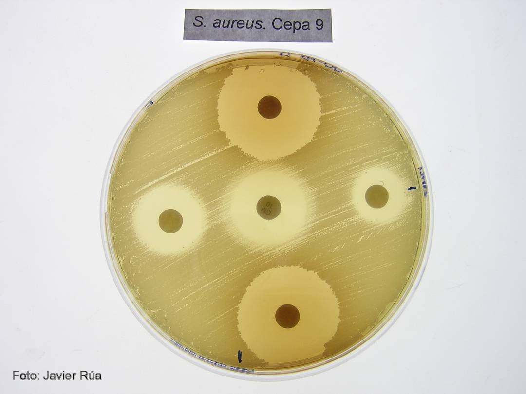
Cultivo de 'Staphylococcus aureus' tratado con los fenoles BHA (derecha) e hidroquinona (arriba).

CSIC/DICYT Un trabajo con participación del Consejo Superior de Investigaciones Científicas (CSIC) ha asignado a una enzima presente en todos los organismos vivos una nueva función en la transferencia de genes virulentos entre bacterias, un proceso que acaba provocando una infección. Los resultados, que aparecen publicados en el último número de Molecular Cell, y que han empleado como modelo la bacteria Staphylococcus aureus, la más frecuente en las infecciones adquiridas en hospitales, establecen el mecanismo de actuación de estas moléculas.

Las bacterias son capaces de transferir material genético entre sí mediante mecanismos de transferencia horizontal de genes. Cuando estos genes son virulentos, las bacterias que los reciben adquieren la capacidad de provocar enfermedades. “Algunos de los genes que codifican para toxinas y otros factores de virulencia están presentes en unas regiones denominadas islas de patogenicidad. Estas islas se transfieren de unas bacterias a otras utilizando virus que infectan bacterias, los llamados bacteriófagos”, explica el investigador del CSIC José Rafael Penadés, que trabaja en el Instituto de Biomedicina de Valencia.

El equipo de investigadores, formado también por científicos del Centro de Investigación y Tecnología Animal y la Universidad CEU Cardenal Herrera, en Valencia, han descubierto que las enzimas dUTPasas son capaces de despertar a las islas de patogenicidad para que detecten que la bacteria está siendo atacada por un virus. Antes de que la bacteria muera infectada, las islas inician su replicación y se transfieren a otras bacterias inocuas, a las que convierten en virulentas.

“El proceso evolutivo ha hecho que las islas detecten que un virus está infectando a las bacterias, lo que producirá su muerte, y utilicen la presencia del bacteriófago para activarse e iniciar su ciclo. Esto ocurre porque algunas proteínas del fago se unen a un represor que bloquea la isla”, explica el investigador del CSIC.

Proteínas G protooncogénicas
Los resultados confirman que las dUTPasas son moléculas señalizadoras que emplean un mecanismo similar al descrito para una familia de proteínas presentes en células eucariotas: las proteínas G protooncogénicas. “Las dUTPasas son activas como señalizadoras cuando se unen a un nucleótido dUTP. Es entonces cuando cambian su conformación y, una vez cumplida su función, degradan el nucleótido y pasan a estar apagadas. Este mecanismo de encendido y apagado es el mismo que el empleado por los protooncogenes”, destaca Penadés.

Según el investigador del CSIC Alberto Marina, el estudio sugiere por primera vez que las dUTPasas cumplen una función señalizadora no sólo en la mayoría de los virus, sino además en organismos vivos complejos como los eucariotas superiores. “Nuestros resultados aportan una visión completamente nueva del mecanismo de actuación de estas enzimas, que depende de una serie de características presentes en las enzimas de los bacteriófagos de Staphylococcus aureus y que están también presentes en otras muchas dUTPasas de un gran número de organismos vivos. Todo ello sugiere que el mecanismo descrito es universal”, concluye el investigador del CSIC.

Referencia bibliográfica 
María Ángeles Tormo-Más, Jorge Donderis, María García-Caballer, Aaron Alt, Ignacio Mir-Sanchís, Alberto Marina y José R. Penadés. Phage dUTPases Control Transfer of Virulence Genes by a Proto-Oncogenic G Protein-like Mechanism. Molecular Cell. DOI: 10.1016.