Mostrando entradas con la etiqueta Biomedicina. Mostrar todas las entradas

viernes, 5 de julio de 2013

Now You Can Build Google’s $1M Artificial Brain on the Cheap

ORIGINAL: Wired
By Daniela Hernandez
06.17.13

Andrew Ng. Photo: Ariel Zambelich/Wired
Andrew Ng wants to bring deep learning — an emerging computer science field that seeks to mimic the human brain with hardware and software — into the DIY era.

Last year at Google he built a computerized brain that worked as a cat detector. It used a roughly 1-billion-connection network trained on 1,000 computers to teach itself how to spot cat videos on YouTube
. While this worked well, Ng says, some researchers walked away thinking, “If I don’t have 1,000 computers, is there still any hope of my making progress on deep learning?The system cost roughly $1 million.

I was quite dismayed at this, particularly given that there are now a few other computer science research areas where a lot of the cutting-edge research is done only within giant companies,” he recalls. “Others simply don’t have the resources to do similar work.

On Monday, he’s publishing a paper that shows how to build the same type of system for just $20,000 using cheap, but powerful, graphics microprocessors, or GPUs. It’s a sort of DIY cookbook on how to build a low-cost neural network. He hasn’t yet decided whether the code for the model will be open sourced, but the new paper gives enough detail for people with enough coding brawn to build their own faux brains.

I hope that the ability to scale up using much less expensive hardware opens up another avenue for everyone around the world,” he says. “That’s the reason I’m excited — you can now build a 1-billion-connection model with $20,000 worth of hardware. It opens up the world for researchers to improve the performance of speech recognition and computer vision.

Down the line, this research on souped-up versions of neural networks running on GPUs could give rise to more powerful — and financially lucrative — GPU-based applications at large tech companies.

Built by companies such as Nvidia and AMD, GPUs power the graphics card on your PC or video game console. But about a decade ago, computer scientists started to realize that they were also really good for doing certain types of mathematical calculations.

GPUs are so incredibly powerful,” says David Anderson, a computer scientist at Berkeley. “Programs that previously ran on supercomputers, we’re now realizing we can rewrite to run on GPUs at a fraction of the price.” His team at Berkeley recently rejigged the volunteer-parallel-computing platform, BOINC, to be able to run on GPUs. BOINC helps scientists analyze astronomical and biomedical data.

Already universities and companies like Google, Shazam, Salesforce, Baidu and imgix are using these graphical chips to meet their ever-expanding computing needs to perform tasks as varied as voice recognition, quantum chemistry, and molecular modeling.

For this new research, Ng’s team also built a super-sized, 11-billion-connection version of the cat detector for roughly $100,000. He wants to build a high-performance computer that will allow researchers who don’t have the deep pockets of some of these companies and universities to do research on deep learning. It’s a bit like what Apple and Microsoft did for personal computing or what cheaper sequencing hardware did for genomics. Both democratized technologies that were inaccessible to many.

The Google Cat experiment ran on 1,000 computers with 16,000 CPUs. Ng’s group distributed their beefed-up, low-cost model, including the database of images on which it was trained, across 64 Nvidia GPUs on 16 computers and used special hardware to connect them in order to minimize the time required for these different modules to communicate with one another.

Ng is excited about this progress, but he admits there’s still work to be done. The new model is not that much smarter – or faster – than the original cat detector even though its neural net has a whopping 11 billion connections, or 10 times as many as its predecessor.

Plus, there are questions as to how easily Ng’s new model could be ported to other applications given that his group had to device specialized hardware and software to make it work.

The infrastructure seems to be particular to their specific unsupervised learning algorithm. The useful algorithms for training these networks, like the supervised algorithms that we use, and the one Google uses to train their photo-tagger are much harder to parallelize,” wrote NYU’s Yann LeCun, one of the pioneers of deep learning, in an email interview.

There are also issues with using GPUs that need to be worked out. Although Google, is trailblazing into the GPU space, most large technology companies have not invested heavily in graphics chips because using them in the cloud can be complicated. CPUs are better at sharing computing resources and can switch easily between several jobs, but the technology to do that on GPUs is not yet mature, says Ng. Plus running jobs on GPUs also requires specialized code.

[GPUs] are simply being co-opted by machine learning and AI researchers for a different purpose. So it’s not exactly a natural fit,” wrote Bruno Olshausen, a computational neuroscientist and the director of the Redwood Center for Theoretical Neuroscience at the University of California, Berkeley, in an email. “If we really want to make progress in building intelligent machines, then we will need to direct our efforts to build new types of hardware that are specifically adapted for neural computation.” Olshausen is currently working on this problem as part of an ongoing multi-university research project.

jueves, 20 de junio de 2013

Amazing moment deaf three-year-old boy hears for the first time

ORIGINAL: Minds
by Genelle Aldred.

Deaf three-year-old Grayson hears his father's voice for the first time after surgery in the US. Report 


viernes, 15 de marzo de 2013

Emory Integrated Genomics Core expands accessibility, service

ORIGINAL: Emory News
Woodruff Health Sciences Center
March 14, 2013

The staff of the Emory Integrated Genomics Core wants to make ambitious genomics research easier for the non-expert.
Do you want to read all 22,000 genes in someone's DNA? Do you want to know the identities of the bacteria in their intestines, or the DNA-bound proteins in their white blood cells? The staff of the Emory Integrated Genomics Core wants to make ambitious genomics research easier for you

Emory's two genomics core facilities have united under one roof. The Emory Integrated Genomics Core (EIGC) is a consolidation of the Cancer Genomics Shared Resource of the Winship Cancer Institute and the GRA Genomics Core of the School of Medicine. The goal of this effort is to create a top-tier genomics resource that is widely available to the Emory research community, integrating cutting-edge genomics technologies with downstream bioinformatics analysis

The EIGC's new scientific director is Michael Zwick, associate professor of human genetics. Zwick will oversee the operations, strategic planning, and expansion of the core"s services. The facility will also serve as the genomics platform for Winship's National Cancer Institute-designated Cancer Center Support Grant.

'Make genomics easier to use for the non-expert' 

"Talking about genomics is easy, but effectively using genomic technologies is more challenging," Zwick says. "We want to make these resources widely available to the Emory research community. Our mission is to make genomics easier to use for the non-expert.

Zwick came to Emory in 2005 after postdoctoral work at Johns Hopkins and Case Western and service with the U.S Navy's Biological Defense Research Directorate. His research team in Human Genetics uses next-generation sequencing technology to identify genes linked with pediatric disorders, which include autism spectrum disorders, congenital heart defects, and early-onset inflammatory bowel disease. He says that nucleic acid sequencing has become a basic commodity of biomedical research, "like water or electricity." 

He says some of the most popular uses of large-scale nucleic acid sequencing in Emory research currently are: 

  • RNA sequencing (RNA-Seq) of tumors — a scan of which genes are the most or least active within a tumor. 
  • Whole exome sequencing — reading the DNA sequence of all the protein-coding genes in a person's genome. Here is a recent example where a boy's metabolic disorder was diagnosed in 2012
  • Chromatin immunoprecipitation sequencing (ChIP-Seq) — a survey of where regulatory proteins are bound within the cell"s nucleus in a sample of tissue or cultured cells. 
Members of the Emory Integrated Genomics Core 
The integrated core is located on the 7th floor of the Woodruff Memorial Research Building. The new facility will combine basic genomic services and advanced next-generation sequencing technology with downstream bioinformatics analysis. It will include three divisions: 

  • a CLIA division enabling processing of patient samples for clinical trialsmanaged by Malania Wilson
  • a research division managed by R. Ben Isett; and 
  • a computational division providing analytical services, managed by Viren Patel
Key CLIA (Clinical Laboratory Improvement Amendments, federal regulations governing lab tests on humans) services include nucleic acid extractions and biobanking, genotyping services, along with access to next-generation sequencing. 

In-house research services include MiSeq, Taqman, Illumina and Affymetrix genotyping. Some large-scale sequencing research tasks will be outsourced to specialized sites such as HudsonAlpha Institute for Biotechnology, Zwick says. 

"In some cases, we can take advantage of economies of scale and use outsourcing to reduce costs," he says. "This means increased capacity, lower costs, a faster turn around time and less waiting for customers.

The computational division will provide data storage and computing services, and will be compatible with other computational biology applications such as proteomics and biostatistics. The core will use Emory High Performance Computer Cluster, a 768 CPU-core cluster to perform computational analysis. Several servers will be devoted to the Galaxy Project, an open, web-based platform for data intensive biomedical research. 

The core is jointly supported by the School of Medicine and the Winship Cancer Institute. Members of the Executive Committee overseeing the EIGC are Walter Curran, Paul Doetsch, Ray Dingledine, Carolyn Meltzer and Steve Warren.

viernes, 18 de enero de 2013

Descrita una nueva ruta de señalización clave en la transferencia de genes virulentos entre bacterias

ORIGINAL: DCYT

El trabajo, publicado en la revista Molecular Cell, asigna a una enzima una nueva función señalizadora
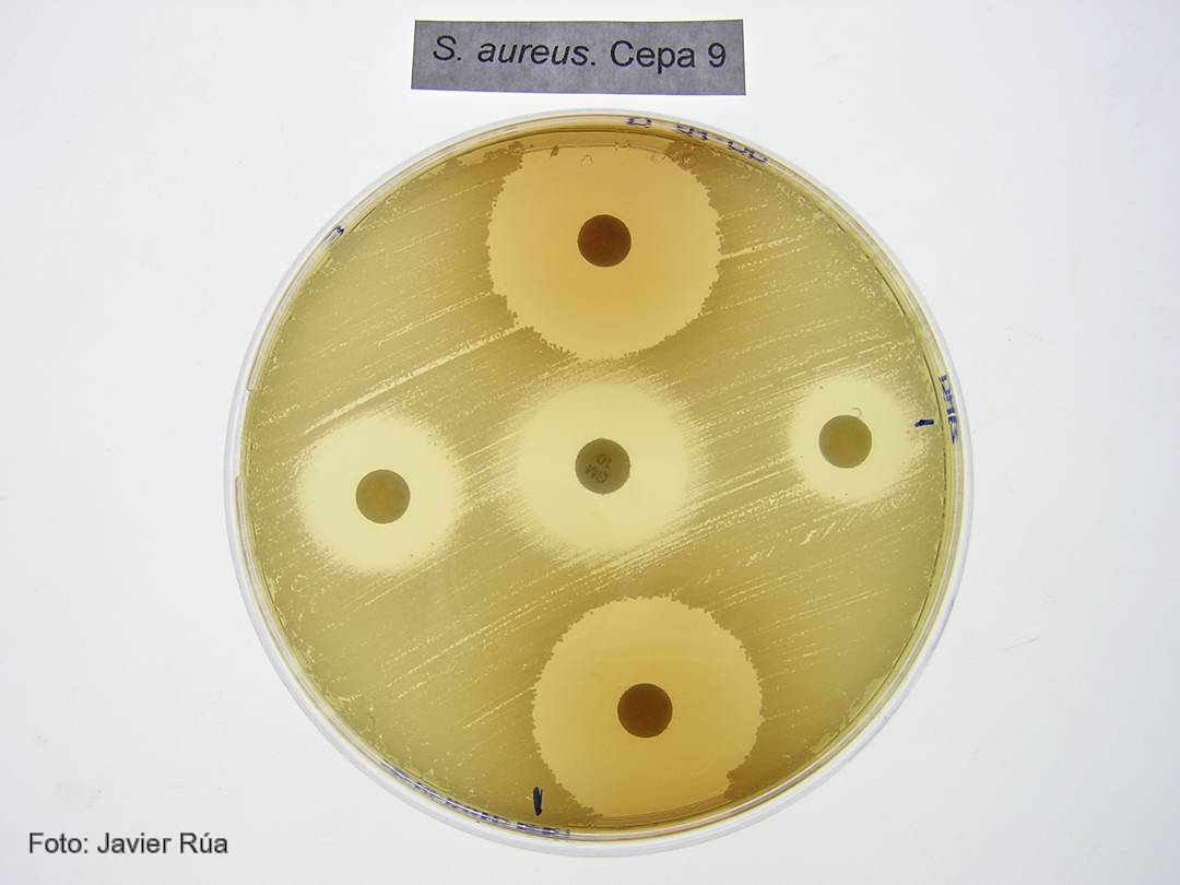
Cultivo de 'Staphylococcus aureus' tratado con los fenoles BHA (derecha) e hidroquinona (arriba).

CSIC/DICYT Un trabajo con participación del Consejo Superior de Investigaciones Científicas (CSIC) ha asignado a una enzima presente en todos los organismos vivos una nueva función en la transferencia de genes virulentos entre bacterias, un proceso que acaba provocando una infección. Los resultados, que aparecen publicados en el último número de Molecular Cell, y que han empleado como modelo la bacteria Staphylococcus aureus, la más frecuente en las infecciones adquiridas en hospitales, establecen el mecanismo de actuación de estas moléculas.

Las bacterias son capaces de transferir material genético entre sí mediante mecanismos de transferencia horizontal de genes. Cuando estos genes son virulentos, las bacterias que los reciben adquieren la capacidad de provocar enfermedades. “Algunos de los genes que codifican para toxinas y otros factores de virulencia están presentes en unas regiones denominadas islas de patogenicidad. Estas islas se transfieren de unas bacterias a otras utilizando virus que infectan bacterias, los llamados bacteriófagos”, explica el investigador del CSIC José Rafael Penadés, que trabaja en el Instituto de Biomedicina de Valencia.

El equipo de investigadores, formado también por científicos del Centro de Investigación y Tecnología Animal y la Universidad CEU Cardenal Herrera, en Valencia, han descubierto que las enzimas dUTPasas son capaces de despertar a las islas de patogenicidad para que detecten que la bacteria está siendo atacada por un virus. Antes de que la bacteria muera infectada, las islas inician su replicación y se transfieren a otras bacterias inocuas, a las que convierten en virulentas.

El proceso evolutivo ha hecho que las islas detecten que un virus está infectando a las bacterias, lo que producirá su muerte, y utilicen la presencia del bacteriófago para activarse e iniciar su ciclo. Esto ocurre porque algunas proteínas del fago se unen a un represor que bloquea la isla”, explica el investigador del CSIC.

Proteínas G protooncogénicas
Los resultados confirman que las dUTPasas son moléculas señalizadoras que emplean un mecanismo similar al descrito para una familia de proteínas presentes en células eucariotas: las proteínas G protooncogénicas. “Las dUTPasas son activas como señalizadoras cuando se unen a un nucleótido dUTP. Es entonces cuando cambian su conformación y, una vez cumplida su función, degradan el nucleótido y pasan a estar apagadas. Este mecanismo de encendido y apagado es el mismo que el empleado por los protooncogenes”, destaca Penadés.

Según el investigador del CSIC Alberto Marina, el estudio sugiere por primera vez que las dUTPasas cumplen una función señalizadora no sólo en la mayoría de los virus, sino además en organismos vivos complejos como los eucariotas superiores. “Nuestros resultados aportan una visión completamente nueva del mecanismo de actuación de estas enzimas, que depende de una serie de características presentes en las enzimas de los bacteriófagos de Staphylococcus aureus y que están también presentes en otras muchas dUTPasas de un gran número de organismos vivos. Todo ello sugiere que el mecanismo descrito es universal”, concluye el investigador del CSIC.

Referencia bibliográfica 
María Ángeles Tormo-Más, Jorge Donderis, María García-Caballer, Aaron Alt, Ignacio Mir-Sanchís, Alberto Marina y José R. Penadés. Phage dUTPases Control Transfer of Virulence Genes by a Proto-Oncogenic G Protein-like Mechanism. Molecular Cell. DOI: 10.1016.

lunes, 17 de diciembre de 2012

Woman Gains Full Mind Control Of Robotic Arm, Just Like in Star Wars

ORIGINAL: Gizmodo
By Jesus Diaz
DEC 17, 2012


This is truly extraordinary: Jan Scheuermann, a 52-year-old quadriplegic woman, has gained full control of a robotic arm. Not just simple commands, but truly complete control with "skill and speed almost similar to that of an able-bodied person."

According to the study—led by the University of Pittsburgh's professor of neurobiology Andrew Schwartzshe achieved this incredible feat after only 13 weeks of training:

The participant was able to move the prosthetic limb freely in the three-dimensional workspace on the second day of training. After 13 weeks, robust seven-dimensional movements were performed routinely. Mean success rate on target-based reaching tasks was 91·6% (SD 4·4) versus median chance level 6·2% (95% CI 2·0-15·3). Improvements were seen in completion time (decreased from a mean of 148 s [SD 60] to 112 s [6]) and path efficiency (increased from 0·30 [0·04] to 0·38 [0·02]). The participant was also able to use the prosthetic limb to do skilful and coordinated reach and grasp movements that resulted in clinically significant gains in tests of upper limb function. No adverse events were reported.

Translation: Jan now has a robotic arm that she can control just like you can control your own arm.

To achieve this, Schwartz's team implanted two 96-channel intracortical microelectrodes in Jan's motor cortex, the part of the brain responsible for controlling the movement of our limbs. The results, according to the scientists, were uncanny. They were surprised by how fast Jan took control of the robotic arm.

Schwartz says that this is "way better than anything that's been demonstrated before." So good that he believes it will change things forever:

I think it really is convincing evidence that this technology is going to be therapeutic for spinal cord injured people. They are doing tasks already that would be beneficial in their daily lives and I think that's fairly conclusive at this point.

It seems that a future in which we can replace a limb with a robotic version, just like Luke Skywalker got a new hand in Star Wars, is nearer than we previously thought. [The Lancet via BBC and El Mundo]

jueves, 11 de octubre de 2012

Light power for nanobiodevices


Scientists in China have created a laser-driven photovoltaic cell that can produce electrical power for nanobiodevices implanted beneath the skin.

Wireless nanobiodevices, such as nanorobots and cardiac pacemakers, are currently limited in their applications by their requirement for power. Nanogenerators that convert mechanical energy into electrical power have been investigated, but the output power is too low for many medical nanobiodevices, and biofuel cells that use chemical energy to provide power are severely limited by the in vivo environment of the devices.

Photovoltaic cells as a power source have previously been limited by low light conversion efficiencies and their lack of biocompatibility. However, Zhigang Chen of Donghua University, Shanghai, and colleagues have improved the conversion efficiency of their dye-sensitised solar cells such that, even through layers of skin, the laser light produces enough power for such devices.

The team improved the cells' efficiency and biocompatibility by changing two of the cells' components - the rare earth nanophosphor (NaYF4:Yb,Er - the part responsible for converting low energy light to high energy light, known as up-conversion) and the electrolyte. "We improved the nanophosphors' up-converting luminescence properties in a one-step synthesis," says Zhigang. They did this by increasing the amount of ligands on the nanophosphor's surface, which decreased surface defects, and by increasing the reaction time, which led to the formation of nanorods. Both actions improved the luminescence efficiency. They found that the cell was almost twice as efficient as previous nanophosphors formed in two steps.

Many components in photovoltaic cells are not biocompatible. One in particular is the organic liquid electrolyte because it can leak and evaporate. The liquid electrolyte can be replaced by a solid electrolyte, but at a cost of reduction in efficiency. Instead, Zhigang replaced the liquid electrolyte with a succinonitrile-based gel electrolyte. The gel is stable up to 80°C and for long periods of time and enhances the device's biocompatibility.

To test the cell under skin, the team covered the cell in a layer of chicken skin and used a laser intensity that is safe for human exposure (720 mW cm-2). They found that the new cell outputs a maximum power of 22.2µW, "which is efficient enough to power many kinds of in vivo devices", says Zhigang.

Richard Brutchey, an expert in inorganic nanomaterials for photovoltaic applications at the University of Southern California, US, says that the work is "a good step forward toward developing implantable bioelectronics, however, an external power source is still required (ie light)". He says that the next step would be to increase the power output to compete with the peak power of implantable fuel cells and to make the device fully biocompatible.

Source: RSC
Top image: Cosmos Magasine

martes, 24 de julio de 2012

Wyss Institute to Receive up to $37 Million from DARPA to Integrate Multiple Organ-on-Chip Systems to Mimic the Whole Human Body

Date: Jul 24, 2012

New instrument would accelerate assessment of drug safety and efficacy, and inform regulatory decision-making

Wyss Institute researchers and a multidisciplinary team of collaborators seek to build and link 10 human organs-on-chips to mimic whole body physiology. The system will incorporate the Institute's Human Lung-on-a-Chip (top) and Human Gut-on-a-Chip (bottom).


BOSTON -- The Wyss Institute for Biologically Inspired Engineering at Harvard University today announced that it has entered into a Cooperative Agreement worth up to $37 million with the Defense Advanced Research Projects Agency (DARPA) to develop an automated instrument that integrates 10 human organs-on-chips to study complex human physiology outside the body. This effort builds on the Institute's past breakthroughs in which Institute researchers engineered microchips that recapitulate the microarchitecture and functions of living organs, such as the lung, heart, and intestine. Each individual organ-on-chip is composed of a clear flexible polymer -about the size of a computer memory stick- that contains hollow microfluidic channels lined by living human cells. Because the microdevices are translucent, they provide a window into the inner-workings of human organs without having to invade a living body.

With this new DARPA funding, Institute researchers and a multidisciplinary team of collaborators seek to build 10 different human organs-on-chips, to link them together to more closely mimic whole body physiology, and to engineer an automated instrument that will control fluid flow and cell viability while permitting real-time analysis of complex biochemical functions. As an accurate alternative to traditional animal testing models that often fail to predict human responses, this instrumented "human-on-a-chip" will be used to rapidly assess responses to new drug candidates, providing critical information on their safety and efficacy.

Several U.S. agencies are working together to help safeguard Americans from deliberate chemical, biological, radiological, and nuclear threats, as well as from emerging infectious diseases, by drastically accelerating the drug development process. As an example, DARPA, the National Institutes of Health (NIH), and the U.S. Food and Drug Administration (FDA) are actively collaborating to develop cutting edge technologies to predict drug safety. The Wyss project was selected under the DARPA Defense Sciences Office (DSO) Microphysiological Systems Program and will be administered through a Cooperative Agreement by the Army Research Office (ARO) and DARPA.
The Lung-on-a-Chip, which makes use of living human cells, mimics the lung's tissue-tissue interface and breathing motions. Watch video to learn more...

This unique platform could help ensure that safe and effective therapeutics are identified sooner, and ineffective or toxic ones are rejected early in the development process. As a result, the quality and quantity of new drugs moving successfully through the pipeline and into the clinic may be increased, regulatory decision-making could be better informed, and patient outcomes could be improved.

Jesse Goodman, FDA Chief Scientist and Deputy Commissioner for Science and Public Health, commented that the automated human-on-chip instrument being developed "has the potential to be a better model for determining human adverse responses. FDA looks forward to working with the Wyss Institute in its development of this model that may ultimately be used in therapeutic development."


Wyss Founding Director, Donald Ingber, M.D., Ph.D., and Wyss Core Faculty member, Kevin Kit Parker, Ph.D., will co-lead this five-year project. Ingber is also the Judah Folkman Professor of Vascular Biology at Harvard Medical School and the Vascular Biology Program at Boston Children's Hospital, and Professor of Bioengineering at Harvard's School of Engineering and Applied Sciences (SEAS). Parker is the Tarr Family Professor of Bioengineering and Applied Physics at SEAS. The organ-on-chip program will also draw on the Institute's leading scientists and engineers, including Geraldine Hamilton, Ph.D., Anthony Bahinski, Ph.D., and Daniel Levner, Ph.D., who have extensive industrial experience in drug development, safety pharmacology, and systems engineering, to accelerate translation of this technology from the lab into the marketplace where it can best help the people who need it most. Other key collaborators participating in the project include John Wikswo, Ph.D., University Professor of Physics at Vanderbilt University, and Andrzej Przekwas, Ph.D., from CFD Research Corporation.

For more information, contact Twig Mowatt
Twig.mowatt@wyss.harvard.edu

###

About the Wyss Institute for Biologically Inspired Engineering at Harvard University 
The Wyss Institute for Biologically Inspired Engineering at Harvard University (http://wyss.harvard.edu) uses Nature's design principles to develop bioinspired materials and devices that will transform medicine and create a more sustainable world. Working as an alliance among Harvard's Schools of Medicine, Engineering, and Arts & Sciences, and in partnership with Beth Israel Deaconess Medical Center, Boston Children's Hospital, Brigham and Women's Hospital, , Dana Farber Cancer Institute, Massachusetts General Hospital, the University of Massachusetts Medical School, Spaulding Rehabilitation Hospital, Tufts University, and Boston University, the Institute crosses disciplinary and institutional barriers to engage in high-risk research that leads to transformative technological breakthroughs. By emulating Nature's principles for self-organizing and self-regulating, Wyss researchers are developing innovative new engineering solutions for healthcare, energy, architecture, robotics, and manufacturing. These technologies are translated into commercial products and therapies through collaborations with clinical investigators, corporate alliances, and new start-ups.

viernes, 1 de junio de 2012

Discovery Uses 'Fracture Putty' to Repair Broken Bone in Days

ORIGINAL: ScienceDaily

ScienceDaily (Feb. 7, 2012) — Broken bones in humans and animals are painful and often take months to heal. Studies conducted in part by University of Georgia Regenerative Bioscience Center researchers show promise to significantly shorten the healing time and revolutionize the course of fracture treatment.

A team of University of Georgia researchers created a new "fracture putty" to speed healing of bone fractures. From left to right are Steve Stice, Jennifer Mumaw, Erin Jordan and John Peroni. (Credit: Image courtesy of University of Georgia)
"Complex fractures are a major cause of amputation of limbs for U.S. military men and women," said Steve Stice, a Georgia Research Alliance Eminent Scholar, animal and dairy scientist in the UGA College of Agricultural and Environmental Sciences and director of the UGA Regenerative Bioscience Center.

"For many young soldiers, their mental health becomes a real issue when they are confined to a bed for three to six months after an injury," he said. "This discovery may allow them to be up and moving as fast as days afterward."

Stice is working with Dr. John Peroni to develop a fast bone healing process. "This process addresses both human and veterinary orthopedic needs," said Peroni, an associate professor of large animal surgery in the UGA College of Veterinary Medicine and a member of the RBC.

Peroni and Stice are leading a large animal research project funded by the U.S. Department of Defense. The project includes scientists and surgeons from the Baylor University College of Medicine, Rice University and the University of Texas, who conducted the early studies.

Engineering new bone

"Healing of critical-size defects is a major challenge to the orthopedic research community," Peroni said. "Large-bone defects must be stabilized and necessitate technologies that induce rapid bone formation in order to replace the missing tissue and allow the individual to return to rapid function. To date, no single material can suffice."

The group they lead is a multidiscipline and multi-institutional group actively working on bone tissue engineering.

"Our group has been working productively together on numerous projects through the last several years," Stice said, "So, a collegial relationship and successful collaborative working relationship is already established."

Between 2009 and 2011, the collaborations received a $1.4 million grant from the DOD for the use of stem cells in fracture healing to be tested in sheep.

"In our experiences with large animal models, following the guidelines established by our animal care and use committee," Stice said, "we have been successful in formulating a product that contains mesenchymal stem cells and allows them to survive in the environment of the fracture long enough to elicit the rapid formation of new bone."

This year, the group showed bone can be generated in sheep in less than four weeks. The speed in which bone is formed is one of the truly unique features of this study.

Fracture putty

To start the bone regeneration process, the RBC used adult stem cells that produce a protein involved in bone healing and generation. They then incorporated them into a gel, combining the healing properties into something Stice calls "fracture putty."

With Peroni's assistance, the Houston-based team used a stabilizing device and inserted putty into fractures in rats. Video of the healed animals at two weeks shows the rats running around and standing on their hind legs with no evidence of injury. The RBC researchers are testing the material in pigs and sheep, too.

"The small-animal work has progressed, and we are making good progress in large animals," he said.

More work is needed to get to human medical trials, but the threat of losing federal funding for biomedical work through the DOD means they will have to find new ways to fund the project.

Next steps

"The next step is to show that we can rapidly and consistently heal fractures in a large animal," Peroni said, "then to convert it to clinical cases in the UGA [College of Veterinary Medicine] clinics where clinicians treat animals with complex fractures all the time."

Once they have something that works for animals, it will be passed over to the DOD for human use.

Peroni, who is chairman of the North American Veterinary Regenerative Medicine Association, is hopeful this material will be promoted to the veterinary and human medical fields through the educational efforts of NAVRMA and the RBC.

However, the RBC isn't the only group working on a faster fix for broken bones.

"Our approach is biological with the putty," Stice said. "Other groups are looking at polymers and engineering approaches like implants and replacements which may eventually be combined with our approach. We are looking at other applications, too, using this gel, or putty, to improve spinal fusion outcomes."

One of the best hopes for the fracture putty is in possible facial cranial replacements, an injury often seen on the battlefield.


The project ends in mid-2012. "By then we are to deliver the system to the DOD," Stice said.

viernes, 25 de mayo de 2012

Glowing Green Orbs & Pink Butterflies Revealed in Winning Bio-Art Images

ORIGINAL: LiveScience
Wynne Parry, LiveScience Senior Writer
22 May 2012

This micrograph shows cells called myoblasts attached to spherical microcarriers, which allow the growth of adult stem cells that have been isolated from skeletal muscle. The stem cells are shown in green. CREDIT: FASEB 2012 Bio-Art Winner - Douglas B. Cowan

A pink butterfly, fluorescent mountains and glowing green orbs surrounded by bubbles are some of the imagery that appears in the winning entries in a "bio-art" competition, which sought to highlight the most artistic portrayals of biomedical research.

This year, 10 images out of about 100 entries were honored in the first Bio-Art Competition, created by the Federation for American Societies for Experimental Biology (FASEB). The winning entries weren't ranked and all were generated by scientists as a byproduct of of biomedical research.

Art wasn't the purpose of research to create artificial stem cell factories, explore the biological basis for psychiatric disease or look at the production of new neurons in the adult brain. Even so, the winning entries included brightly colored and sometimes abstract images.

The winners include one image depicting a scaffold, which resembles the weave of a fabric, upon which cells can grow to form new tissue. Glowing green orbs surrounded by bubbles reveal systems intended to produce muscle stem cells; and images of species of electric fish are trailed by recordings of the discharges.

"Electric fish recognize other members of their own species using the species-specific waveforms of these heartbeatlike discharges," write the team lead by Matthew Arnegard of the Fred Hutchinson Cancer Research Center in Seattle who created the image. (Under the contest guidelines, the image and the statement accompanying it should be visually arresting and clearly communicate a cutting-edge concept in biomedical science.)
One of the winners of the Bio-Art competition. The image above shows foot processes of genetically labeled cells (red) covering walls of capillaries (green) in a mouse kidney. CREDIT: FASEB 2012 Bio-Art Winner - Ivica Grgic and colleagues
Another winning entry depicts what looks like a fluorescent three-dimensional map, but is actually genetically labeled cells covering the walls of capillaries in a mouse kidney. [See Photos of the Winning Bio-Art]

University of Iowa's Li-Hsien Lin captured an image that appears to be a pink butterfly, but is actually a rat spinal cord showing the distribution of different types of enzymes. Understanding how these enzymes work and interact in the nervous system, Lin writes, could lead to better treatments for cardiovascular diseases such as hypertension and heart failure.
This butterfly shaped figure is an image of a rat spinal cord showing the distribution of three types of glutamate and nitric oxide synthesizing enzymes. CREDIT: FASEB 2012 Bio-Art Winner, Li-Hsien Lin
Other honorees included: abstract art created from tissue from a colon biopsy stained for a particular receptor; converging fibers that form the optic nerve in a mouse retina and their attendant immune cells; a 3D image of the limb from a transgenic, embryonic mouse, with bright colors differentiating the muscles, tendons, bones and nerves; and the growth of new neurons in the adult brain.

FASEB is a coalition of biomedical research associations in the United States. For this competition, the organization sought original, laboratory-based images produced by current or former National Institutes of Health-funded investigators, contractors, trainees or members of FASEB constituent societies, according to a news release.

You can follow LiveScience senior writer Wynne Parry on Twitter @Wynne_Parry. Follow LiveScience for the latest in science news and discoveries on Twitter @livescience and on Facebook.