sábado, 30 de julio de 2016

Fantastic Fungi: The Startling Visual Diversity of Mushrooms Photographed by Steve Axford (& New Photos9

Marasmius haematocephalus
To think any one of these lifeforms exists in our galaxy, let alone on our planet, simply boggles the mind. Photographer Steve Axford lives and works in the Northern Rivers area of New South Wales in Australia where he spends his time documenting the living world around him, often traveling to remote locations to seek out rare animals, plants, and even people. But it’s his work tracking down some of the world’s strangest and brilliantly diverse mushrooms and other fungi that has resulted in an audience of online followers who stalk his work on Flickr and SmugMug to see what he’s captured next.

Axford shares via email that most of the mushrooms seen here were photographed around his home and are sub-tropical fungi, but many were also taken in Victoria and Tasmania and are classified as temperate fungi. The temperate fungi are well-known and documented, but the tropical species are much less known and some may have never been photographed before. Mushrooms like the Hairy Mycena and the blue leratiomyces have most likely never been found on the Australian mainland before, and have certainly never been photographed in an artistic way as you’re seeing here.

It was painfully difficult not to include more of Axford’s photography here, so I urge you to explore further. All photos courtesy the photographer. (via Awkward Situationist)

Panus fasciatus

Leratiomyces sp. / Found in Booyong Reserve, Booyong, NSW

Mycena chlorophos


Cyptotrama aspratum or Gold tuft

Schizophyllum commune

Hairy mycena

White Mycena

Mauve splitting waxcap

Marasmius sp. / Marasmius haematocephalus

panus lecomtei

Photo by Steve Axford
Photo by Steve Axford

Photo by Steve Axford

Photo by Steve Axford

Photo by Steve Axford

Photo by Steve Axford

Photo by Steve Axford

Photo by Steve Axford

Photo by Steve Axford

Photo by Steve Axford

Photo by Steve Axford

Photo by Steve Axford

Photo by Steve Axford

Photo by Steve Axford

Photo by Steve Axford

Photo by Steve Axford

Photo by Steve Axford

Photo by Steve Axford
ORIGINAL: Colossal

New Photos of Extremely Unusual Mushrooms and Other Fungi by Steve Axfordby Christopher Jobson on July 29, 2016

Smart bricks will transform how buildings work

ORIGINAL: UWE Bristol

Smart bricks capable of recycling wastewater and generating electricity from sunlight are being developed by a team of scientists from the University of the West of England (UWE Bristol). The bricks will be able to fit together and create 'bioreactor walls' which could then be incorporated in housing, public building and office spaces


The UWE Bristol team is working on the smart technologies that will be integrated into the bricks in this pan European 'Living Architecture' (LIAR) project led by Newcastle University. The LIAR project brings together living architecture, computing and engineering to find a new way to tackle global sustainability issues.

The smart living bricks will be made from bio-reactors filled with microbial cells and algae. Designed to self-adapt to changing environmental conditions the smart bricks will monitor and modify air in the building and recognise occupants.

Each brick will contain Microbial Fuel Cells (MFCs) containing a variety of micro-organisms specifically chosen to 

  • clean water, 
  • reclaim phosphate, 
  • generate electricity and 
  • facilitate the production of new detergents, 
as part of the same process.

The MFCs that will make up the living engine of the wall of smart bricks will be able to sense their surroundings and respond to them through a series of digitally coordinated mechanisms.

Professor Andrew Adamatzky, LIAR Project Director for UWE Bristol, is leading the UWE Bristol team, he said, “The technologies we are developing aim to transform the places where we live and work enabling us co-live with the building.

“A building made from bio-reactors will become a large-scale living organism that addresses all environmental and energy needs of the occupants. Walls in buildings comprised of smart bricks containing bioreactors will integrate massive-parallel computing processors where millions of living creatures sense the occupants in the building and the internal and external environmental conditions.

“Each smart brick is an electrical analogous computer. A building made of such bricks will be a massive-parallel computing processor.”

A photo-bioreactor is a device that can be programmed to utilize a variety of inputs such as 

  • grey water, 
  • microbial consortia (algae and bacteria), 
  • carbon dioxide from the atmosphere, and 
  • different types of nutrient to generate outputs.
These outputs include

  • 'polished' water, 
  • fertiliser, 
  • extractable products (recoverable phosphate), 
  • oxygen, 
  • next generation biodegradable detergents, 
  • electricity, 
  • recoverable biomass, 
  • bio-fluorescence and to a certain extent, 
  • heat.

Professor Ioannis Ieropoulos, Director of the Bristol Bioenergy Centre (BBiC), at the Bristol Robotics Laboratory at UWE Bristol, said, “Microbial Fuel Cells are energy transducers that exploit the metabolic activity of the constituent microbes to break down organic waste and generate electricity. This is a novel application for MFC modules to be made into actuating building blocks as part of wall structures. This will allow us to explore the possibility of treating household waste, generating useful levels of electricity, and have 'active programmable' walls within our living environments.

Rachel Armstrong, Professor of Experimental Architecture at Newcastle University, UK, who is co-ordinating the project, said, “The LIAR project is incredibly exciting – it is bringing together living architecture, computing and engineering to find a new way to tackle global issues, like sustainability.

The €3.2m LIAR (Living Architecture) project is co-ordinated by Newcastle University working with experts from the universities of


The LIAR project has received funding from the European Union's Horizon 2020 research and innovation programme under grant agreement No. 686585

sábado, 16 de julio de 2016

A lab founded by a tech billionaire just unveiled a major leap forward in cracking your brain's code

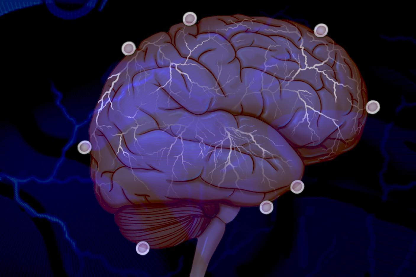
This is definitely not a scene from "A Clockwork Orange." Allen Brain Observatory
As the mice watched a computer screen, their glowing neurons pulsed through glass windows in their skulls.

Using a device called a two-photon microscope, researchers at the Allen Institute for Brain Science could peer through those windows and record, layer by layer, the workings of their little minds.

The result, announced July 13, is a real-time record of the visual cortex — a brain region shared in similar form across mammalian species — at work. The data set that emerged is so massive and complete that its creators have named it the Allen Brain Observatory.

Bred for the lab, the mice were genetically modified so that specific cells in their brains would fluoresce when they became active. Researchers had installed the brain-windows surgically, slicing away tiny chunks of the rodents' skulls and replacing them with five-millimeter skylights.

Sparkling neurons of the mouse visual cortex shone through the glass as images and short films flashed across the screen. Each point of light the researchers saw translated, with hours of careful processing, into data: 
  • Which cell lit up? 
  • Where in the brain? 
  • How long did it glow? 
  • What was the mouse doing at the time? 
  • What was on the screen?
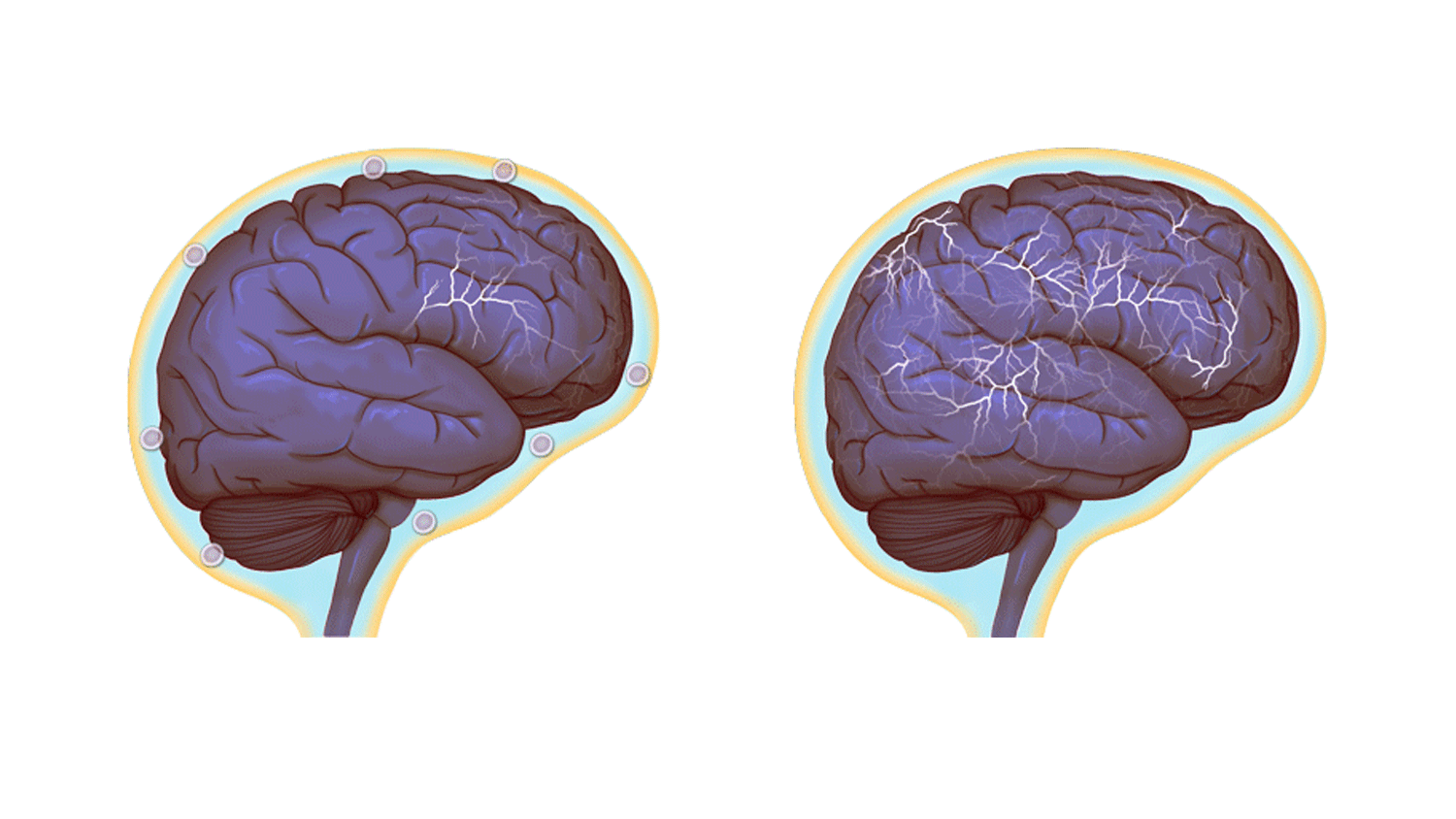
The researchers imaged the neurons in small groups, building a map of one microscopic layer before moving down to the next. When they were finished, the activities of 18,000 cells from several dozen mice were recorded in their database.

"This is the first data set where we're watching large populations of neurons' activity in real time, at the cellular level," said Saskia de Vries, a scientist who worked on the project, at the private research center launched by Microsoft co-founder Paul Allen.


The problem the Brain Observatory wants to solve is straightforward. Science still does not understand the brain's underlying code very well, and individual studies may turn up odd results that are difficult to interpret in the context of the whole brain.

A decade ago, for example, a widely-reported study appeared to find a single neuron in a human brain that always — and only — winked on when presented with images of Halle Berry. Few scientists suggested that this single cell actually stored the subject's whole knowledge of Berry's face. But without more context about what the cells around it were doing, a more complete explanation remained out of reach.

"When you're listening to a cell with an electrode, all you're hearing is [its activity level] spiking," said 
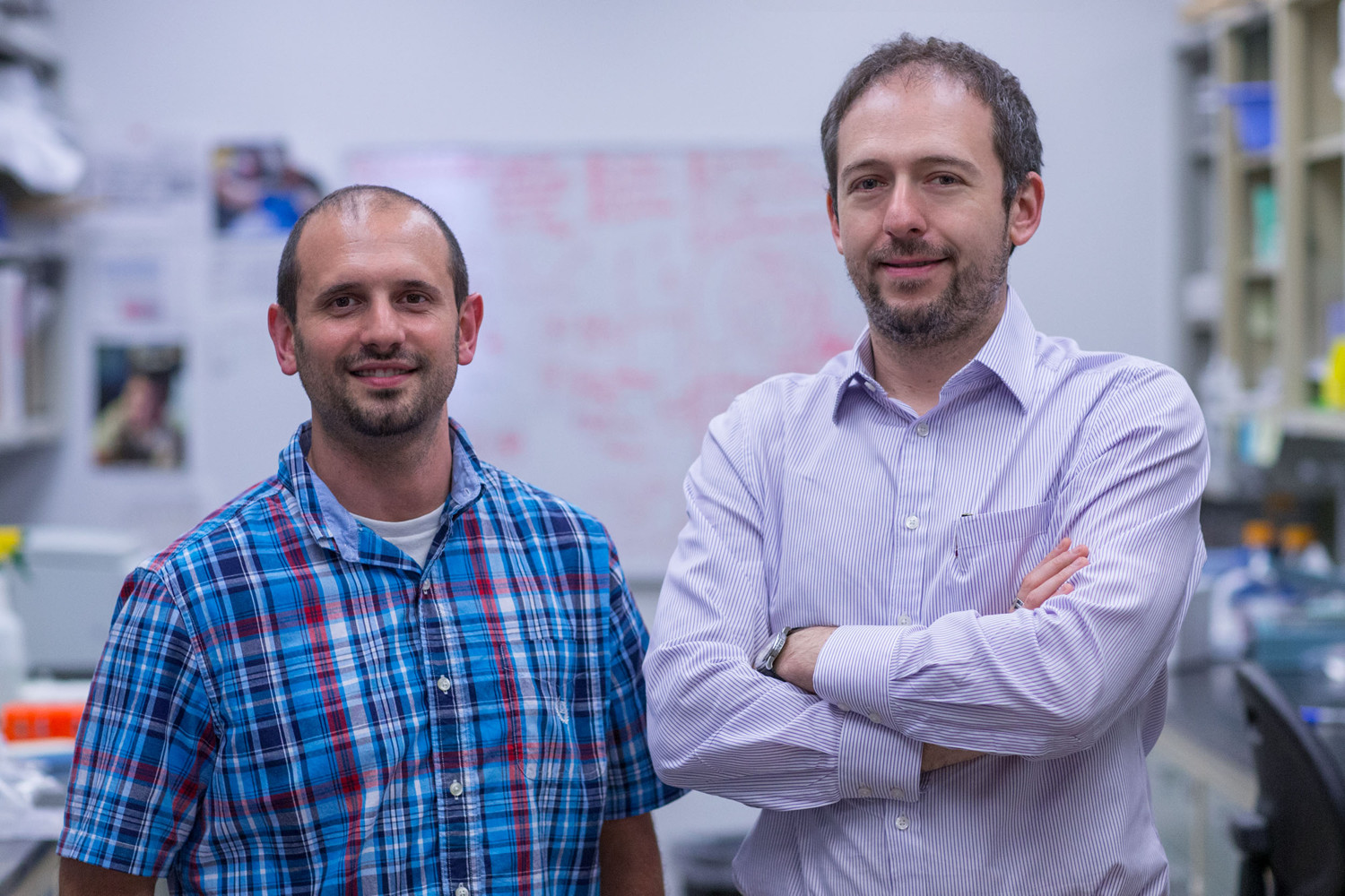
Shawn Olsen, another researcher on the project. "And you don't know where exactly that cell is, you don't know its precise location, you don't know its shape, you don't know who it connects to."

Imagine trying to assemble a complete understanding of a computer given only facts like under certain circumstances, clicking the mouse makes lights on the printer blink.

To get beyond that kind of feeling around in the dark, the Allen Institute has taken what Olsen calls an "industrial" approach to mapping out the brain's activity.

"Our goal is to systematically march through the different cortical layers, and the different cell types, and the different areas of the cortex to produce a systematic, mostly comprehensive survey of the activity," Olsen explained. "It doesn't just describe how one cell type is responding or one particular area, but characterizes as much as we can a complete population of cells that will allow us to draw inferences that you couldn't describe if you were just looking at one cell at a time."

In other words, this project makes its impact through the grinding power of time and effort.

A visualization of cells examined in the project. Allen Brain Observatory
Researchers showed the mice moving horizontal or vertical lines, light and dark dots on a surface, natural scenes, and even clips from Hollywood movies.

The more abstract displays target how the mind sees and interprets light and dark, lines, and motion, building on existing neuroscience. Researchers have known for decades that particular cells appear to correspond to particular kinds of motion or shape, or positions in the visual field. This research helps them place the activity of those cells in context.

One of the most obvious results was that the brain is noisy, messy, and confusing.

"Even though we showed the same image, we could get dramatically different responses from the same cell. On one trial it may have a strong response, on another it may have a weak response," Olsen said.

All that noise in their data is one of the things that differentiates it from a typical study, de Vries said.

"If you're inserting an electrode you're going to keep advancing until you find a cell that kind of responds the way you want it to," he said. "By doing a survey like this we're going to see a lot of cells that don't respond to the stimuli in the way that we think they should. We're realizing that the cartoon model that we have of the cortex isn't completely accurate."

Olsen said they suspect a lot of that noise emerges from whatever the mouse is thinking about or doing that has nothing to do with what's on screen. They recorded videos of the mice during data collection to help researchers combing their data learn more about those effects.

The best evidence for this suspicion? When they showed the mice more interesting visuals, like pictures of animals or clips from the film "Touch of Evil," the neurons behaved much more consistently.

"We would present each [clip] ten different times," de Vries said. "And we can see from trial to trial many cells at certain times almost always respond — reliable, repeatable, robust responses."

In other words, it appears the mice were paying attention.

Allen Brain Observatory
The Brain Observatory was turned loose on the internet Wednesday, with its data available for researchers and the public to comb through, explore, and maybe critique.

But the project isn't over.

In the next year-and-a-half, the researchers intend to add more types of cells and more regions of the visual cortex to their observatory. And their long-term ambitions are even grander.

"Ultimately," Olson said,"we want to understand how this visual information in the mouse's brain gets used to guide behavior and memory and cognition."

Right now, the mice just watch screens. But by training them to perform tasks based on what they see, he said they hope to crack the mysteries of memory, decision-making, and problem-solving. Another parallel observatory created using electrode arrays instead of light through windows will add new levels of richness to their data.

So the underlying code of mouse — and human — brains remains largely a mystery, but the map that we'll need to unlock it grows richer by the day.

ORIGINAL: Tech Insider
Jul. 13, 2016

Shocking New Role Found for the Immune System: Controlling Social Interactions

In a startling discovery that raises fundamental questions about human behavior, researchers at the University of Virginia School of Medicine have determined that the immune system directly affects – and even controls – creatures’ social behavior, such as their desire to interact with others.

So could immune system problems contribute to an inability to have normal social interactions? The answer appears to be yes, and that finding could have significant implications for neurological diseases such as autism-spectrum disorders and schizophrenia.

The brain and the adaptive immune system were thought to be isolated from each other, and any immune activity in the brain was perceived as sign of a pathology. And now, not only are we showing that they are closely interacting, but some of our behavior traits might have evolved because of our immune response to pathogens,” explained Jonathan Kipnis, chair of UVA’s Department of Neuroscience. “It’s crazy, but maybe we are just multicellular battlefields for two ancient forces: pathogens and the immune system. Part of our personality may actually be dictated by the immune system.


Evolutionary Forces at Work
It was only last year that Kipnis, the director of UVA’s Center for Brain Immunology and Glia, and his team discovered that meningeal vessels directly link the brain with the lymphatic system. That overturned decades of textbook teaching that the brain was “immune privileged,” lacking a direct connection to the immune system. The discovery opened the door for entirely new ways of thinking about how the brain and the immune system interact.
Normal brain activity, left, and a hyper-connected brain. (Images by Anita Impagliazzo, UVA Health System)
The follow-up finding is equally illuminating, shedding light on both the workings of the brain and on evolution itself. The relationship between people and pathogens, the researchers suggest, could have directly affected the development of our social behavior, allowing us to engage in the social interactions necessary for the survival of the species while developing ways for our immune systems to protect us from the diseases that accompany those interactions. Social behavior is, of course, in the interest of pathogens, as it allows them to spread.

The UVA researchers have shown that a specific immune molecule, interferon gamma, seems to be critical for social behavior and that a variety of creatures, such as flies, zebrafish, mice and rats, activate interferon gamma responses when they are social. Normally, this molecule is produced by the immune system in response to bacteria, viruses or parasites. Blocking the molecule in mice using genetic modification made regions of the brain hyperactive, causing the mice to become less social. Restoring the molecule restored the brain connectivity and behavior to normal. In a paper outlining their findings, the researchers note the immune molecule plays a “profound role in maintaining proper social function.

It’s extremely critical for an organism to be social for the survival of the species. It’s important for foraging, sexual reproduction, gathering, hunting,” said Anthony J. Filiano, Hartwell postdoctoral fellow in the Kipnis lab and lead author of the study. “So the hypothesis is that when organisms come together, you have a higher propensity to spread infection. So you need to be social, but [in doing so] you have a higher chance of spreading pathogens. The idea is that interferon gamma, in evolution, has been used as a more efficient way to both boost social behavior while boosting an anti-pathogen response.

Understanding the Implications
The researchers note that a malfunctioning immune system may be responsible for “social deficits in numerous neurological and psychiatric disorders.” But exactly what this might mean for autism and other specific conditions requires further investigation. It is unlikely that any one molecule will be responsible for disease or the key to a cure. The researchers believe that the causes are likely to be much more complex. But the discovery that the immune system – and possibly germs, by extension – can control our interactions raises many exciting avenues for scientists to explore, both in terms of battling neurological disorders and understanding human behavior.

Postdoctoral researcher Anthony J. Filiano, left, and Jonathan Kipnis, chairman of UVA’s Department of Neuroscience. (Photo by Sanjay Suchak, University Communications)
Immune molecules are actually defining how the brain is functioning. So, what is the overall impact of the immune system on our brain development and function?” Kipnis said. “I think the philosophical aspects of this work are very interesting, but it also has potentially very important clinical implications.

Findings Published
Kipnis and his team worked closely with UVA’s Department of Pharmacology and with Vladimir Litvak’s research group at the University of Massachusetts Medical School. Litvak’s team developed a computational approach to investigate the complex dialogue between immune signaling and brain function in health and disease.

Using this approach we predicted a role for interferon gamma, an important cytokine secreted by T lymphocytes, in promoting social brain functions,” Litvak said. “Our findings contribute to a deeper understanding of social dysfunction in neurological disorders, such as autism and schizophrenia, and may open new avenues for therapeutic approaches.

The findings have been published online by the prestigious journal Nature. The article was written by Filiano, Yang Xu, Nicholas J. Tustison, Rachel L. Marsh, Wendy Baker, Igor Smirnov, Christopher C. Overall, Sachin P. Gadani, Stephen D. Turner, Zhiping Weng, Sayeda Najamussahar Peerzade, Hao Chen, Kevin S. Lee, Michael M. Scott, Mark P. Beenhakker, Litvak and Kipnis.

This work was supported by the National Institutes of Health (grants No. AG034113, NS081026 and T32-AI007496) and the Hartwell Foundation.

ORIGINAL: News UVA
Josh Barneyjdb9a@virginia.edu
July 13, 2016 


Add caption
Add caption
The discovery was made possible by the work of Antoine Louveau, a postdoctoral fellow in Kipnis’ lab. The vessels were detected after Louveau developed a method to mount a mouse’s meninges — the membranes covering the brain — on a single slide so that they could be examined as a whole. After noticing vessel-like patterns in the distribution of immune cells on his slides, he tested for lymphatic vessels and there they were. The impossible existed. “Live imaging of these vessels was crucial to demonstrate their function, and it would not be possible without collaboration with Tajie Harris,” Kipnis noted. Harris is an assistant professor of neuroscience and a member of the Center for Brain Immunology and Glia. Kipnis also saluted the “phenomenal” surgical skills of Igor Smirnov, a research associate in the Kipnis lab whose work was critical to the imaging success of the study.

martes, 12 de julio de 2016

Monkeys In Brazil Entered The Stone Age 700 Years Ago

A CAPUCHIN WIELDING A HAMMER AGAINST A NUT RESTING ON AN ANVIL. UNIVERSITY OF OXFORD
Humanity is no longer the only species on Earth that has entered the Stone Age. It’s been known for some time now that various other primates use stone tools, including chimpanzees, capuchins, and macaques. Just recently, a study revealed that there was enough archaeological evidence to prove that macaques in Thailand have been crafting geological tools for at least half a century.

Now, it seems that capuchins have them beat. Tools in Brazil, undoubtedly made by capuchin hands, have been dated to be at least 700 years old. This means that just as the Renaissance was beginning in Italy, capuchins were crafting little chisels and hammers out of various stones in South America – although, in all likelihood, they had entered the Stone Age long before this.

As the study in the journal Current Biology notes, the field of primate archaeology is relatively nascent. Michael Haslam, the lead author of this research and the head of the Primate Archaeology (Primarch) project at the University of Oxford, is a pioneer in the field. He’s previously uncovered evidence of stone tool use in Thailand by macaques, but this new discovery is far more of a game-changer.

Until now, the only archaeological record of pre-modern, non-human animal tool use comes from a study of three chimpanzee sites in Cote d'Ivoire in Africa, where tools were dated to between 4,300 and 1,300 years old,” Haslam said in a statement. “Here, we have new evidence that suggests monkeys and other primates out of Africa were also using tools for hundreds, possibly thousands of years.

Brazilian capuchins entered the Stone Age at least 700 years ago. University of Oxford

Capuchins are indubitably clever monkeys. Researchers have long observed them using stones as hand-held hammers and anvils to break open hard, shelled food like cashews and seeds, while younglings watch their elders hammer away and learn from observation.

Their geological knowledge was found to be quite astute anvils were four times heavier than the hammers, and the hammers were four times heavier than the average stones nearby. The anvils tended to be made of layered, flat sandstones, whereas the hammers were forged from pointed, angular quartzite.

Whenever a capuchin is full of delicious nuts, it tends to leave its stone tools by a cache of discarded shells, which over time gets buried by sand and soil. After waiting for the capuchins to scuttle off, the researchers sauntered over to these sites and dug into the ground to see if they could find any older tools.

Using distinctive identifying marks on the tools made by the grinding, slamming, hammering action of long-gone capuchins, 69 tools were successfully excavated from a depth of up to 0.7 meters (2.3 feet), and radiocarbon dated using small pieces of charcoal. The oldest tools were 600 to 700 years of age, which means that 100 generations of capuchins – at least – have been using stone tools. 

They think it’s only a matter of time until older tools are found.
There is an even more tantalizing prospect to this discovery. The European invasion didn’t occur until the year 1500, so the capuchin Stone Age predates this by around 200 years. The indigenous populations of Brazil, therefore, may have come across capuchins breaking open cashew nuts native to this particular area.

It is possible,” Haslam notes, “that the that the first humans to arrive here learned about this unknown food through watching the monkeys and their primate cashew-processing industry.” So instead of monkeys or apes mimicking humans, in this case, it may have been the other way around.
Humans living in the Amazon may have educated themselves about certain stone tools from monkeys once upon a time. ANDRE DIB/Shutterstock

ORIGINAL: IFL Science

lunes, 11 de julio de 2016

Meet the First Artificial Animal

Scientists genetically engineered and 3-D-printed a biohybrid being, opening the door further for lifelike robots and artificial intelligence.

CREDIT: Getty Images
If you met this lab-created critter over your beach vacation, you'd swear you saw a baby ray. In fact, the tiny, flexible swimmer is the product of a team of diverse scientists. They have built the most successful artificial animal yet. This disruptive technology opens the door much wider for lifelike robots and artificial intelligence.

Like most disruption, it started with a simple idea. Kit Kevin Parker, PhD, a Harvard professor researching how to build a human heart, saw his daughter entranced by watching stingrays at the New England Aquarium in Boston. He wondered if he could engineer a muscle that could move in the same sinuous, undulating fashion. The quest for a material led to creating an artificial ray with a 3-D-printed rubber body at the School of Engineering and Applied Sciences at Harvard. Scientists from the University of Illinois at Urbana-Champaign, the University of Michigan, and Stanford University's Medical Center joined the team.

They reinforced the soft rubber body with a 3-D-printed gold skeleton so thin it functions like cartilage. Geneticists adapted rat heart cells so they could respond to light by contracting. Then, they were grown in a carefully arranged pattern on the rubber and around the gold skeleton.

The muscular circuitry is one of the most interesting parts of the research, and there's more about it in this video:


The birth of biohybrid beings
The new engineered animal responds to light so well scientists were able to guide it through an obstacle course 15 times its length using strong and weak light pulses.

The study authors write, "Our ray outperformed existing locomotive biohybrid systems in terms of speed, distance traveled, and durability (six days), demonstrating the potential of self-propelled, phototactically activated tissue-engineered robots."

What biohybrid mean for robots and artificial intelligence
Science of this type is fundamental for engineering special-purpose creations such as artificial worms that sniff out and eat cancer. Or bionic body parts for those who have suffered accidents or disease. Imagine having little swimmers in your system that rush to the site of a medical emergency such as a stroke. The promise of sensor-rich soft tissue frees robots to move more easily and yet not be cut off from needed input. Sensitized robot soft tissue could perform without the energy-sucking heaviness of metal or the artificial barrier of hard-plastic exoskeletons.

Thanks to disruptive, cross-disciplinary applied science like this, entrepreneurs in the next few years will be able to play on the border of what life is, what alive means, and what life can be. Expect to see companies use biohybrid beings to commercialize applications that solve some of the largest, and most lucrative, challenges we face today.

ORIGINAL: INC
BY LISA CALHOUN General partner, Valor Ventures@Lisa_Calhoun

sábado, 9 de julio de 2016

Watch This Amazing 3D Bioprinter Make Artificial Bones From Scratch

Image credit: Aether
If 3D printing is already impacting manufacturing today, what breakthroughs could bioprinting — or printing any mix of organic and inorganic materials — achieve tomorrow? In a recent video, a basic prototype of the Aether 1 bioprinter is shown printing two bones connected by a tendon using six materials that include synthetic bone, conductive ink, stem cells and graphene oxide.

While bioprinted organs are still a long way off — this video offers a glimpse into that future.


According to Aether, the printer works with a wide range of materials — organic, non-organic and both — and is flexible about where those materials come from (instead of requiring anything proprietary). It can include 10 different materials in one print (way more than your average bioprinter) and is compatible with other tools like laser cutters and CNC routers. The printer’s universal tool mounts can even be used with pencils and paintbrushes—or whatever else — for making designs on canvas or other materials.

Such a range of possibilities begins to shift the limitations from the hardware to the imagination of the user. What really stands out in this video is the final product — fabricated bones seeded with two kinds of stem cells hooked together with tendons, transistors, and conductive wires. And this is only a test. What else will researchers and makers come up with?

Of course, if it’s not already obvious, their creation isn’t ready for implantation in anyone, though it does show off the machine’s versatility in an eye-catching way. And beyond versatility, the real selling point may be its price. Aether hopes to offer their printer for around $9,000 — while other bioprinters can cost up to $200,000. 

For such bold claims — can they deliver? The announcement and video is only the first in a series which, according to Aether, will show off even greater functionality. Similar price reductions in 3D printers (that don’t bioprint) have met challenges commercially. But falling prices are a key trend to keep track of in any digitally driven technology like 3D printing.

And it should be noted, there are other companies wading into bioprinting too.

An early player, Organovo is currently working with researchers to use 3D printed tissues to test drug toxicity, and last year, they announced a partnership with cosmetics giant L’Oréal to test beauty products on3D printed skin samples (and hopefully lessen reliance on animal testing). Meanwhile, BioBots is making a bioprinter about the same size and cost as Aether’s but with more focused use cases, like 3D printing tissues for research purposes.

We’ll have to wait for more substantial news to see just how all this fits together. This summer will see the first wave of Aether machines donated to universities and researchers while presale beta units are distributed to customers. By fall 2016, they hope to launch retail sales.

What would you use it for?


ORIGINAL: Singularity Hub