Mostrando entradas con la etiqueta cáncer. Mostrar todas las entradas

lunes, 25 de agosto de 2014

Sorting cells with sound waves

Acoustic device that separates tumor cells from blood cells could help assess cancer’s spread.
Illustration: Christine Daniloff/MIT
Researchers from MIT, Pennsylvania State University, and Carnegie Mellon University have devised a new way to separate cells by exposing them to sound waves as they flow through a tiny channel. Their device, about the size of a dime, could be used to detect the extremely rare tumor cells that circulate in cancer patients’ blood, helping doctors predict whether a tumor is going to spread.
This microfluidic device uses sound waves to sorts cells as they flow through the channel, from left to right. Image courtesy of the researchers
Separating cells with sound offers a gentler alternative to existing cell-sorting technologies, which require tagging the cells with chemicals or exposing them to stronger mechanical forces that may damage them.

Acoustic pressure is very mild and much smaller in terms of forces and disturbance to the cell. This is a most gentle way to separate cells, and there’s no artificial labeling necessary,” says Ming Dao, a principal research scientist in MIT’s Department of Materials Science and Engineering and one of the senior authors of the paper, which appears this week in the Proceedings of the National Academy of Sciences.

Subra Suresh, president of Carnegie Mellon, the Vannevar Bush Professor of Engineering Emeritus, and a former dean of engineering at MIT, and Tony Jun Huang, a professor of engineering science and mechanics at Penn State, are also senior authors of the paper. Lead authors are MIT postdoc Xiaoyun Ding and Zhangli Peng, a former MIT postdoc who is now an assistant professor at the University of Notre Dame.

The researchers have filed for a patent on the device, the technology of which they have demonstrated can be used to separate rare circulating cancer cells from white blood cells.

To sort cells using sound waves, scientists have previously built microfluidic devices with two acoustic transducers, which produce sound waves on either side of a microchannel. When the two waves meet, they combine to form a standing wave (a wave that remains in constant position). This wave produces a pressure node, or line of low pressure, running parallel to the direction of cell flow. Cells that encounter this node are pushed to the side of the channel; the distance of cell movement depends on their size and other properties such as compressibility.

However, these existing devices are inefficient: Because there is only one pressure node, cells can be pushed aside only short distances.

The new device overcomes that obstacle by tilting the sound waves so they run across the microchannel at an angle — meaning that each cell encounters several pressure nodes as it flows through the channel. Each time it encounters a node, the pressure guides the cell a little further off center, making it easier to capture cells of different sizes by the time they reach the end of the channel.


ORIGINAL: MIT
Anne Trafton | MIT News Office 
August 25, 2014

domingo, 27 de abril de 2014

Coming Soon: New Smart Biosensor That Directs Cells To Kill Cancer. Surgery Glasses

These biosensors can further be customised to recognise factors of relevance to various patients' needs. 

Monday, April 21, 2014: Biologists at the Northwestern University's McCormick school of engineering and applied science have developed a ground breaking technology that could modify human cells to create therapeutics used in turn to selectively target and destroy tumour cells in the human body without disrupting healthy cells. The unique protein biosensor engineers cells to kill cancer by helping them effectively distinguish between healthy and cancerous cells.

Add caption
While sitting on the surface of a cell, the biosensor can be programmed to sense its immediate environment for specific factors following which it sends a signal to the engineered cell's nucleus. This triggers a gene expression programme within the cell. "Till date, there was no way to engineer cells in a manner that allowed them to sense key pieces of information about their environment, which could indicate whether the engineered cell is in healthy tissue or sitting next to a tumour," Joshua Leonard, an assistant professor at Northwestern University's McCormick school of engineering and applied science was quoted as saying. 

Moreover, the programme is activated only in the vicinity of tumour cells, thereby minimising any side effects. These biosensors can further be customised to recognise factors of relevance to various patients' needs. "In that way, you could programme a cell-based therapy to specify which cells it should kill," Leonard added.

Meanmwhile, a team of scientists at Washington University School of Medicine in St. Louis (WUSTL) and the University of Arizona (UA) have developed a new pair of hi-tech glasses that can help surgeons to detect cancer cells. These glasses will help surgeons to visualise cancer cells which will glow blue when viewed through these glasses during surgeries. Cancer cells are invisible in normal optics even if you are viewing through a high-powered magnifying device. This innovative technology incorporates a custom video, a head mounted display and then inject a blue dye into a patient. This will specifically bind to cancer cells and makes them glow. Doctors can then easily differentiate cancer cells from healthy cells and can make sure that no tumour cells are left over during surgery. It can detect and remove tumours as small as 1mm

Saurabh Singh, EFYTIMES News Network 

ORIGINAL: EFY Times

jueves, 27 de marzo de 2014

2014 Koch Institute Image Award Winners

Last fall, we featured The Koch Institute Image Award galleries in several Cell Picture Shows. This Show furthers the collaboration, as we showcase this year’s winning submissions. Both the Koch Institute Public Galleries and the Cell Picture Show share a similar ethos: recognition and dissemination of the extraordinary imagery produced through life science research. On March 4, 2014, these winning images were unveiled at MIT’s Koch Institute for Integrative Cancer Research in Cambridge, MA. 

We congratulate the 2014 Image Award Winners and are excited to continue to the collaboration between MIT and Cell Press. This collection of stunning images offers a window into the fascinating worlds opened to us by microscopy and other biomedical imaging techniques.

Biopolymer in Bloom
Julio D’Arcy, Erik Dreaden, and Paula Hammond
Hammond Laboratory
MIT Koch Institute
A New Environment for Studying Cell Growth. Measuring cancer cells’ real-time response to external influences can be challenging. Here, engineers have created biocompatible plastic structures onto which cells can adhere and develop as they would inside the body. The electrically conductive nature of the scaffolds allows researchers to measure the properties of the growing cells. By changing the environment or introducing new substances into the system, researchers can figure out which factors promote or discourage cell growth.
Image: This image, taken with a scanning electron microscope, shows the micro- and nano-scale structures of this device

The More the Messier
Kristin Knouse
Amon Laboratory
MIT Koch Institute

Understanding Complicated Cell Division. The mitotic spindle is an array of tracks that partitions chromosomes during cell division. Most normal cells form bipolar spindles, which segregate chromosomes equally into two daughter cells. However, many cancer cells form multipolar spindles, which cause chromosome mis-segregation and genomic instability.
Image: Like many cancer cells, liver cells also form multipolar spindles during cell division. Shown here is a liver cell with a multipolar spindle (green) pulling the chromosomes (blue) in many directions. Further research into cell division in the liver could indicate how this process is exploited or disrupted in cancer, revealing novel avenues for cancer therapy.

Target Practice
Omar F. Khan and Edmond W. Zaia
Langer and Anderson Laboratories
MIT Koch Institute

Improving Gene Therapy with Nanotechnology. How can we turn off the genes that promote the development of cancer? Using specially designed nanoparticles as genetic patches, engineers can deliver customized payloads to a cell’s gel-like cytoplasm, where most cellular activity occurs, and mitigate the effects of cancer-causing genes in the cell’s nucleus.
Image: This image shows nanoparticles (red) in the cytoplasm of cervical tumor cells (green). As researchers learn more about how cells respond to these therapies, they will continue to tweak the patches to determine the appropriate distribution of synthetic and genetic material to best target different types of cancer.

Blood, Heat, and Tumors
Alex Bagley, Jeff Wyckoff, and Sangeeta Bhatia
Bhatia Laboratory
MIT Koch Institute

Improving Drug Delivery with Gold Nanorods. Blood vessels are highways through the body. They can transport drugs to cancer cells, but finding the appropriate ramp to exit the vessel can be tricky.
Image: This image shows a network of blood vessels (green) and collagen (purple) infused with gold nanorods (yellow) inside of a living tumor. When researchers heat the particles with near-infrared light, the blood vessels become leaky, making it easier to deliver a therapeutic cargo to its final destination. Because blood vessels provide a universal transport system, such combination therapy has widespread implications for treatment, regardless of cancer type or specific drug needed

The Bad Seed
Mandar Deepak Muzumdar 
Jacks Laboratory
MIT Koch Institute

Modeling the Growth of a Tumor. Small changes have big effects. Although scientists know that certain gene mutations trigger tumor formation, the subsequent cellular events that drive cancer progression are not well understood. Cell-specific fluorescent marking allows researchers to track mutated cells over the entire course of cancer development.
Image: This image shows mutated (green) and nonmutated (red and yellow) cells in a pancreas. Over time, the green cells will multiply dramatically and form a solid tumor, while the others will not. Comparing properties and behaviors of the different cell types will set the stage for earlier diagnosis, better treatment, and even chemoprevention of deadly cancers.

Rainbow Connections
Zeynep Saygin
Kanwisher Laboratory
MIT Department of Brain & Cognitive Sciences

Mapping Neural Pathways in the Brain. The human brain is massively complex. Neuroimaging techniques such as MRI provide a noninvasive tool for studying its inner workings.
Image: This image shows pathways of nerve fibers through the brain in three dimensions: up/down (blue), front/back (green), and left/right (red). By comparing these maps of connectivity with maps of neural function, researchers can begin to predict how individual brains will respond to different stimuli. That will eventually help them to understand healthy brain development and will enable earlier diagnosis and interventions for conditions such as autism and dyslexia.

Silencing Echoes
Soheil Feizi, Steven Lee (Artist), Daniel Marbach, Muriel Medard, and Manolis Kellis Computational Biology Group
MIT Computer Science and Artificial Intelligence Laboratory

Cleaning Up Networks. Are all connections meaningful? This image visualizes a new algorithm (known as "network deconvolution") for determining important relationships in complex networks. Like a filter on a camera lens, it reveals which links (lines) between interconnected elements (points) are most essential. As the lens passes over each network area, indirect links disappear and direct links become visible. Already tested on large networks mapping gene regulation, protein folding, and academic co-authorship, network deconvolution can be used to identify key drivers of biological, social, and technological systems.

Ganglion Style
Alex Norton for EyeWire
Seung Laboratory
MIT Department of Brain and Cognitive Sciences and MIT Media Lab

Crowdsourcing Science through Online Games. It's all fun and games until somebody maps a neuron! Then it’s time to move on to the next one. The online game EyeWire challenges players, most of whom have no background in neuroscience, to create virtual 3D models of actual neurons using real laboratory data.
Image: The reconstruction seen here shows ganglion cells in the retina. By comparing this gamer-generated map to previously collected data about the neurons’ firing activity, neuroscientists can create a functional model of how vision works. With more than 100,000 players, EyeWire has already helped researchers to uncover how the eye helps us perceive moving stimuli.

Something Fishy
Annie Cavanagh and David McCarthy
School of Pharmacy
University College London

The Secret Lives of Zebrafish. Humans and fish have more in common than you might expect. Since the 1970s, a tropical freshwater minnow known as the zebrafish has been used to study the genetic and physiological development of living organisms. By mapping the zebrafish genome and studying irregularities in their development, researchers have been able to create robust models of how vertebrates develop and identify genetic conditions that lead to diseases such as cancer.
Image: This image shows a false-color scanning electron micrograph of a zebrafish embryo. It appears in the Koch Institute Public Galleries as part of a partnership between the Koch Institute and Wellcome Images.

Collateral Damage
Aprotim Mazumder, Jennifer A. Calvo, and Leona D. Samson
Samson Laboratory
MIT Koch Institute, Department of Biological Engineering, Department of Biology, and Center for Environmental Health Sciences

Investigating the Side Effects of Chemotherapeutics. How much is too much? When treating cancer, it is important to balance a drug’s effectiveness at killing tumor cells with its toxicity to healthy cells elsewhere in the body.
Image: This image of brain tissue shows cerebellar granule neurons (pink), which sustain significant damage when exposed to certain DNA-damaging therapeutics, and surrounding Purkinjee cells (orange), which do not. Researchers are studying these responses to determine the cell properties and repair mechanisms that make different cell types more or less vulnerable to chemotherapy.

ORIGINAL: Cell

miércoles, 26 de marzo de 2014

Biochip quickly tests results of cancer therapy


The inside structure of the biochip is comprised of 3 layers, each testing a key element of the therapy: drug, oxygen, and light. (Credit: Xia Lou/University of Michigan)


University of Michigan right Original Study ("A high-throughput photodynamic therapy screening platform with on-chip control of multiple microenvironmental factors")

A new type of lab-on-a-chip could make it easier to determine the effectiveness of a promising cancer treatment that combines photosensitive drugs, light, and oxygen.

The treatment, known as photodynamic therapy (PDT), uses a unique type of drug, called a photosensitizer, that generates high-energy oxygen when activated by light, in this case an LED laser.

The oxygen reaction destroys cells locally only in the immediate surrounding area, which in this case is the tumor, without damaging other healthy cells in the rest of the body. PDT may also prompt the patient’s immune system to attack the tumor, whereas without treatment it will likely ignore it.

PDT is a fairly complicated therapy because it requires light, oxygen, and the drug, all three of which need to be carefully controlled,” says Xia Lou, a postdoctoral fellow at the University of Michigan. “With chemotherapy, for example, you only control how much of the drug is used.”

The biochip can test the interaction of the drug, light, and oxygen simultaneously, generating results in a fraction of the time of current testing practices.

In cancer research doctors are always looking for better drugs,” adds Lou. “But there has always been a lack in the ability to efficiently test new drugs.

Researchers are also hoping to get more reliable test results than is the norm. We are providing more precise drug test conditions to insure that the results we are getting will more closely match the actual results from cancer treatment.
Test all three components There are two primary challenges in PDT today:
  • One is determining the best treatment plan for the patient, and 
  • the other is testing the efficacy of new and existing drugs.
For example, the location of a tumor in the body will determine the amount of light needed to excite the drug, and will impact the amount of drug that is administered. Also, when testing new drugs, it is again important to know the amount of light needed to excite the oxygen reaction, and the strength of the reaction.

This adds a high level of complexity compared to conventional drug testing that only tests one component, the drug itself.

Current PDT testing methods might take more than 24 hours to acquire just 10 data points.

With our device,” says Lou, “we can finish the testing in 1 hour and have 1,000 data points. The result is a very comprehensive understanding of how the 3 elements need to be manipulated for maximum effect in an individual patient.

The researchers reported the details of their findings in the journal Lab on a Chip. The NSF Engineering Research Center for Wireless Integrated Microsystems, Thermo Fisher Scientific, and the National Institute of Health supported the project.

Source: University of Michigan

ORIGINAL: Futurity
February 28, 2014 

lunes, 24 de marzo de 2014

Revolutionizing Genomics and Personalized Medicine with IBM Watson (Video of Project Launch Event AND DEMO)


The New York Genome Center and IBM Watson Group Announce Collaboration to Advance Genomic Medicine March 19, 2014


IBM NYGC Press Release
IBM Selected as First Technology Partner for Leading Genomic Research Institution; Project aims to Apply Advanced Analytics to Genomic Treatment Options for Brain Cancer Patients
New York, NY (March 19, 2014) – The New York Genome Center (NYGC) and IBM (NYSE: IBM) today announced an initiative to accelerate a new era of genomic medicine with the use of IBM’s Watson cognitive system. IBM and NYGC will test a unique Watson prototype designed specifically for genomic research as a tool to help oncologists deliver more personalized care to cancer patients.

NYGC and its medical partner institutions plan to initially evaluate Watson’s ability to help oncologists develop more personalized care to patients with glioblastoma, an aggressive and malignant brain cancer that kills more than 13,000 people in the U.S. each year. Despite groundbreaking discoveries into the genetic drivers of cancers like glioblastoma, few patients benefit from personalized treatment that is tailored to their individual cancer mutations. Clinicians lack the tools and time required to bring DNA-based treatment options to their patients and to do so, they must correlate data from genome sequencing to reams of medical journals, new studies and clinical records — at a time when medical information is doubling every five years.

This joint NYGC Watson initiative aims to speed up this complex process, identifying patterns in genome sequencing and medical data to unlock insights that will help clinicians bring the promise of genomic medicine to their patients. The combination of NYGC’s genomic and clinical expertise coupled with the power of IBM’s Watson system will enable further development and refinement of the Watson tool with the shared goal of helping medical professionals develop personalized cancer care.

The new cloud-based Watson system will be designed to analyze genetic data along with comprehensive biomedical literature and drug databases. Watson can continually ‘learn’ as it encounters new patient scenarios, and as more information becomes available through new medical research, journal articles and clinical studies. Given the depth and speed of Watson’s ability to review massive databases, the goal of the collaboration is to increase the number of patients who have access to care options tailored to their disease’s DNA.

Since the human genome was first mapped more than a decade ago, we’ve made tremendous progress in understanding the genetic drivers of disease. The real challenge before us is how to make sense of massive quantities of genetic data and translate that information into better treatments for patients,” said Robert Darnell, M.D., Ph.D., CEO, President and Scientific Director of the New York Genome Center. “Applying the cognitive computing power of Watson is going to revolutionize genomics and accelerate the opportunity to improve outcomes for patients with deadly diseases by providing personalized treatment.

First Watson Application in Genomic Research

Watson will complement rapid genome sequencing and is expected to dramatically reduce the time it takes to correlate an individual’s genetic mutations with reams of medical literature, study findings, and therapeutic indications that may be relevant. The intention is to provide comprehensive information to enable clinicians to consider a variety of treatment options that the clinician can tailor to their patient’s genetic mutations. It will also help NYGC scientists understand the data detailing gene sequence variations between normal and cancerous biopsies of brain tumors.

As genomic research progresses and information becomes more available, we aim to make the process of analysis much more practical and accessible through cloud-based, cognitive innovations like Watson,” said Dr. John E. Kelly III, Senior Vice President and Director of IBM Research. “With this knowledge, doctors will be able to attack cancer and other devastating diseases with treatments that are tailored to the patient’s and disease’s own DNA profiles. If successful, this will be a major transformation that will help improve the lives of millions of patients around the world.

The goal is to have the Watson genomics prototype assist clinicians in providing personalized genomic analytics information as part of a NYGC clinical research study. The solution has been under development for the past decade in IBM’s Computational Biology Center at IBM Research.

New York State’s Investment in Genomic Medicine

New York State is at the forefront of advancing medical science and commercialization. Governor Andrew M. Cuomo recently proposed $105 million to fund a partnership between NYGC and the University at Buffalo’s Center for Computational Research to advance genomics research. This investment to enhance the state’s genomic medicine capabilities, together with NYGC’s acquisition of Illumina’s state-of-the-art HiSeq X Ten whole human genome sequencing system, will accelerate the availability of valuable genomic information in New York.

New York State’s investment in cutting-edge innovative industries is creating jobs and growing the economy in Western New York and across our state,” said Governor Cuomo. “This collaboration between the New York Genome Center and IBM will help make the region a new hub for the growing bio-tech industry.

IBM is NYGC’s Founding Technology Member and will advance the organization’s goals of translating genomic research into clinical solutions for serious disease through the collaboration of medicine, science and technology. As biology increasingly becomes an information science, the promise of genomics is closer to reality with the help of data-driven analytics methods and more powerful computing systems. IBM and NYGC’s computational biology experts are renowned for accelerating life sciences discoveries using deep analytical approaches and next generation information technologies.

Learn more about this story at http://ibm.co/1cXTb6u.

To view a Flickr image gallery that illustrates today’s news please click here.

To join the social conversation on Twitter use the hashtag #NYGCWatson.

About the New York Genome Center
The New York Genome Center (NYGC) is an independent, nonprofit at the forefront of transforming biomedical research and clinical care with the mission of saving lives. As a consortium of renowned academic, medical and industry leaders across the globe, NYGC focuses on translating genomic research into clinical solutions for serious disease. Our member organizations and partners are united in this unprecedented collaboration of technology, science, and medicine. We harness the power of innovation and discoveries to improve people’s lives – ethically, equitably, and urgently. Member institutions include: Albert Einstein College of Medicine, American Museum of Natural History, Cold Spring Harbor Laboratory, Columbia University, Cornell University/Weill Cornell Medical College, Hospital for Special Surgery, The Jackson Laboratory, Memorial Sloan-Kettering Cancer Center, Icahn School of Medicine at Mount Sinai, New York-Presbyterian Hospital, The New York Stem Cell Foundation, New York University, North Shore-LIJ, The Rockefeller University, Roswell Park Cancer Institute and Stony Brook University. For more information, visit: www.nygenome.org.

Website: www.nygenome.org
Facebook: www.facebook.com/nygenome
Twitter: @nygenome

About IBM Watson

Named after IBM founder Thomas J. Watson, Watson was developed in IBM’s Research labs and is now being accelerated into market by the new Watson Group. Watson represents a new class of software, services and apps that think, improve by learning, and discover answers and insights to complex questions from massive amounts of Big Data. Watson’s ability to answer complex questions posed in natural language with speed, accuracy and confidence is transforming decision-making across a variety of industries, including health care, financial services and retail. IBM has advanced Watson from a game-playing innovation into a commercial technology. Using natural language processing and analytics, Watson processes information akin to how people think, representing a major shift in an organization’s ability to quickly analyze, understand and respond to Big Data. Now delivered from the cloud and able to power new consumer and enterprise services and apps, Watson is 24 times faster, smarter with a 2,400 percent improvement in performance, and 90 percent smaller – IBM has shrunk Watson from the size of a master bedroom to three stacked pizza boxes. IBM is investing $1 billion to introduce a new class of cognitive computing services, software and apps, and investing $100 million to spur innovation for software application providers to develop a new generation of Watson-powered solutions. Learn more about IBM Watson at www.ibmwatson.com. Learn more about IBM Research at www.research.ibm.com.
Learn more about IBM healthcare at ibm.com/smarterhealthcare.

Media Contacts:

NYGC
Lark-Marie Antón
(646) 977-7044
lanton@nygenome.org

IBM
Christine Vu
(914) 945-2755
vuch@us.ibm.com
Watson Chromosome analysis from a genome sequencing.

Watson Chromosome Pathways.

Watson Drill Down Results Support from a genome sequencing.
Pathways. Work underway using IBM Watson at the New York Genome Center will apply the use of IBM Watson cognitive technology to map genome sequencing results to retrieve insights from medical literature and drug information to find possible treatment options physicians can recommend to their patients. In this image, a cancer mutation is shown on a cell protein pathway from genome sequencing. (Photo credit: IBM)

IBM Watson Analyzes Human Genome (Infographics credit: IBM)

Original Press Realease (Download PDF)

sábado, 22 de marzo de 2014

Advancing brain cancer treatment through genomics

IBM and the New York Genome Center testing Watson prototype on glioblastoma

We have put Watson to work in any number of different ways and in any number of different industries. Healthcare, though, was its first real job. It’s gone to medical school, and even studied health insurance. And now Watson is working with the New York Genome Center to launch a pilot that tackles a new medical challenge – glioblastoma.

Dr. Robert Darnell, MD, PhD, President, CEO and Scientific Director of the New York Genome Center (left) and Dr. Ajay Royyuru, PhD, Director of the Computational Biology Center, IBM Research (right)

The most common kind of brain cancer, glioblastoma annually kills 13,000 people in the US alone. As a cancer of the brain, it’s difficult to take tissue samples, for one, so it can’t be examined like most other kinds of cancers. And it moves quickly. Diagnosis to death is on average only 12 months.

All cancers are a disease of the genome. It’s the genome itself that’s progressively changing from normal to abnormal when someone has cancer. When we can determine which genes start to “go bad,” we can better-determine what specific treatment would work to stop it. Therein lies the challenge: How can we better understand what is happening at a genetic level?

The key to glioblastoma’s genetic code is in the human genome. So while we know our cells’ biochemical pathways, it’s also an overwhelming amount of data – billions of DNA base sequences, plus millions of studies, medical documents and clinical records.

Different kinds of brain cancers manifest in different ways and progression rates, so finding these details about glioblastoma is a molecule-sized needle in the genome haystack.

That’s why my team – with decades of research experience in biology as a data science – and NYGC, with the expertise and resources of a dozen top hospitals and medical schools, are collaborating on a project with Watson in genomics. Our goals with this prototype and ensuing studies are to assist physicians with discovering personalized treatment for patients with glioblastoma.

Watson can read millions of pages of medical literature in seconds. By applying its natural language processing and analytics to the genome, it could find connections between what’s buried in journals about the interaction of certain genes, and where those genes are in the genome. And so, in the same way Watson evaluates and hypothesizes on other medical diagnosis based on electronic health records and a doctor’s evaluation (see a demo), it could evaluate and hypothesize about mutations in a cancer cell’s genome that caused the disease, not based on a wide demographic swath of those with similar characteristics, but for an individual based on their personal genome.

Connecting medical literature to the genome 


Today, we know and have detailed medical literature on the biochemical pathways our genes take. But we don’t know where in the genome these cancerous perturbations happen in that molecular network of interactions. So, we’re loading Watson with genome data from NYGC, along with medical literature to map out where these deviations happen. Watson will be able to see that, in the context of given cancer mutations in the genome, which pathways matter. And in the context of those interactions, suggest evidence of potential treatments.

IBM Watson and New York Genome Center. Video: IBM SocialMedia

This journey takes clinicians from trials, to validating what genomic knowledge improves treatment, to routine analysis that helps patients. Ultimately, we want to see our partners at NYGC and physicians upload genomic data into the Watson Genome on the cloud, where the system could quickly synthesize a personalized report of available evidence of treatment options.

ORIGINAL: IBM Research
By Dr. Ajay Royyuru, Director of IBM Research’s Computational Biology Center

viernes, 14 de marzo de 2014

The DNA of materials

Angela Belcher is a materials scientist who makes things with viruses. She is now using them to attack cancer

IT’S getting a little challenging,” says Angela Belcher. “I feel I am having to make choices now, which I never really wanted to.” But there are only so many hours in the day and she already combines multiple academic disciplines into a repertoire of research that spans an ambition to drive an electric car powered by a virus battery to building better touch-screens for digital devices and lately to giving surgeons new tools to detect and potentially treat minute traces of cancer.

There is more, but as eclectic as her work seems, it is united by a single intriguing idea. Evolution is a great problem solver and over millions of years has produced creatures of incredible breadth and complexity that can survive in the changing world around them. So why not copy the way nature innovates, speed it up and use it to help solve some of the problems researchers are presently working on. And that, in essence, is what Dr Belcher and her colleagues at the Massachusetts Institute of Technology (MIT) do. They rapidly evolve genetically engineered organisms to manufacture new materials and devices.

From the ocean
It began in the 1990s with an abalone shell, the sturdy home of a mollusc with a beautifully decorated mother-of-pearl interior. Dr Belcher was researching her PhD at the University of California, Santa Barbara, and was studying how abalones build their shells. The molluscs produce proteins which combine with ions of calcium and carbonate in seawater. This provides the material for them to make two types of crystals, which they assemble into layers to create an immensely strong composite structure.


Angela Belcher, 2013 winner of the Lemelson-MIT Prize Video: Lemelson-MIT

As she looked out of the window one day while wondering about this, her gaze drifted to a periodic table of elements stuck on the wall. If an abalone has within its DNA the ability to code for the proteins needed to gather the materials to construct a shell, would it be possible to tinker with the DNA sequences in other creatures to gather some of the elements on the periodic table? In particular, Dr Belcher asked herself, could creatures build semiconductors like those used in electronic circuits?

The idea might seem far fetched, but Dr Belcher thought that the reason it had not happened before could be that nature had never been given the opportunity to try. Sea creatures once had soft bodies but started to build shells and bones 500m years ago in a geological period known as the Cambrian. That could have been in response to increasing levels of minerals in the ocean. “It took them 50m years to get good at it,” says Dr Belcher. “Students in a research lab are not that patient. So the process would need to be speeded up.

The leap from evolutionary biology to semiconductors came naturally to Dr Belcher. She did her bachelor’s degree in creative studies and was allowed to combine different sciences. This highly multidisciplinary approach continued with her three PhD supervisors being experts in molecular biology, chemistry and physics. But it caused a bit of a problem with her first grant proposal in 1999, on becoming a professor at the University of Texas, Austin. The project was to find bacteriophages (a type of virus that infects bacteria, not people) that could be genetically engineered to bind to inorganic materials that they would not normally have an affinity for—in particular, semiconductors. If that was possible, then the viruses might be used as a template to grow and assemble electronic circuits, much like an abalone constructs its shell. It was, said one reviewer of her proposal, an “insane” idea.

Nevertheless, Dr Belcher stuck with her research and eventually was funded by the US Army. (Though interested in promoting basic science, the army likes to keep a look-out for potential breakthroughs in electronics which might benefit the increasing amount of technological kit it now uses.) In a paper published in 2000 in Nature, Dr Belcher demonstrated that her idea did indeed work. In 2002 she joined MIT and further scientific papers followed in collaboration with a number of her colleagues. Some of those papers explored making the components of a battery using viruses.

The technique begins by genetically modifying the somewhat basic DNA of a bacteriophage. This can be done to produce small but multiple changes in a billion or so viruses. All these variants, huge in number but individually tiny enough to be contained in a droplet of liquid, are then exposed to the material which the researchers are interested in manufacturing. Any viruses that show an affinity towards the material by attaching to it are gathered up. There may be only one or two in a billion, but when these candidates are used to infect a bacteria, millions of copies with identical DNA are made. The process can then be repeated to refine the characteristics. It is akin to high-speed natural selection. With further genetic modification and by changing the growth conditions, the selected viruses are used to bind with specific materials and assemble battery components.

Having found viruses happy to attach to nanowires of cobalt oxide, the researchers were able to produce a negative anode, one of the two main functioning parts of a battery. Making a positively charged cathode, the other important bit, was more difficult because for a battery to work well the cathode needs to be highly conductive. Nevertheless, Dr Belcher and her colleagues succeeded in getting some viruses to attach to carbon nanotubes, which are very good at conducting electrons. This resulted in a suitable cathode. It was then possible to assemble virus-made components into a small cell battery capable of lighting up an LED. This work was published in Science in 2009, to wide acclaim. Even Barack Obama was given a demonstration of the battery in action.
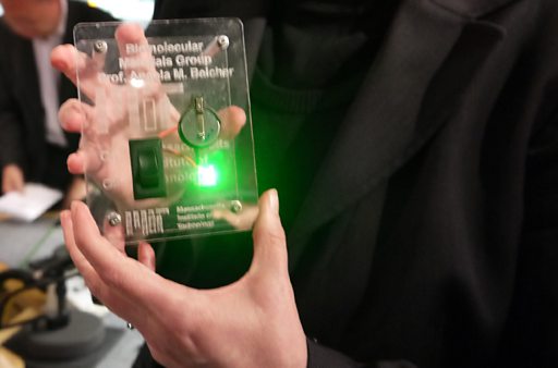
Angela Belcher with Virus Battery. Photo: BBC
Viral motoring
What Dr Belcher would really like to do, however, is to scale the process up so that one day it is possible to produce a virus battery powerful enough to run an electric car. With a paper in Nature Communications in November 2013, that day moved a step closer. The MIT team demonstrated how to use viruses to make a lightweight lithium-air battery, which the researchers think could have an energy density more than twice that of the best lithium-ion cells, the type which are currently used in most electric cars, as well as myriad portable electronic devices. But there is still a long way to go before such a battery could be put into commercial use.
I think it is very appealing that you can grow environmentally friendly battery parts

Many still find the idea strange. “I am sometimes told: ‘what are you doing using a virus to make a battery? These things don’t go together,’” says Dr Belcher. Yet, as she points out, biological processes tend to be non-toxic, low-energy ways to make things, and result in little waste, unlike many conventional manufacturing processes in which batteries are made with noxious materials in energy-intensive factories. “I think it is very appealing that you can grow environmentally friendly battery parts,” she adds.

When scaled up for commercial use, the viruses may take a back seat. This is the case with some of Dr Belcher’s projects which are already in the shops. In 2002, along with Evelyn Hu, a materials scientist then with the University of California, Santa Barbara, Dr Belcher co-founded Cambrios. This is a Silicon Valley company which makes transparent silver nanowires for use in touch screens. The nanowires are contained in inks which can be spread across the screen to provide a grid that produces an electrical signal when touched. The production process itself does not use viruses, but is based on techniques employed by them.


A number of companies are now using Cambrios’s technology in the screens of their smartphones, tablets and televisions. In December 2013 3M, a giant American maker of numerous innovative products, said it would employ Cambrios’s nanowire ink in a film that can be used to make giant, flexible touchscreens.

Image from Silura Technologies
Siluria Technologies, another Californian company, was co-founded by Dr Belcher in 2007. It uses viruses to produce templates for materials that work as catalysts to stimulate novel chemical reactions. Siluria is developing a catalytic process to turn methane, the principal component of natural gas, into petrol (gasoline) for cars. America now has an abundance of natural gas, thanks to the hydraulic fracturing, or “fracking”, of underground rocks. Siluria is building a demonstration plant near Houston to scale up the process and prove its commercial potential.

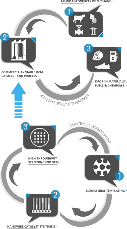
Siluria Process
How our process fits into industry, and how our catalyst materials are produced and continuously improved.


Siluria's scalable OCM process technology is designed to produce drop-in fuels and chemicals from a virtually endless supply of methane. Our catalyst materials make this process technology possible.
At the heart of Siluria's catalyst-development workflow are unique nano-structures inspired by nature. We have combined three highly innovative technologies to create our growing family of commercially viable OCM catalysts.

* Materials are 100% compatible with existing recycling infrastructure.

The same basic virus toolkit is now being used by Dr Belcher and her colleagues in the medical field. Dr Belcher is a member of two MIT faculties: materials science and engineering, and biological engineering. In 2010 she added a third by joining MIT’s Koch Institute for Integrative Cancer Research. She was nervous about this: “Working on cancer is so important. I didn’t want to take up space and not contribute and make a difference.” But she attended tutorials and became more confident by considering cancer as yet another material to work with. Although it is early days, the work looks promising.

The plan is to produce a medical probe which can be used to locate extremely small tumours. One way this is being tried is to get genetically engineered viruses to latch onto carbon nanotubes which glow under light from a laser. These viruses carrying the tubes would be injected into the body, and with light shining on the skin, be capable of glowing up to 10cm or so inside the body. The glow can be detected with a specialised camera.

To get these beacon viruses into the right places, the researchers engineer them to have a second affinity, one that makes them bind to certain kinds of cancer cells. They would then find any tumours, attach to them and glow.

The technique is still experimental and is being tried out in the laboratory on cellular models of ovarian cancer, which can be difficult for surgeons to detect when the tumours are tiny. There is a lot to do, but, says Dr Belcher, “I know we can find very small tumours and that should allow surgeons to remove them.

It also raises an obvious question: if the viruses can find tumours and light them up could they also carry with them some kind of lethal weapon? Not surprisingly, that possibility is being explored, with attempts to engineer cancer-seeking viruses that can carry both an imaging material and a chemotherapy agent.

Dr Belcher’s idea of evolving organisms to help build novel materials and new devices is starting to look like a technique with a large number of potential applications. Already she has to be able to switch from discussing with one group of her colleagues the complexities of binding materials to cancer cells, to debating with others how to clean up industrial wastewater with genetically engineered yeasts (her work is not confined to tinkering with the DNA of viruses). “There are so many areas we would like to be involved in, but we can’t do them all,” Dr Belcher laments. The trouble is, her idea born from an abalone shell seems bound to turn up even more problems which are waiting to be solved.

ORIGINAL: The Economist

martes, 11 de marzo de 2014

Scientists capture first super-res X-rays of living cells

While lower-energy "soft" X-rays can already image living cells, the higher-energy "hard" ones that can view objects as small as a few nanometers haven't been able to -- until now.

 
To avoid damaging the cells, the researchers exposed them for only 0.05 seconds at a time, but still managed to image nanometer-scale structures. Britta Weinhausen/University of Göttingen

Typically, to view super small objects like molecules, the samples must first be dipped in a chemical preservative bath of death that keeps all parts entirely locked in place and thus viewable via very sophisticated tech such as X-ray devices and electron microscopes. The problem, of course, is that those molecules don't behave the same in death as they do in life, so while our current views of life at the nanoscale level are extremely detailed, they're technically speaking views of death, or at the very least, life frozen.

Now scientists at the University of Göttingen in Germany say that, using a new approach with one of the world's most sophisticated X-ray machines, the Petra III, they've been able to view -- however briefly -- actual living cells in their natural environment.

Reporting in the journal Physical Review Letters, the researchers say they grew cancer cells from the adrenal cortex on a silicon nitrate substance that is nearly transparent to X-rays. They then fed those cells nutrients and pumped away their metabolic products so that they could keep the cells alive in as close to a natural environment as possible while still being viewable by the higher-energy (aka "hard" X-ray) Petra III. While lower-energy "soft" X-rays can already image living cells, the resolution isn't as good.

Related stories
3D X-ray provides window into heart health
With X-ray tech, scientists can peer inside cells
Researchers accelerate proton cancer treatment

To avoid killing the cells with the powerful X-ray beams, they exposed the sample in a series of frames that each lasted a mere 0.05 seconds. They then used this same nanodiffraction approach on chemically fixed cells for comparison and found that their cellular structures were noticeably different when viewed on a scale of 30 to 50 nanometers (that is, millionths of a millimeter).

While this initial test was performed using extremely brief and powerful blasts on petri dish cancer cells, the researchers say it offers evidence that we should be able to study living cells at super high resolution without first having to change their molecular behavior -- which could dramatically improve our understanding of life, including diseases and treatments, at the nanoscale level.

ORIGINAL: CNet

martes, 25 de febrero de 2014

Urine test detects not pregnancy but cancer

Paper strip uses nanoparticles to pick up evidence of tumors or blood clots in mice

TEST STRIP A new paper test can pick up signs of cancer and cardiovascular disease in the urine of mice injected with nanoparticles. The top reddish line indicates the test is working and the bottom line reveals the presence of disease. A. Warren

Peeing on a strip of paper could one day reveal signs of cancer and cardiovascular disease. An experimental test uses worm-shaped nanoparticles to spot evidence of tumors and blood clots in mouse urine, researchers report February 24 in the Proceedings of the National Academy of Sciences.

Unlike existing diagnostic tools, the test doesn’t rely on expensive equipment or detection of molecules made by sick people’s bodies. Instead the test looks for synthetic molecules injected into the bloodstream.

It’s brilliant work — a totally different paradigm for detecting disease,” says analytical chemist Andres Martinez of California Polytechnic State University in San Luis Obispo.

Note: To comment, Science News subscribing members must now establish a separate login relationship with Disqus. Click the Disqus icon below, enter your e-mail and click “forgot password” to reset your password. You may also log into Disqus using Facebook, Twitter or Google.

ORIGINAL: ScienceNews
by Meghan Rosen
February 24, 2014

miércoles, 12 de febrero de 2014

Ingenious. Fixing a body’s broken genes is becoming possible


IT SOUNDS like science fiction, and for years it seemed as though it was just that: fiction. But the idea of gene therapy—introducing copies of healthy genes into people who lack them, to treat disease—is at last looking as if it may become science fact.

The field got off to a bad start, with the widely reported death of an American liver patient in 1999. In 2003 some French children who were being treated with it for an immune-system problem called SCID developed leukaemia. Since then, though, things have improved. Indeed one procedure, for lipoprotein lipase deficiency (which causes high levels of blood fats, with all the problems those can bring), has been approved, in Europe, for clinical use.

The most recent success, announced last month in the Lancet, was of an experimental treatment for choroideremia, a type of blindness. This is caused by mutation of the gene for a protein called REP1. Without REP1, the eye’s light receptors degenerate. Robert MacLaren of Oxford University used a virus to deliver working versions of the REP1 gene to the most light-sensitive part of the retina. Five of the six participants in the trial duly experienced an improvement in their sensitivity to light. Two were so improved that they could read more letters than previously on a standard eye chart.

Dr MacLaren’s work complements that of Albert Maguire and Jean Bennett at the University of Pennsylvania, who use gene therapy to treat another eye disease, Leber’s congenital amaurosis. A defective version of a gene called RPE65 means that, in this condition, retinal cells are starved of vitamin A, which also causes blindness. Putting normal copies of RPE65 into the retina leads, as with REP1, to greater light sensitivity and—sometimes—clearer vision.

Drs MacLaren, Maguire and Bennett all use adeno-associated viruses (a type not known to cause illness, and which does not much provoke the immune system) to carry their genetic payloads to the target. Luigi Naldini of the San Raffaele Telethon Institute for Gene Therapy, in Milan, employs a rather scarier vector—one derived from HIV, the virus that causes AIDS—because its life cycle involves it integrating its genes into its host cells’ nuclei.

Last year Dr Naldini and his colleagues reported that, using their safely neutered version of HIV, they had inserted working copies of genes into blood stem cells which lack them, in order to treat metachromatic leukodystrophy (which damages nerves) and Wiskott-Aldrich syndrome (which harms the immune system and reduces blood’s ability to clot). In both cases—though in only a handful of patients, for the diseases are rare—Dr Naldini’s approach either prevented the disease or at least halted its progress.

The rarity of metachromatic leukodystrophy, Wiskott-Aldrich syndrome and many other diseases for which gene therapy might be appropriate means a lot of the applications of this approach are narrow. But a different one—constructing tailored genes and using them to guide the immune system—may have wider application, specifically against cancer.

Michel Sadelain, of the Memorial Sloan-Kettering Cancer Centre, in New York, is one of those at the forefront of a method that works this way. It employs chimeric antigen receptor (CAR) cells, which are engineered versions of T-cells, the part of the immune system that kills body cells, including tumorous ones, which have become threats.

Dr Sadelain’s trick is to take natural T-cells from patients (specifically, leukaemia and lymphoma patients) and add to them genes which turn those cells’ attention to the tumour in question, causing them to seek out its cells and destroy them. He then returns the modified cells to the patient, where they multiply and attack.

The CAR pool

The extra genes in CAR cells are derived in part from monoclonal-antibody genes. These have, in turn, been selected for their affinity to the target tumour. Because CAR cells multiply in the body this is, as Dr Sadelain puts it, like creating a living drug.

Last year Dr Sadelain’s team, and also another group led by Carl June at the University of Pennsylvania, published results showing the promise of CAR cells in treating people with acute lymphoblastic leukaemia. Dr Sadelain’s paper showed that they caused full remission in all five adult patients treated (though two subsequently died of complications, one set of which was unrelated to the treatment); Dr June’s, that they eradicated the cancer from two children. And, at a meeting of the American Society of Hematology held in December, both researchers reported further successes.

There is, moreover, one further technique that might bring gene therapy into the mainstream. Current approaches work by adding genes to affected cells. But it may be possible to modify those cells’ existing, broken genes, using a method called CRISPR-Cas9 editing, a process that takes advantage of a natural antiviral system which chops up genetic material.

CRISPR-Cas9 editing is specific to particular sequences of genetic letters, and can thus be tweaked to do a researcher’s bidding. In a recent edition of Cell, Sha Jiahao of Nanjing Medical University showed how to use it to execute the reverse of gene therapy—creating genetic problems, rather than solving them—in monkeys. His aim was to produce model organisms that might help understanding of diseases in human beings (though making such models out of monkeys is controversial). But the technique might eventually be employed to do running repairs on damaged DNA in people.

That, if it ever happens, is a long way off. In the meantime, the promise of gene therapy can be seen in the fact that it is attracting lawyers. The University of Pennsylvania has licensed its CAR technology to Novartis, a Swiss drugs firm. The pair of them are now fending off a lawsuit brought by competitors including Juno Therapeutics, the creation of three research centres of which Memorial Sloan-Kettering is one. For patients, that suggests gene therapy really is something worth fighting over.

From the print edition: Science and technology


ORIGINAL: The Economist

From the print edition

Feb 8th 2014