Mostrando entradas con la etiqueta CMU. Mostrar todas las entradas

lunes, 20 de agosto de 2018

CMU Engineers Find Innovative Way to Make a Low-Cost 3D Bioprinter

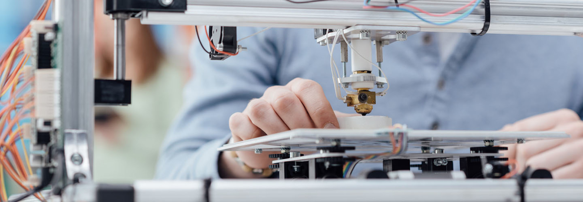
CYANO66/GETTY IMAGES

Starting with a MakerBot 3D printer, researchers tapped open-source hardware and software to build an affordable piece of tech that can print laboratory-grown cells on a large scale.


While 3D printers have already caused quite a buzz in the healthcare field — facilitating difficult surgeries and opening the door to low-cost prosthetics — the concept of bioprinting on a large scale has eluded the industry for the most part. But a recent breakthrough from Carnegie Mellon University’s College of Engineering could change all that.


Bioprinting, or printing laboratory-grown cells in order to form living structures, has the ability to profoundly transform healthcare.


The approach could revolutionize regenerative medicine, enabling the production of complex tissues and cartilage that would potentially support, repair or augment diseased and damaged areas of the body,Science Daily reports.

While researchers from the Massachusetts Institute of Technology and elsewhere have been digging into how to facilitate low-cost bioprinting, these options are often limited in scale or availability. The new open-source and low-cost solution from CMU, which makes use of a standard desktop 3D printer, could open the door to printing biomaterials, like artificial human tissue, and fluids on a larger scale, according to a new paper released by CMU.

Bioprinting has historically been limited in volume, so essentially the goal is to just scale up the process without sacrificing detail and quality of the print,” Kira Pusch, an author of the paper and a recent graduate of CMU’s Materials Science and Engineering undergraduate program, tells CMU’s news site. “What we’ve created is a large volume syringe pump extruder that works with almost any open source fused deposition modeling (FDM) printer. This means that it’s an inexpensive and relatively easy adaptation for people who use 3-D printers.

Open-Source Tools Lead to a ‘Democratizing’ Bioprinter
What makes the CMU bioprinting method unique is a technique the lab developed called Freeform Reversible Embedding of Suspended Hydrogels (FRESH) 3D bioprinting that is designed to specifically print “soft and living materials,” Adam Feinberg, another author of the paper and an associate professor of materials science and biomedical engineering at CMU, tells Robotics Tomorrow. The technique essentially prints the tissue in a gel that is later carefully melted away to ensure the cells remain viable.

Feinberg notes that the technique is capable of printing a wide range of cells “including collagen and other extracellular matrix proteins,” representing most tissue in the body.

Usually there’s a trade-off, because when the systems dispense smaller amounts of material, we have more control and can print small items with high resolution, but as systems get bigger, various challenges arise,” Feinberg, who is also a member of the Bioengineered Organs Initiative at Carnegie Mellon, tells CMU’s news site. “The [large-volume extruder (LVE)] 3-D bioprinter allows us to print much larger tissue scaffolds, at the scale of an entire human heart, with high quality.

The lab began its journey toward large-scale and low-cost bioprinting after it purchased a MakerBot 3D printer. Over the course of six years, researchers modified the printer using open-source hardware and software. In the spirit of that endeavor, the team has made its designs for the printer open source, hoping to further collaboration and discovery in the medical field.

Essentially, we’ve developed a bioprinter that you can build for under $500, that I would argue is at least on par with many that cost far more money,” Feinberg tells CMU’s news site. “Most 3-D bioprinters start between $10,000 and $20,000. This is significantly cheaper, and we provide very detailed instructional videos. It’s really about democratizing technology and trying to get it into more people’s hands.


Juliet is the senior web editor for StateTech and HealthTech magazines. In her six years as a journalist she has covered everything from aerospace to indie music reviews — but she is unfailingly partial to covering technology.

miércoles, 18 de octubre de 2017

Stunning AI Breakthrough Takes Us One Step Closer to the Singularity

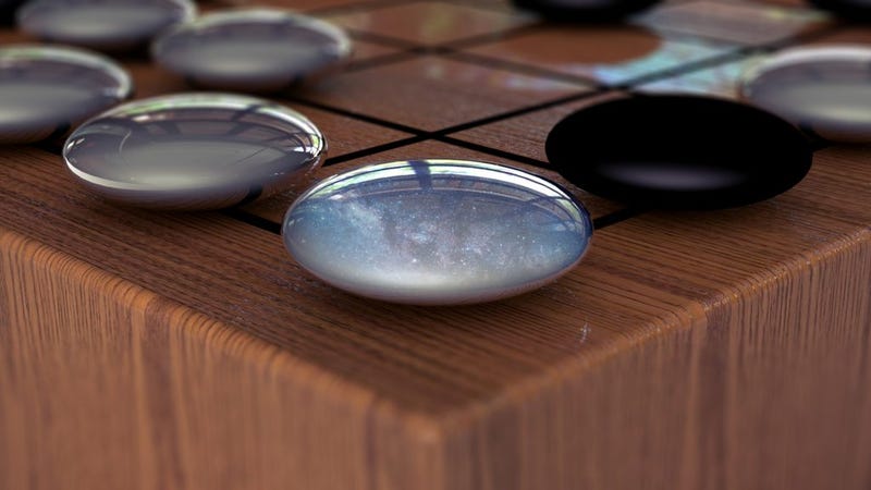
As a new Nature paper points out, “There are an astonishing 10 to the power of 170 possible board configurations in Go—more than the number of atoms in the known universe.” (Image: DeepMind)
Remember AlphaGo, the first artificial intelligence to defeat a grandmaster at Go?
Well, the program just got a major upgrade, and it can now teach itself how to dominate the game without any human intervention. But get this: In a tournament that pitted AI against AI, this juiced-up version, called AlphaGo Zero, defeated the regular AlphaGo by a whopping 100 games to 0, signifying a major advance in the field. Hear that? It’s the technological singularity inching ever closer.

A new paper published in Nature today describes how the artificially intelligent system that defeated Go grandmaster Lee Sedol in 2016 got its digital ass kicked by a new-and-improved version of itself. And it didn’t just lose by a little—it couldn’t even muster a single win after playing a hundred games. Incredibly, it took AlphaGo Zero (AGZ) just three days to train itself from scratch and acquire literally thousands of years of human Go knowledge simply by playing itself. The only input it had was what it does to the positions of the black and white pieces on the board.
  • In addition to devising completely new strategies
  • the new system is also considerably leaner and meaner than the original AlphaGo.
Lee Sedol getting crushed by AlphaGo in 2016. (Image: AP)
Now, every once in a while the field of AI experiences a “holy shit” moment, and this would appear to be one of those moments. Looking back, other “holy shit” moments include:
This latest achievement qualifies as a “holy shit” moment for a number of reasons.

First of all, the original AlphaGo had the benefit of learning from literally thousands of previously played Go games, including those played by human amateurs and professionals. AGZ, on the other hand, received no help from its human handlers, and had access to absolutely nothing aside from the rules of the game. Using “reinforcement learning,” AGZ played itself over and over again, “starting from random play, and without any supervision or use of human data,” according to the Google-owned DeepMind researchers in their study. This allowed the system to improve and refine its digital brain, known as a neural network, as it continually learned from experience. This basically means that AlphaGo Zero was its own teacher.

This technique is more powerful than previous versions of AlphaGo because it is no longer constrained by the limits of human knowledge,” notes the DeepMind team in a release. “Instead, it is able to learn tabula rasa [from a clean slate] from the strongest player in the world: AlphaGo itself.

When playing Go, the system considers the most probable next moves (a “policy network”), and then estimates the probability of winning based on those moves (its “value network”). AGZ requires about 0.4 seconds to make these two assessments. The original AlphaGo was equipped with a pair of neural networks to make similar evaluations, but for AGZ, the Deepmind developers merged the policy and value networks into one, allowing the system to learn more efficiently. What’s more, the new system is powered by four tensor processing units (TPUS)—specialized chips for neural network training. Old AlphaGo needed 48 TPUs.

After just three days of self-play training and a total of 4.9 million games played against itself, AGZ acquired the expertise needed to trounce AlphaGo (by comparison, the original AlphaGo had 30 million games for inspiration). After 40 days of self-training, AGZ defeated another, more sophisticated version of AlphaGo called AlphaGo “Master” that defeated the world’s best Go players and the world’s top ranked Go player, Ke Jie. Earlier this year, both the original AlphaGo and AlphaGo Master won a combined 60 games against top professionals. The rise of AGZ, it would now appear, has made these previous versions obsolete.

The time when humans can have a meaningful conversation with an AI has always seemed far off and the stuff of science fiction. But for Go players, that day is here.

This is a major achievement for AI, and the subfield of reinforcement learning in particular. By teaching itself, the system matched and exceeded human knowledge by an order of magnitude in just a few days, while also developing 
  • unconventional strategies and 
  • creative new moves
For Go players, the breakthrough is as sobering as it is exciting; they’re learning things from AI that they could have never learned on their own, or would have needed an inordinate amount of time to figure out.
[AlphaGo Zero’s] games against AlphaGo Master will surely contain gems, especially because its victories seem effortless,” wrote Andy Okun and Andrew Jackson, members of the American Go Association, in a Nature News and Views article. “At each stage of the game, it seems to gain a bit here and lose a bit there, but somehow it ends up slightly ahead, as if by magic... The time when humans can have a meaningful conversation with an AI has always seemed far off and the stuff of science fiction. But for Go players, that day is here.”
No doubt, AGZ represents a disruptive advance in the world of Go, but what about its potential impact on the rest of the world? According to Nick Hynes, a grad student at MIT’s Computer Science and Artificial Intelligence Laboratory (CSAIL), it’ll be a while before a specialized tool like this will have an impact on our daily lives.

So far, the algorithm described only works for problems where there are a countable number of actions you can take, so it would need modification before it could be used for continuous control problems like locomotion [for instance],” Hynes told Gizmodo. “Also, it requires that you have a really good model of the environment. In this case, it literally knows all of the rules. That would be as if you had a robot for which you could exactly predict the outcomes of actions—which is impossible for real, imperfect physical systems.

The nice part, he says, is that there are several other lines of AI research that address both of these issues (e.g. machine learning, evolutionary algorithms, etc.), so it’s really just a matter of integration. “The real key here is the technique,” says Hynes.

It’s like an alien civilization inventing its own mathematics which allows it to do things like time travel...Although we’re still far from ‘The Singularity,’ we’re definitely heading in that direction.
As expected—and desired—we’re moving farther away from the classic pattern of getting a bunch of human-labeled data and training a model to imitate it,” he said. “What we’re seeing here is a model free from human bias and presuppositions: It can learn whatever it determines is optimal, which may indeed be more nuanced that our own conceptions of the same. It’s like an alien civilization inventing its own mathematics which allows it to do things like time travel,” to which he added: “Although we’re still far from ‘The Singularity,’ we’re definitely heading in that direction.

Noam Brown, a Carnegie Mellon University computer scientist who helped to develop the first AI to defeat top humans in no-limit poker, says the DeepMind researchers have achieved an impressive result, and that it could lead to bigger, better things in AI.

While the original AlphaGo managed to defeat top humans, it did so partly by relying on expert human knowledge of the game and human training data,” Brown told Gizmodo. “That led to questions of whether the techniques could extend beyond Go. AlphaGo Zero achieves even better performance without using any expert human knowledge. It seems likely that the same approach could extend to all perfect-information games [such as chess and checkers]. This is a major step toward developing general-purpose AIs.

As both Hynes and Brown admit, this latest breakthrough doesn’t mean the technological singularity—that hypothesized time in the future when greater-than-human machine intelligence achieves explosive growth—is imminent. But it should cause pause for thought. Once 
  • we teach a system the rules of a game or 
  • the constraints of a real-world problem, 
the power of reinforcement learning makes it possible to simply press the start button and let the system do the rest. It will then figure out the best ways to succeed at the task, devising solutions and strategies that are beyond human capacities, and possibly even human comprehension.

As noted, AGZ and the game of Go represent an oversimplified, constrained, and highly predictable picture of the world, but in the future, AI will be tasked with more complex challenges. Eventually, self-teaching systems will be used to solve more pressing problems, such as protein folding to conjure up new medicines and biotechnologies, figuring out ways to reduce energy consumption, or when we need to design new materials. A highly generalized self-learning system could also be tasked with improving itself, leading to artificial general intelligence (i.e. a very human-like intelligence) and even artificial superintelligence.

As the DeepMind researchers conclude in their study, “Our results comprehensively demonstrate that a pure reinforcement learning approach is fully feasible, even in the most challenging of domains: it is possible to train to superhuman level, without human examples or guidance, given no knowledge of the domain beyond basic rules.

And indeed, now that human players are no longer dominant in games like chess and Go, it can be said that we’ve already entered into the era of superintelligence. This latest breakthrough is the tiniest hint of what’s still to come.

[Nature]

ORIGINAL: Gizmodo 
By George Dvorsky 
2017/10/18

martes, 2 de febrero de 2016

Acoustic tweezers manipulate cells with sound waves


An illustration of the surface acoustic wave generators, with the generated 3-D trapping nodes. The inset indicates a single particle within a 3-D trapping node, which can be manipulated independently along x, y, or z axes.
Technique could enable 3-D printing of cellular structures for tissue engineering.

Engineers at MIT, Penn State University, and Carnegie Mellon University have devised a way to manipulate cells in three dimensions using sound waves. These “acoustic tweezers” could make possible 3-D printing of cell structures for tissue engineering and other applications, the researchers say.

Designing tissue implants that can be used to treat human disease requires precisely recreating the natural tissue architecture, but so far it has proven difficult to develop a single method that can achieve that while keeping cells viable and functional.

The results presented in this paper provide a unique pathway to manipulate biological cells accurately and in three dimensions, without the need for any invasive contact, tagging, or biochemical labeling,” says Subra Suresh, president of Carnegie Mellon and former dean of engineering at MIT. “This approach could lead to new possibilities for research and applications in such areas as regenerative medicine, neuroscience, tissue engineering, biomanufacturing, and cancer metastasis.”

Suresh, Ming Dao, a principal research scientist in MIT’s Department of Materials Science and Engineering, and Tony Jun Huang, a professor of engineering science and mechanics at Penn State, are senior authors of a paper describing the device, published the week of Jan. 25. in the Proceedings of the National Academy of Sciences.

The paper’s lead author is Penn State graduate student Feng Guo. The team also includes Penn State researchers Zhangming Mao, Yuchao Chen, James Lata, Peng Li, Liqiang Ren, Jiayang Liu, Zhiwei Xie, and Jian Yang.

3-D control
The new acoustic tweezers are based on a microfluidic device that the researchers previously developed to manipulate cells in two dimensions. This device produces two acoustic standing waves, which are waves with a constant height. Where the two waves meet, they create a “pressure node” that can trap single cells. By altering the wavelength and another wave property known as the phase, the researchers can move the node and the cell trapped within it.

The research team previously used a similar approach to separate cancer cells from healthy cells, which could be useful for detecting rare tumor cells in a patient’s bloodstream and predicting whether the tumor will spread.

In the new study, the researchers added a third dimension of control: Once the cells are trapped in a horizontal plane, they can be moved up and down by altering the acoustic waves’ power, that is, the rate at which sound energy is emitted. Boosting the power allows the researchers to lift the cells from the surface in a type of “acoustic levitation,” then place them in a specific location, Dao says.

The researchers also developed equations that allow them to accurately predict how changes in the wavelength, phase, and acoustic power will affect cells’ positions.

We now have a good idea of what to expect and how to control the 3-D positioning of the acoustic waves and the pressure nodes, enabling validation of the method as well as system optimization,” Dao says.

“Innovative approach”
In this study, the researchers demonstrated their device on polystyrene particles as well as mouse fibroblast cells. They were able to move the cells, one at a time, into specific positions on a surface and create patterns. They could also stack cells on top of each other.

This is an exceptionally innovative approach of manipulating particles and single cells in 3-D in fluids,” says Taher Saif, a professor of mechanical science and engineering at the University of Illinois at Urbana-Champaign, who was not part of the research team. “Since acoustic energy is used for this manipulation, the approach is noninvasive and the cells maintain their viability. Overall, the method presented will be of significant interest for a broad community, from biologists to bioengineers.

The researchers have filed for a patent on the technology and plan to continue developing it for tissue engineering and other applications.

ORIGINAL: MIT News
Anne Trafton | MIT News Office 
January 25, 2016

domingo, 1 de noviembre de 2015

Carnegie Mellon Researchers Hack & Refine Hardware to 3D Print Soft Tissue and Soon, Heart Muscle

It’s easy to see why 3D printing is so often equated with magic. Who would have ever dreamed we could set up a fairly simple machine, give it some instructions, and watch it fabricate items from the smallest to largest–right in our own homes? That’s enough to blow any time traveler’s mind, but when you look at the progress being made with bioprinting, it’s apparent that we’ve finally entered that future of technology and progress that has been predicted for decades.

Scientists today would most certainly make it clear that bioprinting is not a result of magic, but that of many hours logged in the lab as they work to make it viable in the future for use in the areas of pharmaceuticals and organ transplants. Before concepts can come to fruition, however, the tools must be in place.

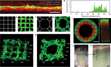
Analysis of the hydrogel filaments and structures fabricated using FRESH
At the Carnegie Mellon University College of Engineering, research in soft material bioprinting may eventually mean that waiting lists for transplants can one day be eliminated, as procedures like repairing the heart may soon become much easier.

Their first concern though was creating the optimum 3D printer for the fabrication of soft materials and tissue. Because the normal 3D printer is used to create models that are generally hard and durable, the research team had to come up with a machine that could make pliable materials. While traditional 3D printing hardware may not work for the type of bioprinting these researchers want to do, all the same benefits that come with 3D printing technology are what propels these researchers to their ultimate goal, with the use of open-source technology and great affordability.

Adam Feinberg and his team in the Regenerative Biomaterials and Therapeutics Group have just released their research in a paper, ‘Three-dimensional printing of complex biological structures by freeform reversible embedding of suspended hydrogels,’ authored by Thomas J. Hinton, Quentin Jallerat, Rachelle N. Palchesko, Joon Hyung Park, Martin S. Grodzicki, Hao-Jan Shue, Mohamed H. Ramadan, Andrew R. Hudson, and also Adam W. Feinberg.

Just published in Science Advances, the researchers outline how they are able to 3D print ‘soft protein and polysaccharide hydrogels’ with a new process they have abbreviated to call FRESH, which stands for freeform reversible embedding of suspended hydrogels.

Add caption
Traditional 3D printers print hard materials, and we’re really trying to move that into a whole new range of soft materials that will eventually allow us to print living things,” says Adam Feinberg, associate professor of Materials Science and Engineering and Biomedical Engineering at Carnegie Mellon University.

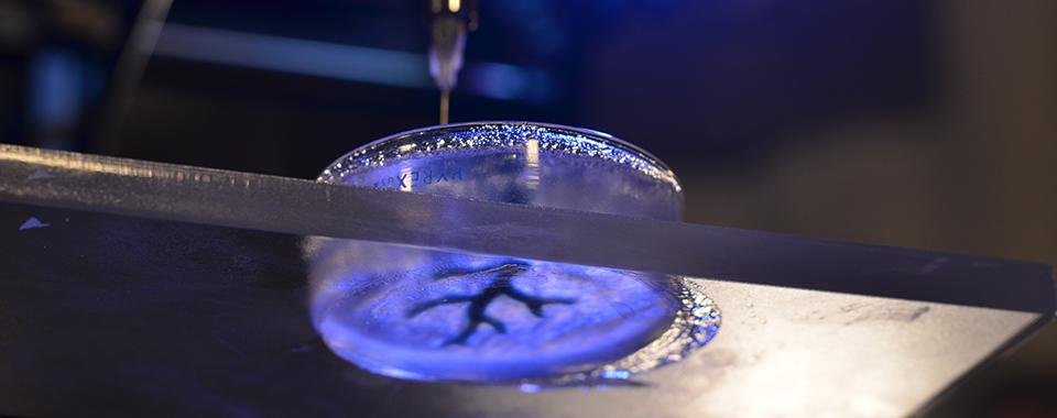
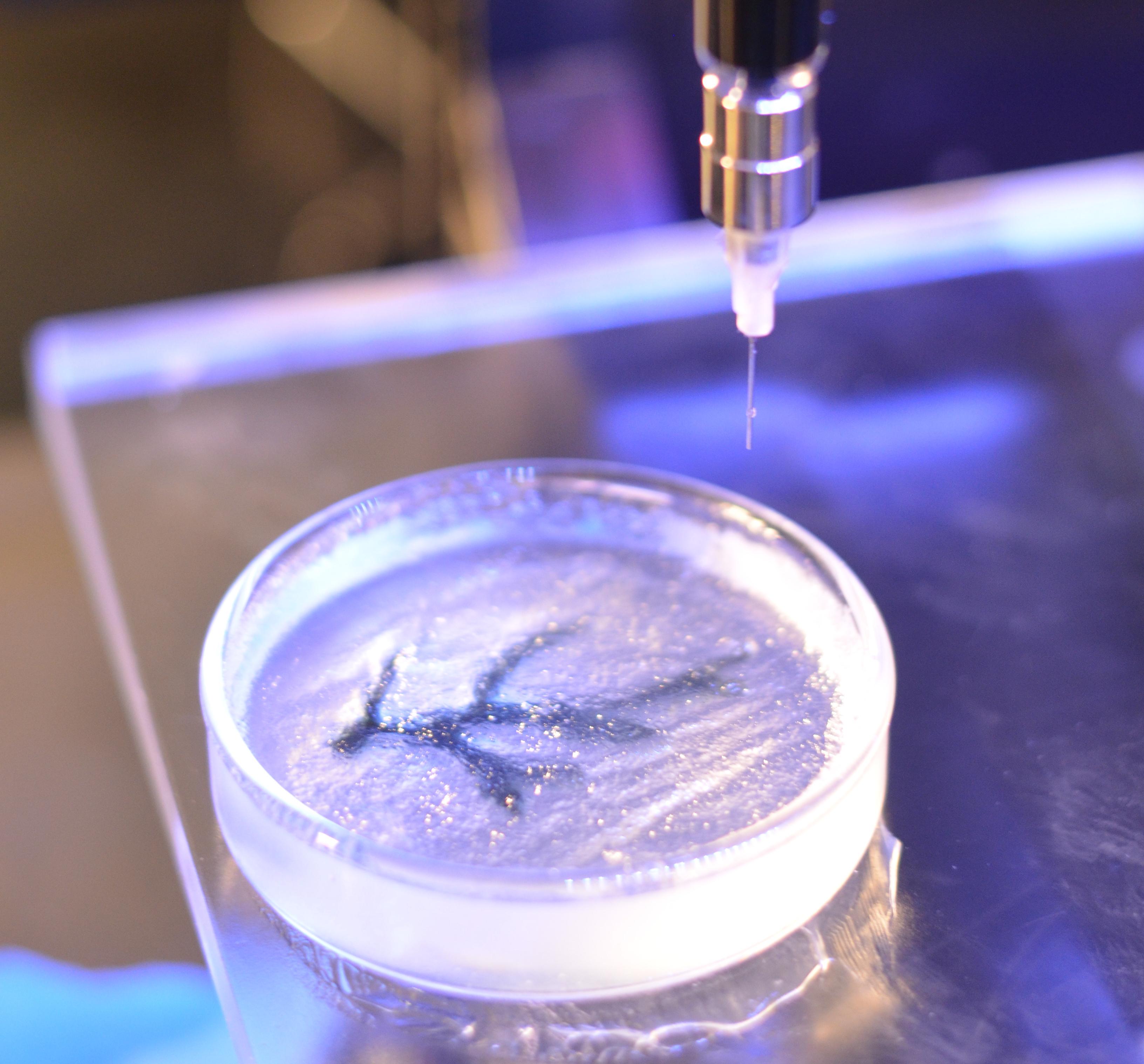
One gel is printed inside of another, which acts as a support and allows for stable printing of layers. The soft materials are 3D printed in a gentle support bath which is heated at the end and allows for removal of the pliant model without damaging or killing it.

The support bath is composed of gelatin micro-particles that act like a Bingham plastic during the print process, behaving as a rigid body at low shear stresses but flowing as a viscous fluid at higher shear stresses,” state the researchers. “This means that, as a needle-like nozzle moves through the bath, there is little mechanical resistance, yet the hydrogel being extruded out of the nozzle and deposited within the bath is held in place. Thus, soft materials that would collapse if printed in air are easily maintained in the intended 3D geometry.
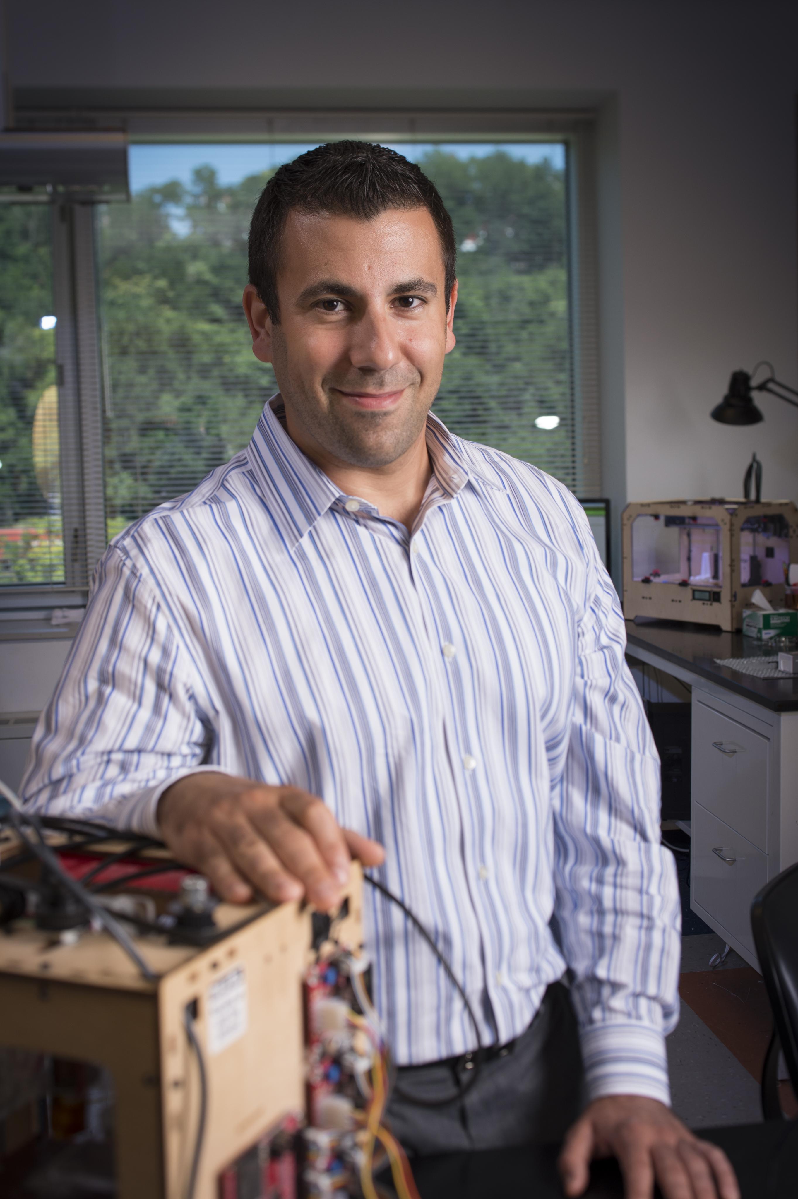
Adam Feinberg
The team has gotten to this point through hacking and building their own 3D printer. While technology for this type of work would generally cost close to $100K, it is the use of open-source software and hardware, resourcefulness, and numerous different innovative approaches that allowed them to make their own machine for exponentially less.

FRESH is built on open-source hardware and software and the gelatin slurry is low cost and readily processed using consumer blenders,” state the researchers. “To emphasize the accessibility of the technology, we implemented FRESH on a $400 3D printer (Printrbot Jr, movie S10) and the STL file to 3D print the custom syringe-based extruder can be downloaded

It should be acknowledged that the direct bioprinting of functional tissues and organs requires further research and development to become fully realized, and a number of companies and academic laboratories are actively working toward this goal.

The researchers see the low cost of their technique, combined with the 3D printing of hydrogels, as one that could transform and significantly expand bioprinting and breakthroughs in a variety of sectors–especially in pharmaceuticals and regenerative therapy.
We’ve been able to take MRI images of coronary arteries and 3-D images of embryonic hearts and 3D bioprint them with unprecedented resolution and quality out of very soft materials like collagens, alginates and fibrins,” said Feinberg.

Their next step as they evolve in this process, is to integrate actual cells from the heart into their 3D printed tissue. This will work in the future as a foundation or scaffold for building actual muscle. We’ll continue to follow this team as they work together in making staggering developments via research, open source software and hardware and 3D printing. Let’s hear your thoughts on the implications of printing such tissue. Discuss in the 3D Printed Soft Tissue forum thread on 3DPB.com.


ORIGINAL: 3DPrint
OCTOBER 26, 2015

martes, 22 de septiembre de 2015

Sensors You Can Swallow Could Be Made of Nutrients and Powered by Stomach Acid

Illustration: Bettinger Group/CMU
The future of ingestible sensors could be a cross between silicon-based circuitry and biodegradable materials, with batteries made of nutrients and running on stomach juices.

That, at least, is the vision of Christopher Bettinger, assistant professor of materials science and biomedical engineering at Carnegie Mellon University. His group is working on edible electronics and ways to power them. Ingestible sensors could provide a gut check for early signs on bacterial infection, look for symptoms of gastrointestinal disorders such as Crohn’s Disease, monitor uptake of medications, and even study the microbiome living inside people.“I think a lot of people hand-wave powering these devices through external RF, but bodies are a pretty good Faraday cageChristopher Bettinger, Carnegie Mellon University

Some ingestible sensors, such as a clear pill containing a camera to examine the GI tracts up close, already exist, but they carry a risk of getting stuck and requiring surgery to remove. And researchers are working on devices made from biocompatible materials such as gelatin and indigo. Bettinger thinks the trick is to make the logic circuits out of silicon, taking advantage of the sophistication of that technology, but to encapsulate them in, say, a biodegradable hydrogel that can squeeze through tight openings. The other parts, such as antennas and batteries, would be made from organic and other bio-safe materials.

If you really want to use these in a clinical setting, we think silicon is pretty good,” says Bettinger, who authored a review article on next-generation devices in the latest issue of Trends in Biotechnology.

One of the main issues is how to supply the sensors with power. “I think a lot of people hand-wave powering these devices through external RF,” he says, “but bodies are a pretty good Faraday cage,” which would prevent radio frequency energy from reaching the sensors. His team has built a battery with a cathode made of melanin—the pigment that colors hair and skin—and an anode made of manganese oxide, a form of a mineral that plays a role in nerve function. The battery has an open design, so that when it hits the stomach, gastrointestinal fluids act as the electrolyte and transport current, much the way the emergency lights of life vests light up when they’re dropped in ocean water. In lab tests, it provided 5 milliwatts of power for up to 20 hours

Various minerals, such as manganese, magnesium, and copper are considered essential nutrients, and could be used to build electronics in amounts smaller than the U.S. Food and Drug Administration “Recommended Daily Allowance”, which should help convince that agency of their safety, Bettinger says. “We think we can go to FDA and say, ‘here’s a battery compound of things that are already in our bodies, plus water,’” he explains. Even silicon, if it interacts with the body, can turn into silicic acid, which has some health benefits.

As for the melanin, Bettinger says, “there’s already more melanin in a serving of squid-ink pasta than will be in our batteries.

The vision of edible electronics may not be far in the future. Proteus Digital Health, of Redwood Shores, Calif., already makes an ingestible sensor that sends data to a patch worn on the skin. Earlier this month they and Otsuka Pharmaceutical, of Tokyo, Japan, filed an application with the FDA for the first combination of a drug with a smart pill. Then hope to sell a pill of Abilify, a drug for mental disorders, with the Proteus sensor embedded within it to monitor drug uptake.

ORIGINAL: IEEE Spectrum
By Neil Savage
Posted 21 Sep 2015

lunes, 25 de agosto de 2014

Sorting cells with sound waves

Acoustic device that separates tumor cells from blood cells could help assess cancer’s spread.
Illustration: Christine Daniloff/MIT
Researchers from MIT, Pennsylvania State University, and Carnegie Mellon University have devised a new way to separate cells by exposing them to sound waves as they flow through a tiny channel. Their device, about the size of a dime, could be used to detect the extremely rare tumor cells that circulate in cancer patients’ blood, helping doctors predict whether a tumor is going to spread.
This microfluidic device uses sound waves to sorts cells as they flow through the channel, from left to right. Image courtesy of the researchers
Separating cells with sound offers a gentler alternative to existing cell-sorting technologies, which require tagging the cells with chemicals or exposing them to stronger mechanical forces that may damage them.

Acoustic pressure is very mild and much smaller in terms of forces and disturbance to the cell. This is a most gentle way to separate cells, and there’s no artificial labeling necessary,” says Ming Dao, a principal research scientist in MIT’s Department of Materials Science and Engineering and one of the senior authors of the paper, which appears this week in the Proceedings of the National Academy of Sciences.

Subra Suresh, president of Carnegie Mellon, the Vannevar Bush Professor of Engineering Emeritus, and a former dean of engineering at MIT, and Tony Jun Huang, a professor of engineering science and mechanics at Penn State, are also senior authors of the paper. Lead authors are MIT postdoc Xiaoyun Ding and Zhangli Peng, a former MIT postdoc who is now an assistant professor at the University of Notre Dame.

The researchers have filed for a patent on the device, the technology of which they have demonstrated can be used to separate rare circulating cancer cells from white blood cells.

To sort cells using sound waves, scientists have previously built microfluidic devices with two acoustic transducers, which produce sound waves on either side of a microchannel. When the two waves meet, they combine to form a standing wave (a wave that remains in constant position). This wave produces a pressure node, or line of low pressure, running parallel to the direction of cell flow. Cells that encounter this node are pushed to the side of the channel; the distance of cell movement depends on their size and other properties such as compressibility.

However, these existing devices are inefficient: Because there is only one pressure node, cells can be pushed aside only short distances.

The new device overcomes that obstacle by tilting the sound waves so they run across the microchannel at an angle — meaning that each cell encounters several pressure nodes as it flows through the channel. Each time it encounters a node, the pressure guides the cell a little further off center, making it easier to capture cells of different sizes by the time they reach the end of the channel.


ORIGINAL: MIT
Anne Trafton | MIT News Office 
August 25, 2014

viernes, 27 de junio de 2014

Welcome to the Claytronics Project


Collaborative Research in Programmable Matter Directed by Carnegie Mellon and Intel

This project combines modular robotics, systems nanotechnology and computer science to create the dynamic, 3-Dimensional display of electronic information known as claytronics.

Our goal is to give tangible, interactive forms to information so that a user's senses will experience digital environments as though they are indistinguishable from reality.

Claytronics is taking place across a rapidly advancing frontier. This technology will help to drive breathtaking advances in the design and engineering of computing and hardware systems

Our research team focuses on two main projects:
  • Creating the basic modular building block of claytronics known as the claytronic atom or catom, and
  • Designing and writing robust and reliable software programs that will manage the shaping of ensembles of millions of catoms into dynamic, 3-Dimensional forms.
Realizing the vision of claytronics through the self-assembly of millions of catoms into synthetic reality will have a profound effect on the experience of users of electronic information. This promise of claytronic technology has become possible because of the ever increasing speeds of computer processing predicted in Moore's Law

This website will introduce you to the ideas that are driving claytronics, the research team that is working to make it happen, and the hardware and software projects that enable the building of claytronic ensembles.

Development of this powerful form of information display represents a partnership between the School of Computer Sciences of Carnegie Mellon University, Intel Corporation at its Pittsburgh Laboratory and FEMTO-ST Institute. As an integral part of our philosophy, the Claytronics Project seeks the contributions of scholars and researchers worldwide who are dedicating their efforts to the diverse scientific and engineering studies related to this rich field of nanotechnology and computer science. 

To understand the future of claytronics, watch the concept video [.mov] created by Carnegie Mellon's Entertainment Technology Center.

Use the links to the left to see a list of publications, some videos and photos documenting our progress, a partial list of talks we have given, and people working on the project.


ORIGINAL: CMU


viernes, 20 de diciembre de 2013

Computer Searches Web 24/7 To Analyze Images and Teach Itself Common Sense


A computer program called the Never Ending Image Learner (NEIL) is running 24 hours a day at Carnegie Mellon University, searching the Web for images, doing its best to understand them on its own and, as it builds a growing visual database, gathering common sense on a massive scale.

NEIL leverages recent advances in computer vision that enable computer programs to identify and label objects in images, to characterize scenes and to recognize attributes, such as colors, lighting and materials, all with a minimum of human supervision. In turn, the data it generates will further enhance the ability of computers to understand the visual world.

But NEIL also makes associations between these things to obtain common sense information that people just seem to know without ever saying — that cars often are found on roads, that buildings tend to be vertical and that ducks look sort of like geese. Based on text references, it might seem that the color associated with sheep is black, but people — and NEIL — nevertheless know that sheep typically are white.

Images are the best way to learn visual properties,” said Abhinav Gupta, assistant research professor in Carnegie Mellon’s Robotics Institute. “Images also include a lot of common sense information about the world. People learn this by themselves and, with NEIL, we hope that computers will do so as well.”

A computer cluster has been running the NEIL program since late July and already has analyzed three million images, identifying 1,500 types of objects in half a million images and 1,200 types of scenes in hundreds of thousands of images. It has connected the dots to learn 2,500 associations from thousands of instances.

The public can now view NEIL’s findings at the project website, www.neil-kb.com.

The research team, including Xinlei Chen, a Ph.D. student in CMU’s Language Technologies Institute, and Abhinav Shrivastava, a Ph.D. student in robotics, will present its findings on Dec. 4 at the IEEE International Conference on Computer Vision in Sydney, Australia

One motivation for the NEIL project is to create the world’s largest visual structured knowledge base, where objects, scenes, actions, attributes and contextual relationships are labeled and catalogued.

What we have learned in the last 5-10 years of computer vision research is that the more data you have, the better computer vision becomes,” Gupta said.

Some projects, such as ImageNet and Visipedia, have tried to compile this structured data with human assistance. But the scale of the Internet is so vast — Facebook alone holds more than 200 billion images — that the only hope to analyze it all is to teach computers to do it largely by themselves.

Shrivastava said NEIL can sometimes make erroneous assumptions that compound mistakes, so people need to be part of the process. A Google Image search, for instance, might convince NEIL that “pink” is just the name of a singer, rather than a color.

People don’t always know how or what to teach computers,” he observed. “But humans are good at telling computers when they are wrong.

People also tell NEIL what categories of objects, scenes, etc., to search and analyze. But sometimes, what NEIL finds can surprise even the researchers. It can be anticipated, for instance, that a search for “apple” might return images of fruit as well as laptop computers. But Gupta and his landlubbing team had no idea that a search for F-18 would identify not only images of a fighter jet, but also of F18-class catamarans.

As its search proceeds, NEIL develops subcategories of objects – tricycles can be for kids, for adults and can be motorized, or cars come in a variety of brands and models. And it begins to notice associations – that zebras tend to be found in savannahs, for instance, and that stock trading floors are typically crowded.

NEIL is computationally intensive, the research team noted. The program runs on two clusters of computers that include 200 processing cores.

This research is supported by the Office of Naval Research and Google Inc.

Related People 

Abhinav Gupta
Abhinav Shrivastava

Related Labs 
Computer Graphics Lab


ORIGINAL: Carnegie Mellon
Computer Graphics Lab
November 20, 2013

martes, 10 de diciembre de 2013

Skin pigment could power safe, implantable battery

Melanin could be used for more than tanning
(Image: Shelbi Lynn Awabdy/Getty)
Your body may use it to catch a tan, but now the skin pigment melanin has been repurposed for the first time to make batteries. These may one day offer a safer way to power electronic devices that can be swallowed or inserted into the human body for drug delivery or internal monitoring.

Rechargeable lithium-ion batteries are widely used in electronics because they are very efficient and can hold their charge for long periods. But because they contain lithium, these batteries are potentially toxic if used long-term inside the body. So Christopher Bettinger at Carnegie Mellon University in Pittsburgh, Pennsylvania, wanted to find a way to build batteries from biological materials.

"If we could safely ingest devices, then we could overcome a lot of the issues we have with current implanted devices, such as infection and inflammation," says Bettinger. "So we started with substances that are biologically derived and occur in the human body naturally, like sodium, water and melanin."

Skin power

To make the bio-battery, Bettinger and his team engineered positively charged anodes out of a mixture containing high levels of melanin, the substance that creates pigment in humans and many other animals. They then introduced sodium ions and loaded the anodes into a steel mesh structure. Melanin's uniform chemical structure means it can pack in plenty of ions, which is key to determining how much charge a battery can hold. This battery could discharge for up to 5 hours, although at a lower power output than standard batteries.

Other biomaterials such as plant matter have been tested as potential electrodes, but they require extra chemical modifications to hold a charge. By contrast, melanin can be used in its natural form, and it is very possible that it could be simply harvested from human skin, says Bettinger. However, using a source with a much higher density of the pigment – the ink sac of a squid, for example – would be more efficient.

"This paper describes a really clever route to producing batteries out of biodegradable materials," says John Rogers at the University of Illinois at Urbana-Champaign. Although the current version isn't fully biodegradable, it shows how batteries could one day be made to dissolve harmlessly in the body.

"The idea is that such technologies could be used as power sources for systems that go into the body, monitor a wound-healing process, deliver therapy as necessary and then naturally disappear after the wound is completely healed. Similar sorts of systems might be designed to treat cancerous tumours, or to treat bone fractures or torn ligaments."

The researchers also found that natural melanin is better at holding charge than synthetic versions. But the melanin battery is not as efficient as the lithium-ion variety, partly because natural melanin is usually found in very dense granules, says Bettinger. Finding a way to make it spongy would help it soak up more sodium ions and pack in even more charge.

Journal reference: PNAS, DOI: 10.1073/pnas.1314345110


ORIGINAL: New Scientist
by Nicola Guttridge
21:10 09 December 2013 

sábado, 23 de noviembre de 2013

The Impact of Brain and Mind Research

Understanding the brain is a grand challenge of science, and in April 2013, President Obama announced the federal BRAIN Initiative, whose goal is to create dramatic improvements in our understanding of brain function and dysfunction. Modeled loosely on the Human Genome Project, this initiative will require the development of new technologies, models, and computational approaches.


With so much at stake, what role can CMU and Pittsburgh play in this initiative? 
This panel of experts will discuss
  • the opportunities and challenges posed by the BRAIN Initiative, including 
  • the potential of this work to bring about revolutionary changes in our understanding of the brain; 
  • in our ability to understand, diagnose and treat brain disorders; and 
  • in the development of models that mimic brain functions.
Opening Remarks: Subra Suresh, Carnegie Mellon University

Moderator: Michael Tarr, Carnegie Mellon University Panelists:

Nathan Urban, Carnegie Mellon University
Marlene Behrmann, Carnegie Mellon University

Tom Mitchell, Carnegie Mellon University
Emery Brown, Massachusetts Institute of Technology, Harvard Medical School
Philip Rubin, Executive Office of the President of the United States
The Impact of Brain and Mind Research
ORIGINAL: CMU
Cèilidh Weekend, McConomy Auditorium, University Center, Carnegie Mellon University
September 28, 2013

lunes, 23 de septiembre de 2013

CMU's Autonomous Car Doesn't Look like a Robot

ORIGINAL: IEEE Spectrum
By Evan Ackerman
Posted 9 Sep 2013 | 14:28 GMT


The future of automobile autonomy isn't going to involve cars covered in cameras and radar and lasers. It's going to be all invisible, and CMU is already there.
The 2011 Cadillac SRX in the picture above is an autonomous car. Carnegie Mellon University had it drive itself 33 miles last week on public roads, from from Cranberry, Pa. to Pittsburgh International Airport. At first glance, you probably wouldn't be able to tell that the car is self-driving, because self-driving cars looked like this just five years ago:
CMU's BOSS
That's CMU's BOSS competing in the DARPA Urban Challenge in 2007, with who knows how many sensors mounted all over it. And even Google's autonomous cars have that signature Velodyne LIDAR mounted on top of them:

Google's autonomous car

By contrast, CMU's SRX relies entirely on automotive-grade radars, lidars, and cameras. You can see them if you look closely in the picture at the top of this article (there's one above the windshield, for example), but you do have to look closely. Inside, there are some extra buttons and screens, but all of the computers are stuffed under the floor in the trunk. And despite the lack of giant and complicated and expensive sensor systems, the car is still able to achieve the level of autonomy that we all want it to, as CMU's Raj Rajkumar explains:


"This car is the holy grail of autonomous driving because it can do it all — from changing lanes on highways, driving in congested suburban traffic and navigating traffic lights."

In addition to controlling the steering, speed and braking, the autonomous systems also detect and avoid obstacles in the road, including traffic cones and barrels, as well as pedestrians and bicyclists, pausing until they are safely out of the way. The systems provide audible warnings of obstacles and communicate vehicle status to its passengers using a human-like voice.

It's unfortunate that while the technology for all of this is arguably mostly ready, society (socially and legally) just isn't yet. You can buy cars with adaptive cruise control and lane departure warnings, which could hypothetically let the car drive itself, at least under some specific circumstances. And despite the fact that even a bad autonomous (or semi-autonomous) car would still save lives overall, there's no legal infrastructure in place to make it possible for manufacturers to implement such technology without undue risk of being sued into oblivion the first time something goes wrong.

Via [ CMU ]

domingo, 5 de mayo de 2013

Researchers create edible battery


Researchers create edible battery

Stimulating damaged tissue, bio-sensing gastric health, targeting drug delivery and more could soon be as easy as popping your morning multivitamin — thanks to the joint work of Carnegie Mellon University's Christopher Bettinger and Jay Whitacre. The two cutting-edge researchers have combined forces to develop edible electronic devices — no larger than ordinary pills — to improve medical care.

Bettinger has been developing pioneering biodegradable electronic materials for medical use, but had a few nagging concerns. "Two questions kept coming up," he explained. "First, how were we going to power these devices? Second, if they're degradable and temporary, then what was the best way to integrate them with the human body?"

Whitacre, associate professor of materials science and engineering and engineering and public policy, had created a revolutionary low-cost, non-toxic sodium ion battery. "I had claimed my device was so non-toxic that you 'could eat the battery,'" explained Whitacre. "Chris came into my office and asked, 'Can you really eat it?' The answer is yes and the rest is history — my edible battery chemistry with his need for low level power in a digestible form were a great match."

"We thought the innovation from that battery could be a great segue to medical materials," added Bettinger. "So we leveraged its advantages in a different setting."

With post-doctoral researchers Young Jo Kim and Sang-Eun Chun as part of the team, they devised a tiny, biocompatible battery in edible form that a patient could 'take' once a day.

The shape-memory polymer conceived in Bettinger's lab starts small when swallowed, then expands in the body where needed. The battery materials created in Whitacre's lab are commonly available and inexpensive — necessary for a daily device. The battery materials pass right through the system while the binding materials naturally biodegrade.

Unlike an implanted device, it's minimally invasive, and as with any orally-taken 'pill,' doesn't require sterilization. And if that weren't enough, the battery activates itself when wet, so the casing can be designed to absorb water at a pre-determined rate meaning "actual initiation would be passively engineered in the device itself," says Bettinger. While specific applications are still in the future, it's a remarkable way to "lay the groundwork."

Source and top image showing a graphic demonstration of the device: Carnegie Mellon University

sábado, 4 de agosto de 2012

New online game to design RNA molecules: advancing nanotechnology?


(Credit: EteRNA)
As we pointed out a few months ago, the greater complexity of folding rules for RNA compared to its chemical cousin DNA gives RNA a greater variety of compact, three-dimensional shapes and a different set of potential functions than is the case with DNA, and this gives RNA nanotechnology a different set of advantages compared to DNA nanotechnology as a road towards atomically precise manufacturing. Proteins have even more complex folding rules and an even greater variety of structures and functions. We also noted here that online gamers playing Foldit topped scientists in redesigning a protein to achieve a novel enzymatic activity that might be especially useful in developing molecular building blocks for molecular manufacturing. Now KurzweilAI.net brings news of an online game that allows players to design RNA molecules “New videogame lets amateur researchers mess with RNA.

EteRNA, an online game with more than 38,000 registered users, allows players to design molecules of ribonucleic acid — RNA — that have the power to build proteins or regulate genes.

EteRNA players manipulate nucleotides, the fundamental building blocks of RNA, to coax molecules into shapes specified by the game.

Those shapes represent how RNA appears in nature while it goes about its work as one of life’s most essential ingredients.

EteRNA was developed by scientists at Stanford and Carnegie Mellon universities, who use the designs created by players to decipher how real RNA works. The game is a direct descendant of Foldit — another science crowdsourcing tool disguised as entertainment — which gets players to help figure out the folding structures of proteins.

The game’s elite players compete for a unique and wondrous prize: the chance to have RNA designs of their own making brought to life. Every two weeks, four to 16 player-designed molecules are picked to be synthesized in an RNA lab at Stanford.

The chance to win this reward has proven highly motivating for EteRNA‘s players. They carefully study the data that the lab provides on how the synthesized molecules behave when ushered into existence, then use their observations to refine their next designs. In doing so, they — like their Foldit-playing peers — have helped scientists take advantage of the human brain’s unparalleled talent for recognizing patterns and solving puzzles.

But EteRNA players have also done something much more profound: By scrutinizing their creations, learning from their triumphs and mistakes, and using their accumulated wisdom to develop new hypotheses, they aren’t just building better RNA molecules; they’re discovering fundamental aspects of biochemistry that no one — not even the world’s top RNA researchers — knew before. And in doing so, they are blurring the line that separates gamer from scientist …

The article goes on to discuss a growing movement among EteRNA players to synthesize their RNA molecules themselves, linking online scientific game-playing and crowd-sourced molecular design to Open Science and DIY Biotechnology. The EteRNA web site provides tutorials to get new users started and instant feedback. Could games like Foldit and EteRNA represent new crowd-sourced paths to the more rapid development of atomically precise manufacturing?
—James Lewis, PhD