lunes, 28 de marzo de 2016

New dawn: Chinese scientists move step closer to creating ‘artificial sun’ in quest for limitless energy via nuclear fusion

Chinese scientists were able to heat plasma to three times the temperature of the core of our sun for a record-breaking 102 seconds as they progressed the search to derive energy from nuclear fusion. Photo: Wikipedia
In a doughnut-shaped chamber in eastern China, scientists have been able to produce hydrogen gas more than three times hotter than the core of the Sun using nuclear fusion - and maintain this temperature for 102 seconds.

The breakthrough puts China one step ahead in the global race to harness a new, artificial kind of solar energy for clean and unlimited energy, the researchers claim. This has become a pressing concern as more of the earth’s natural reserves are rapidly depleting.

The experiment was conducted last week on a magnetic fusion reactor at the Institute of Physical Science in Hefei, capital of Jiangsu province, according to a statement on the institute’s website on Wednesday.

The reactor, officially known as the Experimental Advanced Superconducting Tokamak (EAST), was able to heat a hydrogen gas - a hot ionised gas called a plasma - to about 50 million Kelvins (49.999 million degrees Celsius). The interior of our sun is calculated to be around 15 million Kelvins.

Chinese scientists came a step closer to creating an artificial sun by heating hydrogen gas to 50 million Kelvins for a record time. Photo: Hefei Institute of Physical Science, Chinese Academy of Sciences
According to this thermodynamic scale, absolute zero occurs at zero degrees (equivalent to minus 273.15 degrees Celsius), a point at which all molecular movement stops.

The temperature reached in Hefei was at the other end of the scale, and roughly the same as a mid-sized thermonuclear explosion. The goal of the experiment was to approximate the nuclear fusion conditions that occur deep inside the sun.

Although at least one other experiment in the last decade claims to have produced a hotter temperatures than this, it has never been duplicated and was unable to match the endurance - over one and a half minutes - of the Chinese test.

Meanwhile, physicists in Japan and Europe have been able to reach the same temperature as the Chinese team, but not for longer than a minute due to concerns of provoking a reactor meltdown.

The EAST was invented by Soviet scientists to control nuclear fusion for power generation.

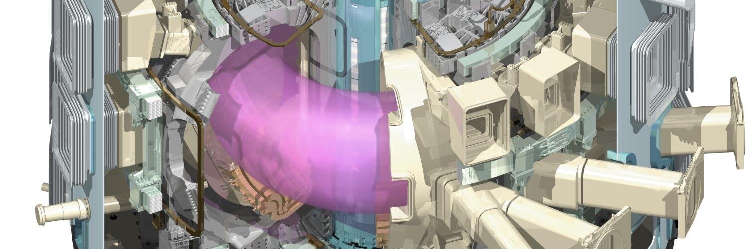
The EAST tokomak device in Hefei. In order to ‘fuse’ two hydrogen atoms to produce energy, it needs to heat the hydrogen plasma for 100 million Kelvins. Photo: Chinese Academy of Sciences
As a tokamak device, it uses a powerful magnetic field to confine plasma in the shape of a torus - imagine a large spinning doughnut -for safety reasons due to the phenomenally high temperatures being generated. The atoms are effectively held floating in place by superconducting magnets.

But controlling hydrogen gas in such a hot and volatile state is a formidable challenge, and one that most of the tokomak devices built over the last 60 years have not been able to sustain for more than 20 seconds.

The scientists in Hefei worked “day and night” to achieve the record level of endurance, according to the institute, which serves as a subsidiary of the Chinese Academy of Sciences.

The team claimed to have solved a number of scientific and engineering problems, such as precisely controlling the alignment of the magnet, and managing to capture the high-energy particles and heat escaping from the “doughnut”.

Inside the ‘doughnut’ (EAST). The metallic walls cannot come into direct contact with the plasma or it will melt or evaporate immediately. The scientists used a powerful magnetic field to keep the hot hydrogen gas suspended in place. Photo: Chinese Academy of Sciences
But they still missed their mark, which was to reach 100 million Kelvins for over 1,000 seconds (nearly 17 minutes), they said, adding that it would still take years to build a commercially viable plant that could operate in a stable manner for several decades.

Unlike the process of nuclear fission that fuels thermal power stations around the world today by splitting the atoms of fissile materials such as uranium, fusion reactions work by “fusing” two light atomic nuclei - for example, two hydrogen atoms - together to release a huge amount of heat.

This can produce levels of energy three to four times greater than the results of nuclear fission. It also generate almost no radioactive waste.

The problem is the amount of heat created. Whereas nuclear fission only generates a few hundred degrees Celsius, fusion requires at least 100 million degrees Celsius (212 million degrees Fahrenheit).

A researcher involved with the EAST project said data from their experiment may be of use to the International Thermonuclear Experimental Reactor (ITER) that is now under construction in France.
Source: @ITERORG

Meanwhile, another 1-billion-euro (US$1.12 billion) project in Germany dubbed the “stellarator” claimed last December to have achieved another milestone in the nuclear fusion quest by heating plasma to around 1 million degrees Celsius for one-tenth of a second.

A colourised computer image shows the moment the first superhot plasma was created in a separate experiment at the Wendelstein 7-X nuclear fusion research centre at the Max-Planck-Institut for Plasma Physics (IPP) in Greifswald, Germany in December. Photo: EPA


China ranks as a member country of the ITER project, which aims to produce 500 megawatts of fusion power for 400 seconds. But Beijing has expressed frustration with the slow pace of development, according to the same researcher, who asked not be identified.

The multibillion US dollar project was initially scheduled to become operational this year. But due to a series of setbacks, many now suspect it will need at least another decade.

Political infighting among different nations about the project’s budget, personnel appointments and other issues are hampering the pace of the project,” said the researcher.

If this chaotic situation continues, other projects in countries like the United States and China may overtake this collective, international effort.

Stephen Chen. chen.binglin@scmp.com
05 February, 2016, 1:45pm


"AI & The Future Of Civilization" A Conversation With Stephen Wolfram

"AI & The Future Of Civilization" A Conversation With Stephen Wolfram [3.1.16]
Stephen Wolfram
What makes us different from all these things? What makes us different is the particulars of our history, which gives us our notions of purpose and goals. That's a long way of saying when we have the box on the desk that thinks as well as any brain does, the thing it doesn't have, intrinsically, is the goals and purposes that we have. Those are defined by our particulars—our particular biology, our particular psychology, our particular cultural history.

The thing we have to think about as we think about the future of these things is the goals. That's what humans contribute, that's what our civilization contributes—execution of those goals; that's what we can increasingly automate. We've been automating it for thousands of years. We will succeed in having very good automation of those goals. I've spent some significant part of my life building technology to essentially go from a human concept of a goal to something that gets done in the world.

There are many questions that come from this. For example, we've got these great AIs and they're able to execute goals, how do we tell them what to do?...

STEPHEN WOLFRAM, distinguished scientist, inventor, author, and business leader, is Founder & CEO, Wolfram Research; Creator, Mathematica, Wolfram|Alpha & the Wolfram Language; Author, A New Kind of Science. Stephen Wolfram's EdgeBio Page

THE REALITY CLUB: Nicholas Carr

AI & THE FUTURE OF CIVILIZATION
Some tough questions. One of them is about the future of the human condition. That's a big question. I've spent some part of my life figuring out how to make machines automate stuff. It's pretty obvious that we can automate many of the things that we humans have been proud of for a long time. What's the future of the human condition in that situation?


More particularly, I see technology as taking human goals and making them able to be automatically executed by machines. The human goals that we've had in the past have been things like moving objects from here to there and using a forklift rather than our own hands. Now, the things that we can do automatically are more intellectual kinds of things that have traditionally been the professions' work, so to speak. These are things that we are going to be able to do by machine. The machine is able to execute things, but something or someone has to define what its goals should be and what it's trying to execute.

People talk about the future of the intelligent machines, and whether intelligent machines are going to take over and decide what to do for themselves. What one has to figure out, while given a goal, how to execute it into something that can meaningfully be automated, the actual inventing of the goal is not something that in some sense has a path to automation.

How do we figure out goals for ourselves? How are goals defined? They tend to be defined for a given human by their own personal history, their cultural environment, the history of our civilization. Goals are something that are uniquely human. It's something that almost doesn't make any sense. We ask, what's the goal of our machine? We might have given it a goal when we built the machine.

The thing that makes this more poignant for me is that I've spent a lot of time studying basic science about computation, and I've realized something from that. It's a little bit of a longer story, but basically, if we think about intelligence and things that might have goals, things that might have purposes, what kinds of things can have intelligence or purpose? Right now, we know one great example of things with intelligence and purpose and that's us, and our brains, and our own human intelligence. What else is like that? The answer, I had at first assumed, is that there are the systems of nature. They do what they do, but human intelligence is far beyond anything that exists naturally in the world. It's something that's the result of all of this elaborate process of evolution. It's a thing that stands apart from the rest of what exists in the universe. What I realized, as a result of a whole bunch of science that I did, was that is not the case.

Research on largest network of cortical neurons to date published in Nature

Robust network of connections between neurons performing similar tasks shows fundamentals of how brain circuits are wired


Even the simplest networks of neurons in the brain are composed of millions of connections, and examining these vast networks is critical to understanding how the brain works. An international team of researchers, led by R. Clay Reid, Wei Chung Allen Lee and Vincent Bonin from the Allen Institute for Brain Science, Harvard Medical School and Neuro-Electronics Research Flanders (NERF), respectively, has published the largest network to date of connections between neurons in the cortex, where high-level processing occurs, and have revealed several crucial elements of how networks in the brain are organized. The results are published this week in the journal Nature.

A network of cortical neurons whose connections were traced from a multi-terabyte 3D data set. The data were created by an electron microscope designed and built at Harvard Medical School to collect millions of images in nanoscopic detail, so that every one of the “wires” could be seen, along with the connections between them. Some of the neurons are color-coded according to their activity patterns in the living brain. This is the newest example of functional connectomics, which combines high-throughput functional imaging, at single-cell resolution, with terascale anatomy of the very same neurons. Image credit: Clay Reid, Allen Institute; Wei-Chung Lee, Harvard Medical School; Sam Ingersoll, graphic artist

This is a culmination of a research program that began almost ten years ago. Brain networks are too large and complex to understand piecemeal, so we used high-throughput techniques to collect huge data sets of brain activity and brain wiring,” says R. Clay Reid, M.D., Ph.D., Senior Investigator at the Allen Institute for Brain Science. “But we are finding that the effort is absolutely worthwhile and that we are learning a tremendous amount about the structure of networks in the brain, and ultimately how the brain’s structure is linked to its function.

Although this study is a landmark moment in a substantial chapter of work, it is just the beginning,” says Wei-Chung Lee, Ph.D., Instructor in Neurobiology at Harvard Medicine School and lead author on the paper. “We now have the tools to embark on reverse engineering the brain by discovering relationships between circuit wiring and neuronal and network computations.” 

For decades, researchers have studied brain activity and wiring in isolation, unable to link the two,” says Vincent Bonin, Principal Investigator at Neuro-Electronics Research Flanders. “What we have achieved is to bridge these two realms with unprecedented detail, linking electrical activity in neurons with the nanoscale synaptic connections they make with one another.

We have found some of the first anatomical evidence for modular architecture in a cortical network as well as the structural basis for functionally specific connectivity between neurons,” Lee adds. “The approaches we used allowed us to define the organizational principles of neural circuits. We are now poised to discover cortical connectivity motifs, which may act as building blocks for cerebral network function.

Lee and Bonin began by identifying neurons in the mouse visual cortex that responded to particular visual stimuli, such as vertical or horizontal bars on a screen. Lee then made ultra-thin slices of brain and captured millions of detailed images of those targeted cells and synapses, which were then reconstructed in three dimensions. Teams of annotators on both coasts of the United States simultaneously traced individual neurons through the 3D stacks of images and located connections between individual neurons.

Analyzing this wealth of data yielded several results, including the first direct structural evidence to support the idea that neurons that do similar tasks are more likely to be connected to each other than neurons that carry out different tasks. Furthermore, those connections are larger, despite the fact that they are tangled with many other neurons that perform entirely different functions.

Part of what makes this study unique is the combination of functional imaging and detailed microscopy,” says Reid. “The microscopic data is of unprecedented scale and detail. We gain some very powerful knowledge by first learning what function a particular neuron performs, and then seeing how it connects with neurons that do similar or dissimilar things.

It’s like a symphony orchestra with players sitting in random seats,” Reid adds. “If you listen to only a few nearby musicians, it won’t make sense. By listening to everyone, you will understand the music; it actually becomes simpler. If you then ask who each musician is listening to, you might even figure out how they make the music. There’s no conductor, so the orchestra needs to communicate.

This combination of methods will also be employed in an IARPA contracted project with the Allen Institute for Brain Science, Baylor College of Medicine, and Princeton University, which seeks to scale these methods to a larger segment of brain tissue. The data of the present study is being made available online for other researchers to investigate.

This work was supported by the National Institutes of Health (R01 EY10115, R01 NS075436 and R21 NS085320); through resources provided by the National Resource for Biomedical Supercomputing at the Pittsburgh Supercomputing Center (P41 RR06009) and the National Center for Multiscale Modeling of Biological Systems (P41 GM103712); the Harvard Medical School Vision Core Grant (P30 EY12196); the Bertarelli Foundation; the Edward R. and Anne G. Lefler Center; the Stanley and Theodora Feldberg Fund; Neuro-Electronics Research Flanders (NERF); and the Allen Institute for Brain Science.
About the Allen Institute for Brain Science

The Allen Institute for Brain Science, a division of the Allen Institute (alleninstitute.org), is an independent, 501(c)(3) nonprofit medical research organization dedicated to accelerating the understanding of how the human brain works in health and disease. Using a big science approach, the Allen Institute generates useful public resources used by researchers and organizations around the globe, drives technological and analytical advances, and discovers fundamental brain properties through integration of experiments, modeling and theory. Launched in 2003 with a seed contribution from founder and philanthropist Paul G. Allen, the Allen Institute is supported by a diversity of government, foundation and private funds to enable its projects. Given the Institute’s achievements, Mr. Allen committed an additional $300 million in 2012 for the first four years of a ten-year plan to further propel and expand the Institute’s scientific programs, bringing his total commitment to date to $500 million. The Allen Institute’s data and tools are publicly available online at brain-map.org.

About Harvard Medical School
HMS has more than 7,500 full-time faculty working in 10 academic departments located at the School’s Boston campus or in hospital-based clinical departments at 15 Harvard-affiliated teaching hospitals and research institutes: Beth Israel Deaconess Medical Center, Boston Children’s Hospital, Brigham and Women’s Hospital, Cambridge Health Alliance, Dana-Farber Cancer Institute, Harvard Pilgrim Health Care Institute, Hebrew SeniorLife, Joslin Diabetes Center, Judge Baker Children’s Center, Massachusetts Eye and Ear/Schepens Eye Research Institute, Massachusetts General Hospital, McLean Hospital, Mount Auburn Hospital, Spaulding Rehabilitation Hospital and VA Boston Healthcare System.

About NERF
Neuro-Electronics Research Flanders (NERF; www.nerf.be) is a neurotechnology research initiative is headquartered in Leuven, Belgium initiated by imec, KU Leuven and VIB to unravel how electrical activity in the brain gives rise to mental function and behaviour. Imec performs world-leading research in nanoelectronics and has offices in Belgium, the Netherlands, Taiwan, USA, China, India and Japan. Its staff of about 2,200 people includes almost 700 industrial residents and guest researchers. In 2014, imec's revenue (P&L) totaled 363 million euro. VIB is a life sciences research institute in Flanders, Belgium. With more than 1470 scientists from over 60 countries, VIB performs basic research into the molecular foundations of life. KU Leuven is one of the oldest and largest research universities in Europe with over 10,000 employees and 55,000 students.

ORIGINAL: Allen Institute
March 28th, 2016

sábado, 26 de marzo de 2016

Medio ambiente debe ser clave en el urbanismo

El experto Charles Waldheim visitó Medellín, Santiago de Chile y Brasilia para conocer más sobre los trabajos locales y, el cruce entre los procesos urbanísticos y el interés ambiental. FOTO CORTESÍA

Con la evolución de la arquitectura el paisaje juega un rol más representativo en las ciudades contemporáneas. Experto Charles Waldheim visitó Medellín para disertar y conocer más sobre el tema.

Medellín junto con Santiago de Chile y Brasilia forma parte de las tres ciudades latinoamericanas escogidas por los expertos de la Universidad de Harvard para plantear discusiones sobre las relaciones entre paisaje y urbanismo.

Charles Waldheim, experto en el tema y profesor de la escuela de diseño de la Universidad de Harvard, participó en Medellín en un encuentro realizado en el Museo de Arte Moderno de Medellín, Mamm y que fue liderado por la Universidad de Harvard y el Centro David Rockefeller para Estudios Latinoamericanos.

El certamen denominado Landscape as Urbanism in the Americas (paisaje como urbanismo en las Américas) fue realizado por la Universidad Eafit, a través de su Centro de Estudios Urbanos y Ambientales, Urbam.

¿Por qué el interés en urbanismo y paisaje?
En mi trabajo como arquitecto encontré limitaciones en la inclusión del urbanismo en profesiones como arquitectura e ingeniería y descubrí que había otros caminos, desde el paisaje en el campo norteamericano. Nosotros importamos el modelo español de construir la ciudad americana de la Ley de Indias, que le da prioridad principalmente a la arquitectura. Pese a que se trata de un modelo muy importante y las ciudades partieron de allí, este opera sacando la ecología, la biología de él. Se trata de un modelo puramente arquitectónico. Y la oposición entre la ciudad y lo que no forma parte de lo construido (los recursos) ha redundado en unas ciudades no tan sanas y sostenibles como podrían serlo”.

¿Cómo ve usted las obras de urbanismo en la ciudad?
Colombia ha formado parte de la discusión internacional en los últimos 10 o 15 años. Y desde la Universidad de Harvard hemos tenido contacto y conocimiento directo con algunos de los proyectos y especialmente con Medellín. En los últimos años aparecieron una seria de oficinas que hacían algo diferente con el paisaje. Una de las prácticas conocidas muestra que el paisaje no está subyugado a otra disciplina, juega un papel primordial”.

¿Qué reconocimientos urbanísticos hará usted en la ciudad?
Vamos a recorrer Parques del Río y las piscinas del complejo acuático de los juegos suramericanos, entre otros, donde la naturaleza y el componente ecológico o biológico juega un destacado papel, pero me gustaría ver y confirmar si ese componente biológico va más allá de la función estética”.

¿Cuál es el papel diferente al decorativo?
Que limpie el agua, el aire, sea un buen lugar para que las especies habiten y puedan generar comida”.

En Medellín se mencionó que Parques del Río podría mejorar la calidad de vida de la ciudad. ¿Cómo incide el paisaje en la calidad de vida de los habitantes?
Sin lugar a dudas generará un cambio y un bienestar en el medio ambiente, pero hay dos condiciones

  • una de ellas, es que habitualmente la calidad de la vivienda alrededor de estos lugares no está en el mismo nivel del parque, del espacio público. Pero puede aparecer la posibilidad de que el diseñador pueda controlar la ecología con el diseño de la vivienda: los dos estén conectados. 
  • Otro elemento presente en Latinoamérica está relacionado con el hecho de que el medio ambiente en el diseño está mas dirigido a lo estético, a la apariencia. Lo estético es importante pero, si tuviera más de biología, biodiversidad sería mucho mejor”.
¿Cómo las personas ubicadas alrededor de Parques del Río pueden conectarse más con él?
Parte del problema es que se entiende la vivienda separada del parque. Se piensa uno disgregado del otro. Debe pensarse junto, que pertenecen al mismo sistema”.

Y qué se recomienda
Planeación, políticas y darle potestad a los diseñadores para que esas relaciones entren a jugar desde el primer momento, para tenerlas en cuenta en los diseños y en la operación. Porque hay una tendencia a entender que cada predio lo desarrolla un arquitecto, un desarrollador de una sola manera y tiende a ser cerrado. Pensarlo de una manera más colectiva, más conectada”.

¿Qué ventaja tiene para el habitante que haya mayor conexión entre el parque y el ciudadano?
Salud pública, si un ciudadano puede caminar por un ambiente no dominado por el auto. También hay calidad de vida. Existe buena evidencia que bajo este sistema se reduce la polución en la ciudad y la calidad del aire. El calor disminuye. Esto también da la oportunidad de que la ciudad consuma lo que produce ella misma, en vez de traer de afuera, insumos que aumentan la huella de carbono”.

¿Por qué está incluida Medellín en ese programa de conferencias y discusiones junto con Santiago de Chile, Chile y Brasilia, Brasil?
En Latinoamérica ha sido lento el acercamiento a las prácticas que involucran el paisaje. Miramos una serie de culturas en Latinoamérica que tuvieran secuelas de paisaje o hubieran manifestaciones alrededor del paisaje a través de escuelas u oficinas. Se quiere mirar y verificar qué está pasando y, las tres ciudades seleccionadas muestran dentro del panorama las prácticas más alternativas en el marco general de la pregunta que nos estamos haciendo. Queremos entender la especificidad de cada cultura y conectar varios nodos, para determinar si la conversación puede ser más grande y que esas personas se conecten como en una red”.

¿Cómo se retroalimenta Medellín?
Se ampliará la discusión que ya se ha tenido a través de una serie de comunicaciones. Además, habrá una página web donde se podrán vincular los proyectos y difundir este tipo de prácticas. También hay un tema de reclutamiento de profesores y estudiantes que puedan ejercer esas nuevas prácticas. Estudiantes que regresen a sus países de origen o profesores que generen la discusión alrededor de esa mirada”.

CONTEXTO DE LA NOTICIA
PARA SABER MÁS
BUSCA SOCIOS EN LATINOAMÉRICA
Charles Waldheim es un arquitecto norteamericano. El profesor de la escuela de diseño de la Universidad de Harvard realiza un trabajo enfocado en las ciudades. Uno de sus intereses particulares está relacionado con investigar cómo funcionan los sistemas naturales en las ciudades. En la Universidad de Harvard organiza grupos estudiantiles y de investigadores para explorar, analizar y estudiar ciertos temas que son del interés de la institución universitaria y del socio estratégico del caso de estudio. Busca socios en universidades, empresas y gobiernos para los proyectos.

Conservation.org's video series "Nature is Speaking"

CONSERVATION INTERNATIONAL PRESENTS "NATURE IS SPEAKING"

STARRING


ORIGINAL: Conservation.org

viernes, 25 de marzo de 2016

JCVI- syn3.0: Minimal Synthetic Bacterial Cell Constructed


THE FIRST MINIMAL SYNTHETIC BACTERIAL CELL IS CONSTRUCTED
Researchers from the J. Craig Venter Institute (JCVI) and Synthetic Genomics, Inc. (SGI) have accomplished the next feat in synthetic biology research—the design and construction of the first minimal synthetic bacterial cell, JCVI-syn3.0.

Using the first synthetic cell, Mycoplasma mycoides JCVI-syn1.0 (created by this same team in 2010), JCVI-syn3.0 was developed through a design, build, and test (DBT) process using genes from JCVI-syn1.0. The new minimal synthetic cell contains only 531,000 base pairs and just 473 genes making it the smallest genome of any self-replicating organism.


A paper describing this research is published in the March 25 print version of the journal, Science by lead author Clyde A. Hutchison, III, Ph.D., senior author J. Craig Venter, Ph.D., and senior team of Hamilton O. Smith, MD, Daniel G. Gibson, Ph.D., and John I. Glass, Ph.D.

J. Craig Venter, Ph.D., Dr. Hamilton O. Smith, M.D., Dan Gibson, Ph.D., Lijie Sun, Ph.D., John Glass, Ph.D., Krishna Kannan, Ph.D., John Gill, and Dr. Clyde A. Hutchison III, Ph.D. Credit: J. Craig Venter Institute

Writing Biological Code
A biological cell is very much like a computer—the genome is the software that encodes the instructions of the cell and the cellular machinery is the hardware that interprets and runs the genome software. Major advances in DNA technologies have made it possible for biologists to now behave as software engineers and rewrite entire genomes to program new biological operating systems.

Project Goals
A major goal in synthetic biology is to have the capacity to predictably design and build DNA that produces a cell with new and improved biological functions that do not already exist in nature. Significant advances have been made in DNA design at the gene and pathway level and in engineering bacteriophage genomes. But, even with all the advances that have been made in genomics and synthetic biology, there is still not a single self-replicating cell in which we understand the function of every one of its genes. Toward this goal, the JCVI/SGI team has been working to understand the gene content of a minimal cell—a cell that has only the machinery necessary for independent life.

Since the generation of the first synthetic cell in 2010, the team found ways to drastically speed up the process of building cells from the bottom up. They developed new tools and semi-automated processes for genome synthesis, including more rapid, more accurate, and more robust methods for going from oligonucleotides (small pieces of DNA) to whole chromosomes. Over the past 10 years whole bacterial chromosome assembly has gone from impossible, to possible in years, to months and now to just weeks with these new methods, which are made available to scientists in this manuscript.

A major outcome of this minimal cell program has been new tools and semi-automated processes for whole genome synthesis. Many of these synthetic biology tools and services are commercially available through SGI-DNA.

THE FIRST MINIMAL SYNTHETIC BACTERIAL CELL IS CONSTRUCTED
J. Craig Venter, Ph.D., Dr. Hamilton O. Smith, M.D., Dan Gibson, Ph.D., Lijie Sun, Ph.D., John Glass, Ph.D., Krishna Kannan, Ph.D., John Gill, and Dr. Clyde A. Hutchison III, Ph.D.  Credit: J. Craig Venter Institute. Download: High Resolution JPEG
J. Craig Venter, Ph.D. and Hamilton O. Smith, M.D.  Credit: J. Craig Venter Institute. Download: High Resolution JPEG
Hamilton O. Smith, M.D. and Clyde A. Hutchison III, Ph.D.  Credit: J. Craig Venter Institute Download: High Resolution JPEG
J. Craig Venter, Ph.D. Credit: J. Craig Venter Institute. Download High Resolution JPEG
Clyde A. Hutchison III, Ph.D. Credit: J. Craig Venter Institute. Download: High Resolution JPEG
John Glass, Ph.D. Credit: J. Craig Venter Institute. Download: High Resolution JPEG
Dan Gibson, Ph.D. Credit: J. Craig Venter Institute. Download: High Resolution JPEG
Ray-Yuan Chuang, Ph.D. Credit: J. Craig Venter Institute. Download: High Resolution JPEG
Electron micrographs of clusters of JCVI-syn3.0 cells magnified about 15,000 times. This is the world’s first minimal bacterial cell. Its synthetic genome contains only 473 genes. Surprisingly, the functions of 149 of those genes are unknown. Credit: Tom Deerinck and Mark Ellisman of the National Center for Imaging and Microscopy Research at the University of California at San Diego Download: High Resolution JPEG
Download: High Resolution JPEG
Four design-build-test cycles produced JCVI-syn3.0. (A) The cycle for genome design, building by means of synthesis and cloning in yeast, and testing for viability by means of genome transplantation. After each cycle, gene essentiality is reevaluated by global transposon mutagenesis. (B) Comparison of JCVI-syn1.0 (outer blue circle) with JCVI-syn3.0 (inner red circle), showing the division of each into eight segments.The red bars inside the outer circle indicate regions that are retained in JCVI-syn3.0. (C) A clusterof JCVI-syn3.0 cells, showing spherical structures of varying sizes (scale bar, 200 nm). Credit: J. Craig Venter Institute. Download: High Resolution PDF
Fig. 7. Comparison of syn1.0 and syn3.0 growth features. (A) Cells derived from 0.2 µm–filtered liquid cultures were diluted and plated on agar medium to compare colony size and morphology after 96 hours (scale bars, 1.0 mm). (B) Growth rates in liquid static culture were determined using a fluorescent measure (relative fluorescent units, RFU) of double-stranded DNA accumulation over time (minutes) to calculate doubling times (td). Coefficients of determination (R2) are shown. (C) Native cell morphology in liquid culture was imaged in wet mount preparations by means of differential interference contrast microscopy (scale bars, 10 µm). Arrowheads indicate assorted forms of segmented filaments (white) or large vesicles (black). (D) Scanning electron microscopy of syn1.0 and syn3.0 (scale bars, 1 µm). The picture on the right shows a variety of the structures observed in syn3.0 cultures. Credit: J. Craig Venter Institute. Download: High Resolution PDF

ORIGINAL: J Craig Venter Institute

miércoles, 23 de marzo de 2016

Creating 3-D tissue and its potential for regeneration

Bioprinting technique may provide potential for tissue repair and regenerative medicine


Researchers are one step closer to embedding vascular networks into thick human tissues, which could result in tissue repair and regeneration — and ultimately even replacement of whole organs.

A team at the Wyss Institute for Biologically Inspired Engineering at Harvard University and the Harvard John A. Paulson School for Engineering and Applied Sciences (SEAS) has invented a method for 3-D bioprinting thick vascularized tissue constructs. The vasculature network enables fluids, nutrients, and cell growth factors to be perfused uniformly throughout the tissue.

The advance was reported Monday in the journal Proceedings of the National Academy of Sciences.

This latest work extends the capabilities of our multi-material bioprinting platform to thick human tissues, bringing us one step closer to creating architectures for tissue repair and regeneration,” says the study’s senior author, Jennifer A. Lewis, who is a Wyss core faculty member and the Hansjörg Wyss Professor of Biologically Inspired Engineering at SEAS.

Printing Vascular Tissue


Printing vessel vasculature is essential for sustaining functional living tissues. Until now, bioengineers have had difficulty building thick tissues, lacking a method to embed vascular networks. Credit: Lewis Lab/ Wyss Institute at Harvard University

In the study, Lewis and her team showed that their 3-D printed, vascularized tissues could thrive and function as living tissue architectures for upwards of six weeks.

To date, scaling up human tissues built of a variety of cell types has been limited by an inability to embed life-sustaining vascular networks. Building on their earlier work, Lewis and her team have now increased the tissue thickness threshold nearly tenfold, setting the stage for future advances in tissue engineering and repair. The method combines vascular plumbing with living cells and an extracellular matrix, enabling the structures to function as living tissues.

As an example of what can be done with the technology, Lewis’ team printed 1-centimeter-thick tissue containing human bone marrow stem cells surrounded by connective tissue. By pumping bone growth factors through supporting vasculature lined with the same endothelial cells found in human blood vessels, the scientists induced the cells to develop into bone cells over the course of one month, according to the study.

This research will help to establish the fundamental scientific understanding required for bioprinting of vascularized living tissues,” said Zhijian Pei, National Science Foundation program director for the Directorate for Engineering Division of Civil, Mechanical, and Manufacturing Innovation, which funded the project. “Research such as this enables broader use of 3-D human tissues for drug safety and toxicity screening and, ultimately, for tissue repair and regeneration.

Lewis’ novel 3-D bioprinting method uses a customizable, printed silicone mold to house the printed tissue structure. Inside this mold, layers of vascular channels made of pluronic (a material that liquefies at refrigerator temperature) and living stem cells are interdigitated like locking fingers. A cellular matrix is poured around this structure, and solidifies. The entire device is then refrigerated until the pluronic turns to liquid and is sucked out by a vacuum. This creates channels through which liquid containing endothelial cells, oxygen, nutrients, and growth factors — basically, simulated blood — can flow.

The bioprinted material can be used to create living tissue cultures as well as to drive directed tissue growth such as differentiating stem cells. To achieve a variety of tissue shapes, thicknesses, and composition, the shape of the printed silicone chip can be customized and the printable cellular material can be tuned to include a wide variety of cell types. In other words, this new method creates a fully controllable, living 3-D tissue environment, researchers say.

Having the vasculature prefabricated within the tissue allows enhanced cell functionality at the deep core of the tissue, and gives us the ability to modulate those cell functions through the use of perfusable substances such as growth factors,” said David Kolesky, a graduate researcher at the Wyss Institute and SEAS and one of the study’s first authors.

Jennifer and her team are shifting the paradigm in the field of tissue engineering based on their unique bioprinting approach,” said Wyss Institute Director Donald Ingber. “Their ability to build living 3-D vascularized tissues from the bottom up provides a potential way to form macroscale functional tissue replacements that can be surgically connected to the body’s own blood vessels to provide immediate perfusion of these artificial tissues, and thus, greatly increase their likelihood of survival. This would overcome many of the problems that held back tissue engineering from clinical success in the past.

Ingber is also the Judah Folkman Professor of Vascular Biology at Harvard Medical School and the vascular biology program at Boston Children’s Hospital, and professor of bioengineering at SEAS. In addition to Lewis and Kolesky, other team members on the new study include co-first authors Kimberly Homan, research associate at the Wyss Institute, and Mark Skylar-Scott, postdoctoral fellow at the Wyss Institute.

The work was supported by the National Science Foundation and the Wyss Institute for Biologically Inspired Engineering at Harvard University.

Adapted from a Wyss Institute press release written by Kat J. McAlpine, Wyss Institute Communications.

ORIGINAL: Harvard Gazzette
March 8, 2016

martes, 22 de marzo de 2016

A powerful message delivered to us by Leonardo DiCaprio and Janine Benyus.

"Biomimicry" - A film produced by Leonardo DiCaprio
Biomimicry is the practice of looking deeply into nature for solutions to engineering, design and other challenges in creating a long-term, sustainable world. In this episode of "This Planet:"

"Biomimicry" features science writer Janine Benyus, showing how mimicking nature solves some of our most pressing problems, from reducing carbon emissions to saving water. Then watch two short videos about applications of the biomimicy principles
Cockroach Robots To The Rescue!" demonstrates what happens when a tiny robot is taught to move like a cockroach.
"Lotus 7.0" explores a work of art that mimics nature, by Dutch artist Dan Roosegaarde.
  • “Biomimicry” was directed by Leila Conners, produced by Mathew Schmid and Bryony Schwan, with executive producers Roee Sharon Peled and George DiCaprio and sponsored by Leonardo DiCaprio via Tree Media
  • Cockroach Robots To The Rescue!” was produced by Roxanne Makasdjian and Stephen McNally, UC Berkeley Poly-PEDAL Lab. 
  • "Lotus 7.0” was produced by LeCube.com.
ORIGINAL: LinkTV

lunes, 14 de marzo de 2016

IKEA To Use Mushroom-Based Packaging That Will Decompose In A Garden Within Weeks

The furniture retailer is looking at using biodegradable myceliumfungi packaging” as part of its efforts to reduce waste and increase recycling.


It’s no secret polystyrene is devastating to the environment. But, do you know how exactly that is so? According to a fact-sheet provided by Harvard, polystyrene – which is made from petroleum, a non-sustainable, non-renewable, heavily polluting and fast-disappearing commodity – is not biodegradable, as it takes thousands of years to break down. In addition, it is detrimental to wildlife that ingests it.

Despite this well-known data, humans continue to toss more than 14 million tons of the stuff into landfills every year, according to the French ministry of ecology.

Sadly, until every individual decided to “be the change” and live consciously, styrofoam pollution will continue to be a problem. In fact, it’s already estimated that by 2050, 99% of birds on this planet will have plastic in their guts.

This is unacceptable. Thankfully, the Swedish company Ikea clearly agrees.

Aware of the environmental devastation polystyrene creates, the furniture retailer is looking to use the biodegradable mycelium “fungi packaging” as part of its efforts to reduce waste and increase recycling.
Credit: Ecovative
Mycelium is the part of a fungus that effectively acts as its roots, reports National Post. It grows in a mass of branched fibers, attaching itself to the soil or whatever surface it is growing on.

The American company Ecovative is responsible for developing the alternative styrofoam. Mushroom Packaging, as it’s called, is created by letting the mycelium grow around clean agricultural waste, such as corn stalks or husks. Over a few days, the fungus fibers bind the waste together, forming a solid shape. It is then dried to prevent it from growing any further.


Credit: Ecovative
The ingenious, eco-friendly packaging is truly a revolutionary invention, and it is one Ikea is intent on utilizing.

Joanna Yarrow, head of sustainability for Ikea in the U.K., relayed to the press that Ikea is looking to introduce the mycelium packaging because a lot of products that traditionally come in polystyrene cannot be recycled with ease or at all.

Mushroom Packaging, on the other hand, can be disposed of simply by throwing it in the garden where it will biodegrade within weeks.

Credit: Ecovative
The great thing about mycelium is you can grow it into a mould that then fits exactly. You can create bespoke packaging,” said Yarrow.

The mushroom-based packaging was invented in 2006 and is manufactured in Troy, New York. Already, Ecovative is selling its product to large companies, including Dell – which uses the packaging to cushion large computer servers. In addition, it is working with a number of companies in Britain.

Credit: Ecovative
In the past, Ikea launched a vegetarian substitute for meatballs as a more eco-friendly alternative to the Swedish dish served in its cafes. The incentive to do so wasn’t purely to please more consumers but to reduce carbon emissions caused by supporting animal agriculture.

This article was written by: By Amanda Froelich and first appeared on True Activist

This article (Ikea To Use Mushroom-Based Packaging That Will Decompose In A Garden Within Weeks!) is free and open source. You have permission to republish this article under a Creative Commons license with attribution to the author and TrueActivist.com

2016/03/02