Mostrando entradas con la etiqueta biosensor. Mostrar todas las entradas

viernes, 3 de junio de 2016

Bumblebees' Little Hairs Can Sense Flowers' Electric Fields

Scientists say bumblebees can sense flowers' electric fields through the bees' fuzzy hairs.
Jens Meyer/AP

AUDIO 

Flowers generate weak electric fields, and a new study shows that bumblebees can actually sense those electric fields using the tiny hairs on their fuzzy little bodies.

"The bumblebees can feel that hair bend and use that feeling to tell the difference between flowers," says Gregory Sutton, a Royal Society University Research Fellow at the University of Bristol in the United Kingdom.

People used to think that perceiving natural electric fields was something that animals only did in water. Sharks and eels can do it, for example. The platypus and spiny anteaters were the only land critters known to have electroreceptive organs, but these have to be submerged in water in order to work.

Then, a few years ago, Sutton and his colleagues showed that bumblebees could sense electric fields in the air.

"There is, all the time, a background electric field in the atmosphere," says Sutton. "Any plant that's connected to the ground will generate its own electric field just by interactions with the atmosphere."

He wondered if bumblebees could sense those electric fields and use them in some way. So his team tested that idea with the help of a bunch of almost identical artificial flowers.

The scientists took half of the flowers and put 30 volts on them, then filled them with sugar water. The other flowers were filled with a bitter liquid. "And the bees will eventually learn to go to the ones that are charged to 30 volts," says Sutton.

When they turned off the voltage, the bees lost the ability to differentiate between the flowers and began to forage randomly, showing that the bees really were relying on those electric fields.

But how were the bumblebees able to sense them? That's what the researchers tracked in their latest study, described in the Proceedings of the National Academy of Sciences.

"We used a laser beam that could measure small motions of an antenna or a hair, and that's how we measured how much the air and the antenna moved in response to an electric field," says Sutton.

They also stuck a very fine electrode wire into the nerve at the socket of the bottom of a hair to record the activity of nerve cells there.

"They've got these really fuzzy hairs all over their body, and when they approach something with an electric field, that electric field will bend the hairs on their body," says Sutton. And that bending generates a nerve signal.

The results suggest that bumblebees can sense an electric field produced by a flower that's up to 55 centimeters (nearly 22 inches) away. But that's under ideal conditions in the lab — Sutton says 10 centimeters or so (about 4 inches) is more likely in the real world.

"I'm very excited by this because these little mechanically sensitive hairs are common all over the insect world," says Sutton. "I think this might be something we see in more insects than just bumblebees."

"Basically this just adds to the long list of incredible things that bees can do," says Robert Gegear, who studies pollinating insects at Worcester Polytechnic Institute in Worcester, Mass.

He says it's unclear if bees really use electric fields in the real world, where flowers have a ton of other compelling features, like color and smell.

"And so the one question I have is: 'What is the functional relevance?' — not just from the bee side but from the plant side as well," says Gegear.

For all we know, Gegear says, bumblebees may detect electric fields for something that has nothing to do with flowers, like navigation or communication.

ORIGINAL: NPR
May 30, 20163:00 PM ET

jueves, 24 de septiembre de 2015

Chatting With Bacteria To Save The World

Top Image Source: Stocktrek Images/Getty
The most recent E. coli epidemic in the U.S. struck late last year, when 33 people in Arizona, California and Nevada suffered from abdominal cramps, nausea and diarrhea after eating grab-and-go chicken salads. For two patients, the infection produced toxic substances that killed their red blood cells, which then clogged and damaged the tiny blood vessels in their kidneys, crucial for filtering out waste products and regulating blood pressure. Since treatments like dialysis often lead to a full recovery, no one died; but the conditon can lead to potentially fatal kidney failure, or long-term kidney damage that may require medication or dietary changes to keep blood pressure low.
The next step is to use a microprocessor to convert the light pulses emitted by bacteria into speech.
Foodborne illnesses affect 48 million — or 1 in 6 — Americans, and kill 3,000 each year, according to CDC estimates. Usually a quick sniff test or glance at the expiration date can reveal whether or not a food item is past its prime. But it’s tricky with E. coli contamination, which is impossible to detect by smell, taste or appearance alone.

But what if an alarm system could alert us to contamination?
It’s a possibility, thanks to research led by Manuel Porcar, a synthetic biology researcher at the University of Valencia in Spain. His group engineered harmless strains of E. colibacteria to emit different colors of light depending on its environment, from temperature to pH.
What’s next?
Convert the light waves into actual speech. That means we could add these engineered bacteria to food packaging, and if they detect enivronmental conditions that indicate contamination, they can tell us — literally — to avoid eating the package’s contents.

“It seems like science fiction,” Porcar says. “But it’s a simple idea, and it worked well.”

Manuel Porcar. Source: Via Twitter
And food safety is just one application. For example, pharmacists can place a sample of a drug containing the engineered bacteria in a special machine outfitted with a microprocessor, so the bacteria can let them know whether they made the drug correctly by producing proteins that emit different colors of fluorescent light depending on the amount of a certain ingredient. Distillers can use the engineered bacteria in a similar way to determine whether their alcohol is ready to bottle.

”The amount of light the bacteria emitted went up or down depending on their comfort level.”

The project, published online in Letters in Applied Microbiology, was Porcar and his students’ entry to the 2012 International Genetically Engineered Machine (iGEM) competition, in which undergraduate student teams build biological systems from a library of DNA sequences that encode specific biological parts.

One of Porcar’s students asked a simple yet tantalizing question: Can we talk to bacteria through light pulses?

To find out, the team engineered four strains of E. coli to produce proteins that emit different colors and amounts of fluorescent light depending on environmental factors considered crucial for survival. They designed
  • one strain to glow cyan under low glucose conditions, 
  • another to glow red with increasing temperature, and 
  • a third to glow green with decreasing oxygen levels. Finally, they designed 
  • a fourth strain to fluoresce yellow under low-nitrogen conditions
Sure enough, when the researchers tweaked the environment in which the E. coli bacteria were growing, the amount of light they emitted went up or down depending on their comfort level. For example, exposing the heat-sensitive strain to pulses of increasing temperatures caused it to glow red more brightly each time.

Are bacteria happy, are they stressed, will they refuse to obey?
The next step is to use a microprocessor to convert vocal questions into light pulses that stimulate the engineered E. coli to produce fluorescent light-emitting proteins. Then the microprocessor would convert that light into vocal responses, depending on its wavelength. So if the microprocessor detects wavelengths that result in bright red light, “the machine would say, ‘I’m very warm. Please refresh me,’” Porcar explained.

So far, the researchers have designed a microprocessor that can convert speech into light pulses, and vice-versa, but they haven’t integrated it into a complete system. Porcar has no plans to continue the project and, as far he knows, no one else has taken up the charge. But Victor de Lorenzo, a microbiologist at the Spanish National Center for Biotechnology, is engineering cells to command each other to perform sophisticated computations. These cells can then serve as building blocks for circuits to perform even more complex tasks, such as cleaning up toxic metals.
Schematic drawing of the Microbial Thermoelectric Cell (Auto-CAD).All dimensions are given in mm. Source: PLOS ONE
Nonetheless, Porcar’s study — the first-ever attempt to communicate with bacteria — highlights the importance of regular “check-ins” with bacteria to optimize their performance. “On one hand, the domesticated biological object must follow predictably the orders of the master,” de Lorenzo says. “But we have thus far not cared about the other direction — how bacteria feel while responding to our orders. Are they happy, are they stressed, will they refuse to obey?”

Today, Porcar is continuing to investigate bacteria’s potential. His group has developed a device that converts the heat that bacteria emit — for example, when they digest sugar during alcohol production — into electricity to power small electronics.

We might be unable to make bacteria behave exactly as we want by rational design.

But Porcar’s work also raises the controversial question of whether engineering principles can be applied to living systems— a central tenet of synthetic biology often trumpeted by the popular media. “The main reason is that, in my opinion, cells are not machines because they’re not designed,” he says. “They arise from natural selection and evolution.”

If living systems really were machines, then each part should behave independently of each other. But Porcar thinks the opposite is true. A major limitation of the “talking bacteria” project was that growing different strains together failed to provide readouts of multiple environmental conditions; for example, the strain designed to sense oxygen could no longer do so.

Porcar is testing his hypothesis for this year’s iGEM entry. The results might vastly change the way scientists approach synthetic biology. “We might be unable to make bacteria behave exactly as we want by rational design,” he says. Porcar thinks “rational design plus some room for fine-tuning with natural selection” might be more effective.

Typically, scientists insert one specific DNA sequence — encoding an anti-malarial protein, for example — into bacteria, allowing them to replicate, forming clones. Procar instead suggests allowing bacteria to naturally accumulate mutations in their DNA over the course of a few weeks, perhaps with the help of UV radiation, generating different variants of the protein and growing them with the malaria-causing parasite to select which one works best.

Porcar is challenging and stretching the way we think of bacteria. More than just cogs in a machine, they’re living systems themselves, meaning that our best chance of benefiting from them may be working with them — and even asking them how they’re doing.

* Editor’s note: An earlier version of this article did not adequately credit a source, The New Scientist blog.

MELISSA PANDIKA

OZY AUTHOR

Melissa Pandika is a lab rat-turned-journalist with an eye to all things science, medicine and more. Likes distance running, snails, late-night Korean BBQ + R&B slow jams.

ORIGINAL: OZY
BY MELISSA PANDIKA
MAY 19 2014

martes, 22 de septiembre de 2015

Sensors You Can Swallow Could Be Made of Nutrients and Powered by Stomach Acid

Illustration: Bettinger Group/CMU
The future of ingestible sensors could be a cross between silicon-based circuitry and biodegradable materials, with batteries made of nutrients and running on stomach juices.

That, at least, is the vision of Christopher Bettinger, assistant professor of materials science and biomedical engineering at Carnegie Mellon University. His group is working on edible electronics and ways to power them. Ingestible sensors could provide a gut check for early signs on bacterial infection, look for symptoms of gastrointestinal disorders such as Crohn’s Disease, monitor uptake of medications, and even study the microbiome living inside people.“I think a lot of people hand-wave powering these devices through external RF, but bodies are a pretty good Faraday cage—Christopher Bettinger, Carnegie Mellon University

Some ingestible sensors, such as a clear pill containing a camera to examine the GI tracts up close, already exist, but they carry a risk of getting stuck and requiring surgery to remove. And researchers are working on devices made from biocompatible materials such as gelatin and indigo. Bettinger thinks the trick is to make the logic circuits out of silicon, taking advantage of the sophistication of that technology, but to encapsulate them in, say, a biodegradable hydrogel that can squeeze through tight openings. The other parts, such as antennas and batteries, would be made from organic and other bio-safe materials.

“If you really want to use these in a clinical setting, we think silicon is pretty good,” says Bettinger, who authored a review article on next-generation devices in the latest issue of Trends in Biotechnology.

One of the main issues is how to supply the sensors with power. “I think a lot of people hand-wave powering these devices through external RF,” he says, “but bodies are a pretty good Faraday cage,” which would prevent radio frequency energy from reaching the sensors. His team has built a battery with a cathode made of melanin—the pigment that colors hair and skin—and an anode made of manganese oxide, a form of a mineral that plays a role in nerve function. The battery has an open design, so that when it hits the stomach, gastrointestinal fluids act as the electrolyte and transport current, much the way the emergency lights of life vests light up when they’re dropped in ocean water. In lab tests, it provided 5 milliwatts of power for up to 20 hours. 

Various minerals, such as manganese, magnesium, and copper are considered essential nutrients, and could be used to build electronics in amounts smaller than the U.S. Food and Drug Administration “Recommended Daily Allowance”, which should help convince that agency of their safety, Bettinger says. “We think we can go to FDA and say, ‘here’s a battery compound of things that are already in our bodies, plus water,’” he explains. Even silicon, if it interacts with the body, can turn into silicic acid, which has some health benefits.

As for the melanin, Bettinger says, “there’s already more melanin in a serving of squid-ink pasta than will be in our batteries.”

The vision of edible electronics may not be far in the future. Proteus Digital Health, of Redwood Shores, Calif., already makes an ingestible sensor that sends data to a patch worn on the skin. Earlier this month they and Otsuka Pharmaceutical, of Tokyo, Japan, filed an application with the FDA for the first combination of a drug with a smart pill. Then hope to sell a pill of Abilify, a drug for mental disorders, with the Proteus sensor embedded within it to monitor drug uptake.

ORIGINAL: IEEE Spectrum
By Neil Savage
Posted 21 Sep 2015

lunes, 15 de junio de 2015

Harvard Creates Half-Man Cyborg Flesh

 nanoelectric-scaffold-culture-640x460 


Share This article
28.7K1.6K100281

Bioengineers at Harvard University have created the first examples of cyborg tissue: 
  • Neurons, 
  • heart cells, 
  • muscle, and 
  • blood vessels 
that are interwoven by nanowires and transistors.

These cyborg tissues are half living cells, half electronics. As far as the cells are concerned, they’re just normal cells that behave normally — but the electronic side actually acts as a sensor network, allowing a computer to interface directly with the cells. In the case of cyborg heart tissue, the researchers have already used the embedded nanowires to measure the contractions (heart rate) of the cells.

To create cyborg flesh, you start with a three-dimensional scaffold that encourages cells to grow around them. These scaffolds are generally made of collagen, which makes up the connective tissue in almost every animal. The Harvard engineers basically took normal collagen, and wove nanowires and transistors into the matrix to create nanoelectric scaffolds (nanoES). The neurons, heart cells, muscle, and blood vessels were then grown as normal, creating cyborg tissue with a built-in sensor network.
Cardiac cells, with a nanoelectroic electrode highlighted
So far the Havard team has mostly grown rat tissues, but they have also succeeded in growing a 1.5-centimeter (0.6in) cyborg human blood vessel. They’ve also only used the nanoelectric scaffolds to read data from the cells — but according to lead researcher Charles Lieber, the next step is to find a way of talking to the individual cells, to “wire up tissue and communicate with it in the same way a biological system does.”
A computer chip, containing a sample of nanoES tissue
Suffice it to say, if you can use a digital computer to read and write data to your body’s cells, there are some awesome applications. If you need a quick jolt of adrenaline, you would simply tap a button on your smartphone, which is directly connected to your sympathetic nervous system. You could augment your existing physiology with patches — a patch of nanoelectric heart cells, for example, that integrates with your heart and reports back if you experience any problems. When we eventually put nanobots into our bloodstream, small pulses of electricity emitted by the cells could be used as guidance to damaged areas. In the case of blood vessels and other organs, the nanoelectric sensor network could detect if there’s inflammation, blockage, or tumors.

Realistically, though, we’re a long way away from such applications
. In the short term, though, these cyborg tissues could be used to create very accurate organs-on-a-chip — lab-grown human organs that are encased within computer chips and then used to test drugs or substance toxicity, without harming a single bunny or bonobo.

Read: Nanotech: will it kill us all?, and Stanford’s wireless, implantable “Innerspace” medical device

Research paper: doi:10.1038/nmat3404 (paywalled)


ORIGINAL: ExtremeTech
August 29, 2012 at 6:54 am

domingo, 27 de abril de 2014

Coming Soon: New Smart Biosensor That Directs Cells To Kill Cancer. Surgery Glasses

These biosensors can further be customised to recognise factors of relevance to various patients' needs. 

Monday, April 21, 2014: Biologists at the Northwestern University's McCormick school of engineering and applied science have developed a ground breaking technology that could modify human cells to create therapeutics used in turn to selectively target and destroy tumour cells in the human body without disrupting healthy cells. The unique protein biosensor engineers cells to kill cancer by helping them effectively distinguish between healthy and cancerous cells.

Add caption
While sitting on the surface of a cell, the biosensor can be programmed to sense its immediate environment for specific factors following which it sends a signal to the engineered cell's nucleus. This triggers a gene expression programme within the cell. "Till date, there was no way to engineer cells in a manner that allowed them to sense key pieces of information about their environment, which could indicate whether the engineered cell is in healthy tissue or sitting next to a tumour," Joshua Leonard, an assistant professor at Northwestern University's McCormick school of engineering and applied science was quoted as saying. 

Moreover, the programme is activated only in the vicinity of tumour cells, thereby minimising any side effects. These biosensors can further be customised to recognise factors of relevance to various patients' needs. "In that way, you could programme a cell-based therapy to specify which cells it should kill," Leonard added.

Meanmwhile, a team of scientists at Washington University School of Medicine in St. Louis (WUSTL) and the University of Arizona (UA) have developed a new pair of hi-tech glasses that can help surgeons to detect cancer cells. These glasses will help surgeons to visualise cancer cells which will glow blue when viewed through these glasses during surgeries. Cancer cells are invisible in normal optics even if you are viewing through a high-powered magnifying device. This innovative technology incorporates a custom video, a head mounted display and then inject a blue dye into a patient. This will specifically bind to cancer cells and makes them glow. Doctors can then easily differentiate cancer cells from healthy cells and can make sure that no tumour cells are left over during surgery. It can detect and remove tumours as small as 1mm. 

Saurabh Singh, EFYTIMES News Network 

ORIGINAL: EFY Times

jueves, 16 de enero de 2014

Google Unveils Smart Contact Lens That Lets Diabetics Measure Their Glucose Levels


This isn’t Google Glass in a contact lens, but it may just be Google’s first step in this direction. The company’s Google[x] lab just teased a smart contact lens on its blog that is meant to help diabetics measure their glucose levels.

The company says it is currently testing prototypes of this contact lens that use a tiny wireless chip and a miniaturized glucose sensor. These chips are embedded in between two soft layers of lens material.

In its announcement, Google notes that scientists have long looked into how certain body fluids can help them track glucose levels.Tears, it turns out, work very well, but given that most people aren’t Hollywood actors and can cry on demand, using tears was never really an option.

According to Google, the sensor can take about one reading per second, and it is working on adding tiny LED lights to the lens to warn users when their glucose levels cross certain thresholds. The sensors are so small that they “they look like bits of glitter.”

Google says it is working with the FDA to turn these prototypes into real products and that it is working with experts to bring this technology to market. These partners, the company says, “will use our technology for a smart contact lens and develop apps that would make the measurements available to the wearer and their doctor.”


[image via recode]

ORIGINAL: Tech Crunch

jueves, 5 de diciembre de 2013

Sensor-Laden Prosthetic Hand Gives Wearer a Sense of Touch

Prosthetic limbs now offer wide ranges of motion, but few provide detailed feedback to their wearers.
For all the advances we’ve made in prostheses in recent years—from artificial legs that closely mimic a natural gait to lifelike prosthetic arms that can twist and grip on command—they’re still largely one-way devices. Wearers can command their limbs to do something, but they receive little to no feedback in return. Picking up a cherry may be simple. Not crushing it in the process? Well, that’s a little more difficult.

Researchers may be on the cusp of changing that, though. Scientists and engineers at the Cleveland Veterans Affairs Medical Center and Case Western Reserve University have developed a prosthetic hand that returns a sense of touch via 20 sensors that are tied into the wearer’s nervous system.

David Talbot, reporting for Technology Review:
At the heart of the technology is a custom version of an interface known as a cuff electrode. Three nerve bundles in the arm—radial, median, and ulnar—are held in the seven-millimeter cuffs, which gently flatten them, putting the normally round bundles in a more rectangular configuration to maximize surface area.

Then a total of 20 electrodes on the three cuffs deliver electrical signals to nerve fibers called axons from outside a protective sheath of living cells that surround those nerve fibers. This approach differs from other experimental technologies, which penetrate the sheath in order to directly touch the axons. These sheath-penetrating interfaces are thought to offer higher resolution, at least initially, but with a potentially higher risk of signal degradation or nerve damage over the long term. And so they have not been tested for longer than a few weeks.

While there have been other prosthetic hands that provide feedback to the user through neural interfaces, the large number of touch-sensitive points appears to be a first. Researchers have been testing the new hand with a local former factory worker, Igor Spetic, who had his hand amputated after it was crushed in a drop forge. In tests seen in a YouTube video, you can see Spetic struggle to pluck a cherry from its stem. With an older prosthesis, he crushes fruit after fruit. But with the newer, sensor-laden device, Spetic nails the task nearly every time.

Igor Spetic tries to pluck cherries from their stems with two types of prosthesis—without crushing the fruit.


Spetic still hasn’t worn the device outside of the lab, but results appear promising, especially given how long he’s had the cuff electrodes embedded in his arm. We’re not quite ready to start doling out Luke Skywalker-like prosthetic hands, but we’re not far off, either.

ORIGINAL: PBS Nova
05 Dec 2013

jueves, 25 de julio de 2013

NYU-Poly Nano Scientists Reach the Holy Grail in Label-Free Cancer Marker Detection: Single Molecules

ORIGINAL: Polytechnic Institute of New York University
July 24, 2013

Add caption
BROOKLYN, N.Y.—Just months after setting a record for detecting the smallest single virus in solution, researchers at the Polytechnic Institute of New York University (NYU-Poly) have announced a new breakthrough: They used a nano-enhanced version of their patented microcavity biosensor to detect a single cancer marker protein, which is one-sixth the size of the smallest virus, and even smaller molecules below the mass of all known markers. This achievement shatters the previous record, setting a new benchmark for the most sensitive limit of detection, and may significantly advance early disease diagnostics. Unlike current technology, which attaches a fluorescent molecule, or label, to the antigen to allow it to be seen, the new process detects the antigen without an interfering label.
Stephen Arnold, university professor of applied physics and member of the Othmer-Jacobs Department of Chemical and Biomolecular Engineering, published details of the achievement in Nano Letters, a publication of the American Chemical Society.

In 2012, Arnold and his team were able to detect in solution the smallest known RNA virus, MS2, with a mass of 6 attograms. Now, with experimental work by postdoctoral fellow Venkata Dantham and former student David Keng, two proteins have been detected: a human cancer marker protein called Thyroglobulin, with a mass of just 1 attogram, and the bovine form of a common plasma protein, serum albumin, with a far smaller mass of 0.11 attogram. “An attogram is a millionth of a millionth of a millionth of a gram,” said Arnold, “and we believe that our new limit of detection may be smaller than 0.01 attogram.”

This latest milestone builds on a technique pioneered by Arnold and collaborators from NYU-Poly and Fordham University. In 2012, the researchers set the first sizing record by treating a novel biosensor with plasmonic gold nano-receptors, enhancing the electric field of the sensor and allowing even the smallest shifts in resonant frequency to be detected. Their plan was to design a medical diagnostic device capable of identifying a single virus particle in a point-of-care setting, without the use of special assay preparations.

At the time, the notion of detecting a single protein—phenomenally smaller than a virus—was set forth as the ultimate goal.

“Proteins run the body,” explained Arnold. “When the immune system encounters virus, it pumps out huge quantities of antibody proteins, and all cancers generate protein markers. A test capable of detecting a single protein would be the most sensitive diagnostic test imaginable.”

To the surprise of the researchers, examination of their nanoreceptor under a transmission electron microscope revealed that its gold shell surface was covered with random bumps roughly the size of a protein. Computer mapping and simulations created by Stephen Holler, once Arnold’s student and now assistant professor of physics at Fordham University, showed that these irregularities generate their own highly reactive local sensitivity field extending out several nanometers, amplifying the capabilities of the sensor far beyond original predictions. “A virus is far too large to be aided in detection by this field,” Arnold said. “Proteins are just a few nanometers across—exactly the right size to register in this space.”

The implications of single protein detection are significant and may lay the foundation for improved medical therapeutics. Among other advances, Arnold and his colleagues posit that the ability to follow a signal in real time—to actually witness the detection of a single disease marker protein and track its movement—may yield new understanding of how proteins attach to antibodies.

Arnold named the novel method of label-free detection “whispering gallery-mode biosensing” because light waves in the system reminded him of the way that voices bounce around the whispering gallery under the dome of St. Paul’s Cathedral in London. A laser sends light through a glass fiber to a detector. When a microsphere is placed against the fiber, certain wavelengths of light detour into the sphere and bounce around inside, creating a dip in the light that the detector receives. When a molecule like a cancer marker clings to a gold nanoshell attached to the microsphere, the microsphere’s resonant frequency shifts by a measureable amount.

The research has been supported by a grant from the National Science Foundation (NSF). This summer, Arnold will begin the next stage of expanding the capacity for these biosensors. The NSF has awarded a new $200,000 grant to him in collaboration with University of Michigan professor Xudong Fan. The grant will support the construction of a multiplexed array of plasmonically enhanced resonators, which should allow a variety of protein to be identified in blood serum within minutes.

The publication in Nano Letters marks the 100th journal-paper published since the 1978 founding of NYU-Poly’s Microparticle Photophysics Laboratory for BioPhotonics, directed by Arnold.

Useful NSF Web Sites:
NSF Home Page: http://www.nsf.gov
NSF News: http://www.nsf.gov/news/
For the News Media: http://www.nsf.gov/news/newsroom.jsp
Science and Engineering Statistics: http://www.nsf.gov/statistics/
Awards Searches: http://www.nsf.gov/awardsearch/

viernes, 28 de septiembre de 2012

Researchers Create Living ‘Neon Signs’ Composed of Millions of Glowing Bacteria

ORIGINAL: Biology.UCSD.edu
December 18, 2011
By Kim McDonald

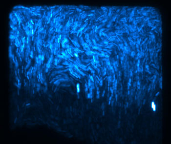
Thousands of fluorescent E. coli bacteria

 make up a biopixel. Hasty Lab, UC San Diego
In an example of life imitating art, biologists and bioengineers at UC San Diego have created a living neon sign composed of millions of bacterial cells that periodically fluoresce in unison like blinking light bulbs.

Their achievement, detailed in this week’s advance online issue of the journal Nature, involved attaching a fluorescent protein to the biological clocks of the bacteria, synchronizing the clocks of the thousands of bacteria within a colony, then synchronizing thousands of the blinking bacterial colonies to glow on and off in unison.

A little bit of art with a lot more bioengineering, the flashing bacterial signs are not only a visual display of how researchers in the new field of synthetic biology can engineer living cells like machines, but will likely lead to some real-life applications.

Using the same method to create the flashing signs, the researchers engineered a simple bacterial sensor capable of detecting low levels of arsenic. In this biological sensor, decreases in the frequency of the oscillations of the cells’ blinking pattern indicate the presence and amount of the arsenic poison.

Because bacteria are sensitive to many kinds of environmental pollutants and organisms, the scientists believe this approach could be also used to design low cost bacterial biosensors capable of detecting an array of heavy metal pollutants and disease-causing organisms. And because the senor is composed of living organisms, it can respond to changes in the presence or amount of the toxins over time unlike many chemical sensors.

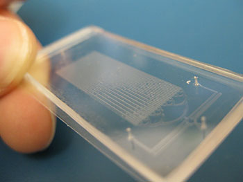
Tiny microfluidic chips allow the researchers to
 synchronize the bacteria to fluoresce 
or blink in unison
“These kinds of living sensors are intriguing as they can serve to continuously monitor a given sample over long periods of time, whereas most detection kits are used for a one-time measurement,” said Jeff Hasty, a professor of biology and bioengineering at UC San Diego who headed the research team in the university’s Division of Biological Sciences and BioCircuits Institute. “Because the bacteria respond in different ways to different concentrations by varying the frequency of their blinking pattern, they can provide a continual update on how dangerous a toxin or pathogen is at any one time.”

“This development illustrates how basic, quantitative knowledge of cellular circuitry can be applied to the new discipline of synthetic biology,” said James Anderson, who oversees synthetic biology grants at the National Institutes of Health’s National Institute of General Medical Sciences, which partially funded the research. “By laying the foundation for the development of new devices for detecting harmful substances or pathogens, Dr. Hasty’s new sensor points the way toward translation of synthetic biology research into technology for improving human health.”

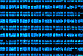
The smaller chips contain about
500 blinking bacterial colonies or biopixels
The development of the techniques to make the sensor and the flashing display built on the work of scientists in the Division of Biological Sciences and School of Engineering, which they published in two previous Nature papers over the past four years. In the first paper, the scientists demonstrated how they had developed a way to construct a robust and tunable biological clock to produce flashing, glowing bacteria. In the second paper, published in 2010, the researchers showed how they designed and constructed a network, based on a communication mechanism employed by bacteria, that enabled them to synchronize all of the biological clocks within a bacterial colony so that thousands of bacteria would blink on and off in unison.

“Many bacteria species are known to communicate by a mechanism known as quorum sensing, that is, relaying between them small molecules to trigger and coordinate various behaviors,” said Hasty, explaining how the synchronization works within a bacterial colony. “Other bacteria are known to disrupt this communication mechanism by degrading these relay molecules.”

But the researchers found the same method couldn’t be used to instantaneously synchronize millions of bacteria from thousands of colonies.