Mostrando entradas con la etiqueta EPFL. Mostrar todas las entradas

miércoles, 16 de mayo de 2018

This Hard-to-Destroy Drone Goes From Rigid to Flexible When It Crashes

Image: EPFL Don't worry, the drone is fine!
Anyone who’s ever flown a drone of any sort will tell you that sooner or later, you’re going to crash it. The question is how exactly you will go about doing this, and how much of the drone will be functional after it’s happened. Most flying animals somewhat frustratingly don’t have this problem: Birds and insects run into things occasionally (or all the time, for small bugs), and just shrug it off and keep on going, thanks to their biological design, which includes both stiffness and flexibility. Now roboticists at the EPFL, in Lausanne, Switzerland, are relying on these same qualities to design a highly resilient quadrotor that’s impressively difficult to destroy.

There are three primary strategies for designing drones with impact resistance.
  1. The first is to just protect the propellers by surrounding them with the frame of the drone or with individual propeller guards. Most commercial drones have something like this. With this level of protection, you’re less likely to injure people, but since the prop protection is rigid, you’re more likely to injure the drone itself if (I mean, when) you crash it. The EPFL quadrotor uses a flexible frame that locks in place with magnets. When a collision occurs, the frame breaks away from the magnets, and once the energy is dissipated, elastic bands pull the frame back together and you’re good to go.
  2. The second level of impact protection is to design your drone in a way that it can absorb energy from the crash without breaking into pieces. One way of doing that is to decouple the frame of the drone by, say, using flexible, elastic couplers. This gives you a “squishy” drone, which is very effective at handling impacts, but it’s also squishy in-flight, which causes all kinds of structural and stability problems
  3. The most impressive level of impact protection that we’ve seen in drones is the brute force approach of just surrounding the entire thing with a flexible, rotating cage. Flyability has made a compelling business case for using drones with protective cages, and for some applications, it’s fantastic. You do, however, pay a penalty, since the cage can increase the overall size of the drone by upwards of 60 percent, meaning that it’s safer to run into things, but you’re also much more likely to run into things. The cage adds mass as well, leading to a drone that can’t lift as much or fly as far.

EPFL’s idea is a compromise of sorts: They’ve managed to create a drone with a frame that’s rigid right up until it smashes into something, at which point it turns flexible:

The inspiration for this design came from insect wings. To fly, insects need wings with high stiffness, but flexibility is critical for absorbing shock, and wasps do it with a special joint that allows the entire wing to “reversibly crumple” during a collision, as the EPFL researchers explain:

Wasp wings display dual stiffness, that is the ability to reversibly transition between rigid and soft states, which provides mechanical resilience without impairing flight performances. The wings of wasps contain a flexible resilin joint... This design allows the wing tip to slightly flex during flight (rigid state), but reversibly crumple… during collisions (soft state). If the dual-stiffness behavior is impaired... the rigid wings undergo severe tear during collision. Therefore, this design provides crash resilience by effectively preventing wing overload during collisions without compromising flight capabilities.
Image: EPFL The quadcopter has two main parts: the external frame and a central case. They are held together by magnetic joints, each with two magnets and a spring (inset).
The quadrotor that the researchers came up with uses a flexible frame that locks in place around the core of the quadrotor with magnets. When a collision occurs, the frame breaks away from the magnets, absorbing the energy of the collision. Once the energy is dissipated, elastic bands pull the frame back into its original configuration, and the magnets snap together again, and you’re good to go.

The uniqueness of the proposed design lies in the fact that the frame is rigid during flight, but softens during collisions. This allows combination of the advantages of both rigid and soft systems: stability and rapid response to user commands during flight, leading to flight performance equivalent to a drone equipped with a standard rigid frame, and crash resilience like a soft system. The experiments showed a satisfying survivability of the frame of the drone, that withstood roughly 50 collisions with no permanent damage.

The researchers, who are members of EPFL’s Laboratory of Intelligent Systems, suggest that an approach like this could be useful for all kinds of robots, not just drones. Most robots are rigid for various performance reasons (like precision), but the ability to be flexible when necessary without using intrinsically soft materials could could come in handy for grasping, locomotion, or simply as an added safety measure for human-robot interaction tasks.

S. Mintchev, Member, IEEE, S. de Rivaz and D. Floreano, Senior Member, IEEE
Insect-Inspired Mechanical Resilience for Multicopters,” by S. Mintchev, S. de Rivas, and D. Floreano from EPFL was published in the 25 January 2017 issue of IEEE Robotics and Automation Letters.

[ EPFL ]

ORIGINAL: IEEE Spectrum
By Evan Ackerman
Posted 9 Mar 2017

jueves, 5 de marzo de 2015

The first photo of light as both particle and wave

Since the days of Einstein, scientists have been trying to directly observe how light behaves both as a particle and a wave at the same time. Now, the first-ever snapshot of this dual nature has been captured, potentially opening up a new route towards quantum computing.

Scientists at École Polytechnique Fédérale de Lausanne (EPFL) have designed an experiment that makes use of the way electrons interact with light to take a photograph of its dual nature. (Image: © Fabrizio Carbone/EPFL.)

When UV light hits a metal surface, it causes an emission of electrons. Albert Einstein explained this ‘photoelectric’ effect by proposing that light – thought to only be a wave – is also a stream of particles.

But no experiment has ever been able to capture both of these ‘split personalities’ of light at the same time. The closest researchers of quantum mechanics have come is seeing either wave or particle, but always at different times.

Now, researchers at École Polytechnique Fédérale de Lausanne (EPFL) in Switzerland and Trinity College and the Lawrence Livermore National Laboratory in the US have done an experiment with a clever twist: using electrons to image light. The team is the first to capture a single snapshot of light behaving simultaneously as both a wave and a stream of particles particle. The breakthrough work, spearheaded by Fabrizio Carbone at EPFL, was published today in the journal Nature Communications.

A new take on a classic effect 
The experiment is set up like this: A pulse of laser light is fired at a tiny metallic nanowire. The laser adds energy to the charged particles in the nanowire, causing them to vibrate. Light travels along this tiny wire in two possible directions, like cars on a highway. When waves travelling in opposite directions meet each other, they form a new wave that looks like it’s standing in place. Here, this standing wave becomes the source of light for the experiment, radiating around the nanowire.
The imaging was done at EPFL’s ultrafast energy-filtered transmission electron microscope – one of only two in the world. (Photo: EPFL.)
This is where the experiment’s trick comes in: The scientists shot a stream of electrons close to the nanowire, using them to image the standing wave of light. As the electrons interacted with the confined light on the nanowire, they either sped up or slowed down. Using an ultrafast microscope to image the position where this change in speed occurred, Carbone’s team could now visualise the standing wave, which acts as a fingerprint of the wave-nature of light.

While this phenomenon shows the wave-like nature of light, it also simultaneously demonstrates its particle aspect. As the electrons pass close to the standing wave of light, they ‘hit’ the light’s particles – the photons – thereby either accelerating or slowing down their speed. This change in speed appears as an exchange of energy ‘packets’ (quanta) between electrons and photons. The very occurrence of these energy packets shows that the light on the nanowire behaves as a particle.

New route towards quantum computing?

“This experiment demonstrates that, for the first time ever, we can film quantum mechanics – and its paradoxical nature – directly,” says Carbone. In addition, the importance of this pioneering work can extend beyond fundamental science and to future technologies. As Carbone explains: “Being able to image and control quantum phenomena at the nanometre scale like this opens up a new route towards quantum computing.”

Adapted from article by Nik Papageorgiou, EPFL Mediacom  

ORIGINAL: Technologist.eu
Technologist Online
Mar 2, 2015

lunes, 19 de enero de 2015

A Bendable Implant Taps the Nervous System without Damaging It

Swiss researchers allow rats to walk again with a rubbery electronic implant.

Why It Matters

Neuroscientists need new materials to restore movement to paralyzed people.

An implant made of silicone and gold wires is as stretchy as human tissue.

Medicine these days entertains all kinds of ambitious plans for reading off brain signals to control wheelchairs, or using electronics to bypass spinal injuries.
But most of these ideas for implants that can interface with the nervous system run up against a basic materials problem: wires are stiff and bodies are soft.

That motivated some researchers at the École Polytechnique Fédérale, in Lausanne, Switzerland, to design a soft, flexible electronic implant, which they say has the same ability to bend and stretch as dura mater, the membrane that surrounds the brain and spinal cord.

The scientists, including Gregoire Courtine, have previously showed that implants can allow mice with spinal injuries to walk again. They did this by sending patterns of electrical shocks to the spinal cord via electrodes placed inside the spine (see “Paralyzed Rats Take 1,000 Steps, Orchestrated by Computer”). But the rigid wires ended up damaging the mice’s nervous systems.

So Courtine joined electrical engineer Stéphanie Lacour (see “Innovators Under 35, 2006: Stéphanie Lacour”) to come up with a new implant they call “e-dura.” It’s made from 
  • soft silicone, 
  • stretchy gold wires, and 
  • rubbery electrodes flecked with platinum, 
  • as well as a microchannel through which the researchers were able to pump drugs.
The work builds on ongoing advances in flexible electronics. Other scientists have built patches that match the properties of the skin and include circuits, sensors, or even radios (see “Stick-On Electronic Tattoos”).

What’s new is how stretchable electronics are merging with a widening effort to invent new ways to send and receive signals from nerves (see “Neuroscience’s New Toolbox”). “People are pushing the limits because everyone wants to precisely interact with the brain and nervous system,” says Polina Anikeeva, a materials scientist at MIT who develops ultrathin fiber-optic threads as a different way of interfacing with neural tissue.

The reason metal or plastic electrodes eventually cause damage, or stop working, is that they cause compression and tissue damage. A stiff implant, even if it’s very thin, will still not stretch as the spinal cord does. “It slides against the tissue and causes a lot of inflammation,” says Lacour. “When you bend over to tie your shoelaces, the spinal cord stretches by several percent.

The implant mimics a property of human tissue called viscoelasticity—somewhere between rubber and a very thick fluid. Pinch the skin on your hand with force and it will deform, but then flow back into place.

Using the flexible implant, the Swiss scientists reported today in the journal Science that they could overcome spinal injury in rats by wrapping it around the spinal cord and sending electrical signals to make the rodent’s hind legs move. They also pumped in chemicals to enhance the process. After two months, they saw few signs of tissue damage compared to conventional electrodes, which ended up causing an immune reaction and impairing the animal’s ability to move.

The ultimate aim of this kind of research is an implant that could restore a paralyzed person’s ability to walk. Lacour says that is still far off, but believes it will probably involve soft electronics. “If you want a therapy for patients, you want to ensure it can last in the body,” she says. “If we can match the properties of the neural tissue we should have a better interface.”

ORIGINAL:
Tech Review
By Antonio Regalado 
January 8, 2015

jueves, 6 de febrero de 2014

This Bionic Hand Allows Amputee to 'Feel' Again

Image credit: alexpb

It seems like every other day we read about some far-out, new technology that makes us scratch our heads and say, "What the heck?" In this series, we'll take a look at all types of crazy new gadgets, apps and other technologies -- and the entrepreneurs dreaming them up.

One thing's for sure: no "bionic man" has ever been able to do this before.

In 2004, Dennis Aabo Sørensen lost his left hand after a firework exploded during a New Year's Eve celebration.

Little did he know that, in order to 'feel' again, all he had to do was wait for prosthetic technology to advance to the stage where electrodes could be surgically implanted in his nerves and connected to a bionic hand.

Nine years later, that day has arrived.

With the help of a high profile team of international robotic experts, Sørensen received said bionic hand, which allowed him to tell the shape and stiffness of objects while blindfolded.

Scientists have been working on the project of touch sensitive prosthetics for years now, but this is said to be the first time that an amputee has experienced real-time touch sensations through a bionic hand. Silvestro Micera -- a researcher who has worked on the project for the past 15 years -- and his team added sensors to the artificial hand, which could detect and measure information about touch, the BBC reported. Using computer algorithms, the researchers converted the electrical signals they emitted into an impulse that sensory nerves could read.

Sørensen, for his part, was in complete awe: "Suddenly you could see my left hand was talking to my brain again and it was magic," he told USA Today, when asked to describe the first moment he could 'feel' again after nine years. "It was surreal. I grabbed the object in my hand and knew it was round. It was a baseball."


Unfortunately, due to safety restrictions (the bionic hand is still a prototype) the sensors were removed from Sørensen's hand after the experiment was completed. But the project's success points to amazing capabilities for prosthetics devices of the future: one day, scientists predict, bionic hands will not only be able to feel, but also detect texture and temperature.

Imagine the ability to feel a previously missing hand closing around an object. And sensory capable bionic arms could also allow amputees to grab things in the dark, as well as perform more nuanced tasks like cracking an egg.

While it could be up to 10 years before sensory-enabled bionic hands like Sørensen's are commercially available, the bionic future looks bright: "These results show the possibilities for amputees," Micera told USA Today, before predicting that the same technology could also be used for prosthetic legs.


ORIGINAL: Entrepreneur
February 6, 2014

domingo, 22 de diciembre de 2013

An Aspiring Scientist’s Frustration with Modern-Day Academia: A Resignation

 
Here is a mind-blowing text that was sent to all EPFL researchers (presumably) by a doctoral student during the week-end. It expresses feelings that are worth to think about.

Just to be crystal-clear:
  • I am not the author of this text.
  • I don’t publish the name of his/her author, since I have no proof that his/her e-mail address was not spoofed. (NOTE: Gene Bunin acknowledges being the author of the letter in his website)
  • I don’t think that the exposed facts are a problematic unique to EPFL, nor to any other Swiss university: to the contrary, this is probably a worldwide phenomenon.
  • Finally, I would like to make very clear that I did not experience the same feelings at all during my (very happy) PhD times at EPFL. So, don’t try to make any parallel with my own experience.
  • Like the author, I don’t have any good idea how to change the system towards a better one.
Still, if you are or have been in the academic world, I think it is worth to invest 10 minutes to read this text.

Dear EPFL,

I am writing to state that, after four years of hard but enjoyable PhD work at this school, I am planning to quit my thesis in January, just a few months shy of completion. Originally, this was a letter that was intended only for my advisors. However, as I prepared to write it I realized that the message here may be pertinent to anyone involved in research across the entire EPFL, and so have extended its range just a bit. Specifically, this is intended for graduate students, postdocs, senior researchers, and professors, as well as for the people at the highest tiers of the school’s management. To those who have gotten this and are not in those groups, I apologize for the spam.

While I could give a multitude of reasons for leaving my studies – some more concrete, others more abstract – the essential motivation stems from my personal conclusion that I’ve lost faith in today’s academia as being something that brings a positive benefit to the world/societies we live in. Rather, I’m starting to think of it as a big money vacuum that takes in grants and spits out nebulous results, fueled by people whose main concerns are not to advance knowledge and to effect positive change, though they may talk of such things, but to build their CVs and to propel/maintain their careers. But more on that later.

Before continuing, I want to be very clear about two things: 

First, not everything that I will say here is from my personal firsthand experience. Much is also based on conversations I’ve had with my peers, outside the EPFL and in, and reflects their experiences in addition to my own.  
Second, any negative statements that I make in this letter should not be taken to heart by all of its readers. It is not my intention to demonize anyone, nor to target specific individuals. I will add that, both here and elsewhere, I have met some excellent people and would not – not in a hundred years – dare accuse them of what I wrote in the previous paragraph. However, my fear and suspicion is that these people are few, and that all but the most successful ones are being marginalized by a system that, feeding on our innate human weaknesses, is quickly getting out of control.

I don’t know how many of the PhD students reading this entered their PhD programs with the desire to actually *learn* and to somehow contribute to science in a positive manner. Personally, I did. If you did, too, then you’ve probably shared at least some of the frustrations that I’m going to describe next.

(1) Academia: It’s Not Science, It’s Business
I’m going to start with the supposition that the goal of “science” is
  • to search for truth, 
  • to improve our understanding of the universe around us, and 
  • to somehow use this understanding to move the world towards a better tomorrow
At least, this is the propaganda that we’ve often been fed while still young, and this is generally the propaganda that universities that do research use to put themselves on lofty moral ground, to decorate their websites, and to recruit naïve youngsters like myself.
I’m also going to suppose that in order to find truth, the basic prerequisite is that you, as a researcher, have to be brutally honest – first and foremost, with yourself and about the quality of your own work. Here one immediately encounters a contradiction, as such honesty appears to have a very minor role in many people’s agendas. Very quickly after your initiation in the academic world, you learn that being “too honest” about your work is a bad thing and that stating your research’s shortcomings “too openly” is a big faux pas. Instead, you are taught to “sell” your work, to worry about your “image”, and to be strategic in your vocabulary and where you use it. Preference is given to good presentation over good content – a priority that, though understandable at times, has now gone overboard. The “evil” kind of networking (see, e.g.,http://thoughtcatalog.com/2011/networking-good-vs-evil/) seems to be openly encouraged. With so many business-esque things to worry about, it’s actually surprising that *any* scientific research still gets done these days. Or perhaps not, since it’s precisely the naïve PhDs, still new to the ropes, who do almost all of it.

jueves, 25 de julio de 2013

Biocombustible a partir de algas de aguas residuales

ORIGINAL: Noticias De La Ciencia
Jueves, 25 julio 2013

Investigadores de la Universidad de Antioquia (Colombia) y la Escuela Politécnica de Lausanne (EPFL, en Suiza) se unieron en un proyecto para producir biomasa de microalgas y captar biológicamente CO2 y así generar, posteriormente, metano para utilizarlo como combustible.Las algas son capaces de utilizar las sales disueltas, presentes en las aguas residuales, y así aumentar la biomasa para después, mediante un tratamiento hidrotérmico, convertir esta biomasa en Metano” asegura Alejandro Acosta, investigador del Grupo de Biotransformación de la Universidad de Antioquia.

En Colombia se realiza el estudio relacionado con el uso de las aguas residuales del sector industrial y doméstico para el cultivo y aumento de la biomasa de microalgas; en Suiza se encargan de tomar esa biomasa y convertirla en gas metano al emplear una tecnología de gasificación hidrotérmica catalítica denominada SunCHem, desarrollada por EPFL.

Ya se han realizado pruebas con otros materiales orgánicos diferentes a las algas provenientes, principalmente, de materiales maderables. Pero la velocidad de crecimiento es muy baja cuando se compara con la producción que podríamos lograr utilizando microalgas”, afirma Acosta.

Los ensayos experimentales se realizaron con microalgas cultivadas en la Planta de Tratamiento San Fernando, al Sur de Medellín. Ésta realiza dos procesos de tratamiento convencionales; con la captura de las sales que permanecen en las aguas, se empezará a realizar un tercer proceso de tratamiento que sólo es común en países industrializados.


Microalgas. (Foto: AGENCIENCIA)

Así podrían verterse nuevamente las aguas minimizando el impacto ambiental al río y se aprovechan las microalgas para la generación de un biogás”, explica Acosta.

Además de la generación de combustible, las algas incidirán en el efecto del CO2 en el ambiente pues son fijadoras biológicas de este gas invernadero. (Fuente: AGENCIENCIA/DICYT)


Copyright © 1996-2013 Amazings® / NCYT® | (Noticiasdelaciencia.com / Amazings.com). Todos los derechos reservados.
Depósito Legal B-47398-2009, ISSN 2013-6714 - Amazings y NCYT son marcas registradas. Noticiasdelaciencia.com y Amazings.com son las webs oficiales de Amazings.
Todos los textos y gráficos son propiedad de sus autores. Prohibida la reproducción total o parcial por cualquier medio sin consentimiento previo por escrito.
+ Excepto cuando se indique lo contrario, la traducción, la adaptación y la elaboración de texto adicional de este artículo han sido realizadas por el equipo de Amazings® / NCYT®.

lunes, 25 de marzo de 2013

Nanowire solar cells raises efficiency limit


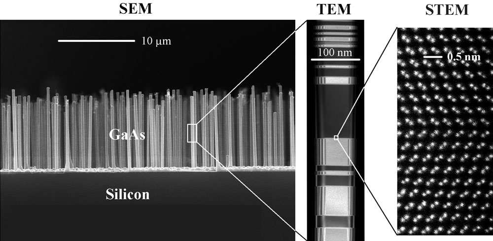
Scientists from the Nano-Science Center at the Niels Bohr Institut, Denmark and the Ecole Polytechnique Fédérale de Lausanne, Switzerland, have shown that a single nanowire can concentrate the sunlight up to 15 times of the normal sun light intensity. The results are surprising and the potential for developing a new type of highly efficient solar cells is great.
Nanowire crystals used as the solar cells. SEM (Scaning Electron Microscope) image of GaAs nanowire crystal grown on a Silicon substrate
- Due to some unique physical light absorption properties of nanowires, the limit of how much energy we can utilize from the sun's rays is higher than previous believed. These results demonstrate the great potential of development of nanowire-based solar cells, says PhD Peter Krogstrup on the surprising discovery that is described in the journal Nature Photonics.

The research groups have during recent years studied how to develop and improve the quality of the nanowire crystals, which is a cylindrical structure with a diameter of about 10,000 part of a human hair. The nanowires are predicted to have great potential in the development not only of solar cells, but also of future quantum computers and other electronic products.

- It turns out that the nanowires naturally concentrate the sun's rays into a very small area in the crystal by up to a factor 15. Because the diameter of a nanowire crystal is smaller than the wavelength of the light coming from the sun it can cause resonances in the intensity of light in and around nanowires. Thus, the resonances can give a concentrated sunlight, where the energy is converted, which can be used to give a higher conversion effeciency of the sun's energy, says Peter Krogstrup, who with this discovery contributes to that the research in solar cell technology based on nanowires get a real boost.
The figure shows that the sun's rays are drawn into a nanowire, which stands on a substrate. At a given wavelength the sunlight is concentrated up to 15 times. Consequently, there is great potential in using nanowires in the development of future solar cells. (credit: Niels Bohr Institute)

New efficiency limit
The typical efficiency limit - the so-called "Shockley-Queisser Limit" - is a limit, which for many years has been a landmark for solar cells efficiency among researchers, but now it seems that it may be increased.

- It's exciting as a researcher to move the theoretical limits, as we know. Although it does not sound like much, that the limit is moved by only a few percent, it will have a major impact on the development of solar cells, exploitation of nanowire solar rays and perhaps the extraction of energy at international level. However, it will take some years years before production of solar cells consisting of nanowires becomes a reality, says Peter Krogstrup who just completed his PhD at the Niels Bohr Institute, University of Copenhagen.

The research is conducted in collaboration with the Laboratory des Matériaux Semiconducteurs, Ecole Polytechnique Fédérale de Lausanne, the Foundation and the company SunFlake A / S. Their scientific findings work support results published in the journal Science in January. Here, a group of researchers from Lund, showed that the sun’s rays was sucked into the nanowires due to the high amount of power that their solar cell produced.

Article in Nature Photonics >>

domingo, 3 de febrero de 2013

The HBP proposal has been submitted!


Vision
Understanding the human brain is one of the greatest challenges facing 21st century science. If we can rise to the challenge, we can gain profound insights into what makes us human, develop new treatments for brain diseases and build revolutionary new computing technologies. Modern computing technology has brought these goals within sight. ICT is ready to give us a completely new understanding of the brain and its diseases; understanding the brain will lead inevitably to radical innovation in computing.

Neuroscience is generating exponentially growing volumes of data and knowledge on specific aspects of the healthy and diseased brain, in different species, at different ages. Yet despite these incredible advances, we still lack a unified understanding of the brain that can span its multiple levels of organisation, from genes to cognition and behaviour. The lack of such an understanding is a huge obstacle for pharmaceutical companies trying to develop drugs for brain diseases. It also explains why neuroscience has yet to significantly impact ICT.

Scientists have been researching isolated aspects of the brain for more than a century but despite incredible progress, it has become obvious that it will take another century or more before we can measure every gene, protein, cell, synapse and circuit in the brain, in all possible conditions and species, at every possible age, in every possible disease. An alternative strategy is to identify data that absolutely has to be measured experimentally, and to predict the rest from what we already know.

This requires a focused plan to integrate and exploit the massive volumes of data and knowledge we already have and the deluge of new data coming from labs all over the world. This will require the development of radically new ICT: new supercomputing technologies to federate and manage the data, to integrate it in computer models and simulations of the brain, to identify patterns and organisational principles that only appear when the data is put together, and to identify gaps to be filled by new experiments.

Therefore, the HBP’s first goal is to build an integrated system of six ICT-based research platforms, providing neuroscientists, medical researchers and technology developers with access to highly innovative tools and services that can radically accelerate the pace of their research. These will include

  • a Neuroinformatics Platform, that links to other international initiatives, bringing together data and knowledge from neuroscientists around the world and making it available to the scientific community; 
  • a Brain Simulation Platform, that integrates this information in unifying computer models, making it possible to identify missing data, and allowing in silico experiments, impossible in the lab; 
  • a High Performance Computing Platform that provides the interactive supercomputing technology neuroscientists need for data-intensive modeling and simulations; 
  • a Medical Informatics Platform that federates clinical data from around the world, providing researchers with new mathematical tools to search for biological signatures of disease; a Neuromorphic Computing Platform that makes it possible to translate brain models into a new class of hardware devices and to test their applications; 
  • a Neurorobotics Platform, allowing neuroscience and industry researchers to experiment with virtual robots controlled by brain models developed in the project. 

The platforms are all based on previous pioneering work by the partners and will be available for internal testing within eighteen months of the start of the project. Within thirty months, the platforms will be open for use by the community, receiving continuous upgrades to their capabilities, for the duration of the project.

The second goal of the project is to trigger and drive a global, collaborative effort that uses the platforms to address fundamental issues in future neuroscience, future medicine and future computing. A significant and steadily growing proportion of the budget will fund research by groups outside the original HBP Consortium, working on themes of their own choosing. Proposals for projects will be solicited through competitive calls for proposals and evaluated by independent peer review.

The end result will be not just a new understanding of the brain but transformational new ICT. As modern computers exploit ever-higher numbers of parallel computing elements, they face a power wall: power consumption rises with the number of processors, potentially to unsustainable levels. By contrast, the brain manages billions of processing units connected via kilometres of fibres and trillions of synapses, while consuming no more power than a light bulb. Understanding how it does this – the way it computes reliably with unreliable elements, the way the different elements of the brain communicate – can provide the key not only to a completely new category of hardware (Neuromorphic Computing Systems) but to a paradigm shift for computing as a whole, moving away from current models of “bit precise” computing towards new techniques that exploit the stochastic behaviour of simple, very fast, low-power computing devices embedded in intensely recursive architectures. The economic and industrial impact of such a shift is potentially enormous.

In short, the goal of the Human Brain Project is to build a completely new ICT infrastructure for future neuroscience, future medicine and future computing that will catalyse a global collaborative effort to understand the human brain and its diseases and ultimately to emulate its computational capabilities.

Accroches
EUROPEAN PARTNERS/


Please note all information in this website reflects the Human Brain Project-Proposal which was submitted 23 October 2012

jueves, 31 de mayo de 2012

Robotic Rehab Helps Paralyzed Rats Walk Again

ORIGINAL: Science Magazine
by Greg Miller on 31 May 2012, 2:06 PM 

Reanimated. In a new study, robot-assisted rehabilitation helped paralyzed rats regain use of their legs. Credit: Courtesy of EPFL. Lausanne, Switzerland
By employing a combination of drugs, electrical stimulation, and robot-assisted rehabilitation, researchers have restored a remarkable degree of voluntary movement in rats paralyzed by a spinal cord injury. After several weeks of treatment, the rodents were able to walk—with some assistance—to retrieve a piece of food, even going up stairs or climbing over a small barrier to get it. The rats' recovery raises hopes that a similar combination strategy could help restore movement in some people with spinal injuries. Indeed, such efforts are already underway.

Spinal injuries cause paralysis because they sever or crush nerve fibers that connect the brain to neurons in the spinal cord that move muscles throughout the body. These fibers, or axons, are the long extensions that convey signals from one end of a neuron to another, and unfortunately, they don't regrow in adults. That's why paralysis from a spinal injury is a lifelong disability. Restoring axons' ability to regrow using growth factors, stem cells, or other therapies has been a longstanding—but frustratingly elusive—goal for researchers.

The new study, which appears in Science today, takes a different approach. Instead of trying to repair the main information superhighway from the brain to the body, Grégoire Courtine, of the Swiss Federal Institute of Technology in Lausanne, and colleagues focused on alternative routes. Most spinal injuries in people do not sever the spinal cord completely, explains Courtine. To approximate this situation in rats, his team made two surgical cuts in the spinal cord, severing all of the direct connections from the brain, but leaving some tissue intact in between the cuts. Then they had the rodents begin a rehab regime intended to bypass the fractured freeway, as it were, by pushing more traffic onto neural back roads and building more of them.

This regime, which began about a week after the rats were injured, lasted about 30 minutes a day. During each session, the researchers injected the animals with a cocktail of drugs to improve the function of rats' neural circuits in the part of the spinal cord involved in leg movements, and they stimulated this area with electrodes. With its spinal cord thus primed for action, a rat was fitted into a harness attached to a robotic device that supported its weight and allowed it to walk forward on its hind legs to the extent that it was able. At first, the rats could not move their legs at all, let alone walk.

But after 2 or 3 weeks, the rodents began taking steps toward a piece of food after a gentle nudge from the robot. By 5 or 6 weeks, they were able to initiate movement on their own and walk to get the food. And after a few additional weeks of intensified rehab, they were able to walk up rat-sized stairs and climb over a small barrier placed in their path. Rats that did not undergo rehab, in contrast, showed no improvement at all. Rats suspended over a moving treadmill that elicited reflex-like stepping movement, did not improve either, suggesting that full recovery depends on making intentional movements, not just any movement.

"It's a really remarkable finding," says Michael Beattie, a neuroscientist at the Brain and Spinal Injury Center at the University of California, San Francisco. Additional experiments in the paper make a compelling case that the rats' recovery is due to new neural connections forming to create a detour around the injury, he says. Beattie notes that Courtine's work suggests that all three components of the rehab strategy—the drugs, the electrical stimulation, and the robot-assisted physical therapy—seem to be necessary to maximize recovery. "I think that it actually provides a lot of hope that this kind of strategy will have a big payoff" in people, Beattie concludes.

A case study published last year reported some recovery of voluntary movements in a man paralyzed in a vehicle accident, after he underwent a combination of electrical stimulation and physical therapy. The new rodent research provides a potential explanation for that patient's recovery, says one of the lead authors of the case study, neuroscientist V. Reggie Edgerton at the University of California, Los Angeles. Edgerton says two more patients are undergoing similar rehab now, and his group hopes to add drug therapy to enhance nerve repair in the future. "We're not there yet," he says. "But the bottom line is, things are still looking good."

As encouraging as the new findings are, Courtine is careful to note the strategy's limitations
For one thing, it wouldn't work if the spinal cord were completely severed
In addition, treated rats could only make voluntary movements while the electrical stimulation was turned on, and the same was mostly true of the patient Edgerton and colleagues worked with. "This is not a cure for spinal cord injury," Courtine says. "It's a promising proof of principle."

Follow ScienceNOW on Facebook and Twitter

lunes, 16 de enero de 2012

El mecanismo que da forma a la vida, desde las ballenas hasta las lombrices de tierra


© CC Genista
Los ratones no tienen la cola en la espalda y las costillas no se desarrollan a partir de las vértebras lumbares. Y por una buena razón. 
Científicos en la EPFL han descubierto el mecanismo que determina la forma que adquieren  muchos animales - incluyendo a los humanos, las ballenas azules, y los insectos.

¿Por qué no crecen los brazos desde el centro de nuestro cuerpo? La cuestión no es tan trivial como parece. Vértebras, extremidades, costillas, coxis ... en sólo dos días, todos estos elementos toman su lugar en el embrión, en el lugar correcto y con la precisión de un reloj suizo. Intrigado por la extraordinaria fiabilidad de este mecanismo, los biólogos se han preguntado cómo funciona. Ahora, los investigadores de la EPFL (Ecole Polytechnique Fédérale de Lausanne) y la Universidad de Ginebra (UNIGE) haber resuelto el misterio. Su descubrimiento se publicará 13 de octubre 2011 en la revista Science.

El embrión se construye una capa a la vez
Durante el desarrollo de un embrión, todo sucede en un momento determinado. En aproximadamente 48 horas, se pasará de la parte superior de la parte inferior, una rebanada a la vez - los científicos llaman a ésto segmentación del embrión. "Estamos hechos de treinta y tantos cortes horizontales", explica Denis Duboule, profesor de la EPFL y UNIGE. "Estos cortes corresponden más o menos el número de vértebras que tenemos".

Cada hora y media, se construye un segmento nuevo. Los genes correspondientes a las vértebras cervicales, las vértebras torácicas, las vértebras lumbares y el coxis se activan en el el momento justo, uno tras otro. "Si el tiempo no es seguido al pié de la letra, se terminará con costillas saliendo de las vértebras lumbares", bromea Duboule. ¿Cómo funcionan los genes para saber cómo se lanzan a la acción de una manera perfectamente sincronizada? "Asumimos que el ADN juega el papel de una especie de reloj. Pero no entendíamos cómo ".

Cuando el ADN funciona como un reloj mecánico
Genes muy específicos, conocidos como "Hox", están involucrados en este proceso. Responsables de la formación de las extremidades y la columna vertebral, tienen una característica notable. "Los genes Hox están situadas exactamente una tras otra en la cadena de ADN, en cuatro grupos. Primero el cuello, el tórax, el lumbar, y así sucesivamente ", explica Duboule. "Este acuerdo único, inevitablemente, tuvo que jugar un papel".

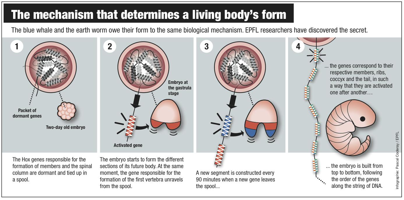
El proceso es sorprendentemente simple. En los primeros momentos del embrión, los genes Hox están latentes, empaquetado como un carrete de hilo enrollado en el ADN. Cuando sea el momento adecuado, la cadena comienza a relajarse. Cuando el embrión comienza a formar los niveles superiores, los genes que codifican la formación de las vértebras cervicales salen de la cola y se activan. Luego es el turno de las vértebras torácicas, y así sucesivamente hasta la rabadilla. La hebra de ADN actúa un poco como una tarjeta perforada  de los equipos antiguos, entrega de instrucciones específicas, ya que cada vez pasa por la máquina.

"Un nuevo gen que sale de la cola de cada noventa minutos, que se corresponde con el tiempo necesario para una nueva capa del embrión que se construirá", explica Duboule. "Se necesitan dos días para que la hebra de ADN se desenroye por completo, lo que es el mismo tiempo que se necesita para todas las capas del embrión se completen." Este sistema es la primera "mecánica" de reloj que se haya descubierto en la genética. Y explica por qué el sistema es tan extraordinariamente preciso.

Este descubrimiento es el resultado de muchos años de trabajo. Bajo la dirección de Duboule y Daniel Noordermeer, el equipo analizó miles de carretes de genes Hox. Con la ayuda del Instituto Suizo de Bioinformática, los científicos fueron capaces de recopilar grandes cantidades de datos y el modelo de la estructura de la cola de impresión y cómo se desenvuelve en el tiempo.
El mecanismo que determina la forma de los cuerpos viviientes. EPFL

La serpiente: una línea de montaje verdadera vertebral
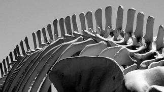
El proceso descubierto de la EPFL es compartida por numerosos seres vivos, desde los seres humanos a algunos tipos de gusanos, de las ballenas azules a los insectos. La estructura de todos estos animales - la distribución de sus vértebras, extremidades y otros apéndices a lo largo de su cuerpo - está programado como una hoja de reproductor de música de piano por la secuencia de los genes Hox a lo largo de la cadena de ADN.

El cuerpo sinuoso de la serpiente es un ejemplo perfecto. Hace unos años, Duboule descubrió en estos animales un defecto en el gen Hox que normalmente detiene el proceso de toma de las vértebras. "Ahora sabemos lo que está pasando. El proceso no se detiene, y el embrión de serpiente sólo sigue haciendo vértebras, todas idénticas, hasta que el proceso sólo pierde fuerza. "

El reloj de Hox es una demostración de la extraordinaria complejidad de la evolución. Una propiedad notable del mecanismo es su extrema estabilidad, explica Duboule. "Los relojes circadianos o menstrual implican una química compleja. De este modo, puede adaptarse a contextos cambiantes, pero en un sentido general son bastante imprecisos. El mecanismo que hemos descubierto debe ser infinitamente más estable y preciso. Hasta el más mínimo cambio acabaría conduciendo a la aparición de una nueva especie. "

---
Esta investigación se lleva a cabo en el Centro Nacional de Competencia en Investigación (NCCR) Frontiers in Genetics. El NCCRs son una iniciativa del gobierno suizo para estimular la investigación y la educación en áreas clave. http://www.frontiers-in-genetics.org