Mostrando entradas con la etiqueta microscopía. Mostrar todas las entradas

miércoles, 8 de octubre de 2014

Stefan Hell (Nobel Prize in Chemistry 2014): STED - Insights into the nanoworld

Ok, so do you wonder what all the fuss is about with the Chemistry Nobel Prize this year : ) -? If so, this film will do the trick & tell you all about it! A quick introduction of Max Planck researcher Stefan Hell & and his pioneering work in the field of ultra-high resolution fluorescence microscopy.


Optical microscopes cannot distinguish between objects that are closer together than about 200 nanometers – about one two hundredth of a hair's breadth. The reason for this is the wave nature of light, the half wavelength of which roughly corresponds to those 200 nanometers. The STED microscopy developed by Stefan Hell is the first optical microscope technology to go beyond this magic barrier, enabling researchers to gain fascinating insights into the nanoworld.

ORIGINAL: What's The Big Deal

jueves, 27 de marzo de 2014

2014 Koch Institute Image Award Winners

Last fall, we featured The Koch Institute Image Award galleries in several Cell Picture Shows. This Show furthers the collaboration, as we showcase this year’s winning submissions. Both the Koch Institute Public Galleries and the Cell Picture Show share a similar ethos: recognition and dissemination of the extraordinary imagery produced through life science research. On March 4, 2014, these winning images were unveiled at MIT’s Koch Institute for Integrative Cancer Research in Cambridge, MA. 

We congratulate the 2014 Image Award Winners and are excited to continue to the collaboration between MIT and Cell Press. This collection of stunning images offers a window into the fascinating worlds opened to us by microscopy and other biomedical imaging techniques.

Biopolymer in Bloom
Julio D’Arcy, Erik Dreaden, and Paula Hammond
Hammond Laboratory
MIT Koch Institute
A New Environment for Studying Cell Growth. Measuring cancer cells’ real-time response to external influences can be challenging. Here, engineers have created biocompatible plastic structures onto which cells can adhere and develop as they would inside the body. The electrically conductive nature of the scaffolds allows researchers to measure the properties of the growing cells. By changing the environment or introducing new substances into the system, researchers can figure out which factors promote or discourage cell growth.
Image: This image, taken with a scanning electron microscope, shows the micro- and nano-scale structures of this device

The More the Messier
Kristin Knouse
Amon Laboratory
MIT Koch Institute

Understanding Complicated Cell Division. The mitotic spindle is an array of tracks that partitions chromosomes during cell division. Most normal cells form bipolar spindles, which segregate chromosomes equally into two daughter cells. However, many cancer cells form multipolar spindles, which cause chromosome mis-segregation and genomic instability.
Image: Like many cancer cells, liver cells also form multipolar spindles during cell division. Shown here is a liver cell with a multipolar spindle (green) pulling the chromosomes (blue) in many directions. Further research into cell division in the liver could indicate how this process is exploited or disrupted in cancer, revealing novel avenues for cancer therapy.

Target Practice
Omar F. Khan and Edmond W. Zaia
Langer and Anderson Laboratories
MIT Koch Institute

Improving Gene Therapy with Nanotechnology. How can we turn off the genes that promote the development of cancer? Using specially designed nanoparticles as genetic patches, engineers can deliver customized payloads to a cell’s gel-like cytoplasm, where most cellular activity occurs, and mitigate the effects of cancer-causing genes in the cell’s nucleus.
Image: This image shows nanoparticles (red) in the cytoplasm of cervical tumor cells (green). As researchers learn more about how cells respond to these therapies, they will continue to tweak the patches to determine the appropriate distribution of synthetic and genetic material to best target different types of cancer.

Blood, Heat, and Tumors
Alex Bagley, Jeff Wyckoff, and Sangeeta Bhatia
Bhatia Laboratory
MIT Koch Institute

Improving Drug Delivery with Gold Nanorods. Blood vessels are highways through the body. They can transport drugs to cancer cells, but finding the appropriate ramp to exit the vessel can be tricky.
Image: This image shows a network of blood vessels (green) and collagen (purple) infused with gold nanorods (yellow) inside of a living tumor. When researchers heat the particles with near-infrared light, the blood vessels become leaky, making it easier to deliver a therapeutic cargo to its final destination. Because blood vessels provide a universal transport system, such combination therapy has widespread implications for treatment, regardless of cancer type or specific drug needed

The Bad Seed
Mandar Deepak Muzumdar 
Jacks Laboratory
MIT Koch Institute

Modeling the Growth of a Tumor. Small changes have big effects. Although scientists know that certain gene mutations trigger tumor formation, the subsequent cellular events that drive cancer progression are not well understood. Cell-specific fluorescent marking allows researchers to track mutated cells over the entire course of cancer development.
Image: This image shows mutated (green) and nonmutated (red and yellow) cells in a pancreas. Over time, the green cells will multiply dramatically and form a solid tumor, while the others will not. Comparing properties and behaviors of the different cell types will set the stage for earlier diagnosis, better treatment, and even chemoprevention of deadly cancers.

Rainbow Connections
Zeynep Saygin
Kanwisher Laboratory
MIT Department of Brain & Cognitive Sciences

Mapping Neural Pathways in the Brain. The human brain is massively complex. Neuroimaging techniques such as MRI provide a noninvasive tool for studying its inner workings.
Image: This image shows pathways of nerve fibers through the brain in three dimensions: up/down (blue), front/back (green), and left/right (red). By comparing these maps of connectivity with maps of neural function, researchers can begin to predict how individual brains will respond to different stimuli. That will eventually help them to understand healthy brain development and will enable earlier diagnosis and interventions for conditions such as autism and dyslexia.

Silencing Echoes
Soheil Feizi, Steven Lee (Artist), Daniel Marbach, Muriel Medard, and Manolis Kellis Computational Biology Group
MIT Computer Science and Artificial Intelligence Laboratory

Cleaning Up Networks. Are all connections meaningful? This image visualizes a new algorithm (known as "network deconvolution") for determining important relationships in complex networks. Like a filter on a camera lens, it reveals which links (lines) between interconnected elements (points) are most essential. As the lens passes over each network area, indirect links disappear and direct links become visible. Already tested on large networks mapping gene regulation, protein folding, and academic co-authorship, network deconvolution can be used to identify key drivers of biological, social, and technological systems.

Ganglion Style
Alex Norton for EyeWire
Seung Laboratory
MIT Department of Brain and Cognitive Sciences and MIT Media Lab

Crowdsourcing Science through Online Games. It's all fun and games until somebody maps a neuron! Then it’s time to move on to the next one. The online game EyeWire challenges players, most of whom have no background in neuroscience, to create virtual 3D models of actual neurons using real laboratory data.
Image: The reconstruction seen here shows ganglion cells in the retina. By comparing this gamer-generated map to previously collected data about the neurons’ firing activity, neuroscientists can create a functional model of how vision works. With more than 100,000 players, EyeWire has already helped researchers to uncover how the eye helps us perceive moving stimuli.

Something Fishy
Annie Cavanagh and David McCarthy
School of Pharmacy
University College London

The Secret Lives of Zebrafish. Humans and fish have more in common than you might expect. Since the 1970s, a tropical freshwater minnow known as the zebrafish has been used to study the genetic and physiological development of living organisms. By mapping the zebrafish genome and studying irregularities in their development, researchers have been able to create robust models of how vertebrates develop and identify genetic conditions that lead to diseases such as cancer.
Image: This image shows a false-color scanning electron micrograph of a zebrafish embryo. It appears in the Koch Institute Public Galleries as part of a partnership between the Koch Institute and Wellcome Images.

Collateral Damage
Aprotim Mazumder, Jennifer A. Calvo, and Leona D. Samson
Samson Laboratory
MIT Koch Institute, Department of Biological Engineering, Department of Biology, and Center for Environmental Health Sciences

Investigating the Side Effects of Chemotherapeutics. How much is too much? When treating cancer, it is important to balance a drug’s effectiveness at killing tumor cells with its toxicity to healthy cells elsewhere in the body.
Image: This image of brain tissue shows cerebellar granule neurons (pink), which sustain significant damage when exposed to certain DNA-damaging therapeutics, and surrounding Purkinjee cells (orange), which do not. Researchers are studying these responses to determine the cell properties and repair mechanisms that make different cell types more or less vulnerable to chemotherapy.

ORIGINAL: Cell

domingo, 23 de marzo de 2014

Magnified Photos Show the Intricate Details of Butterfly Wings

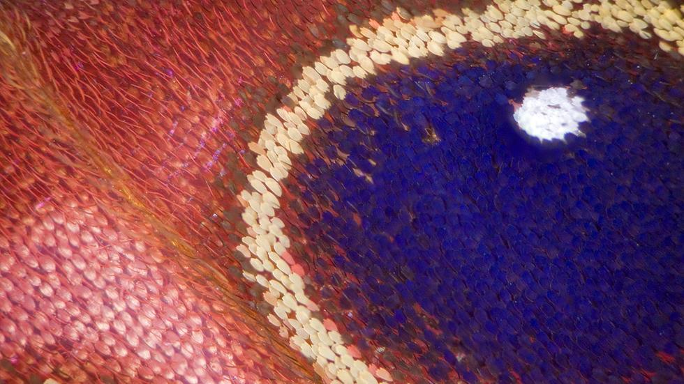
This image shows the colorful sunset moth's wing. (Linden Gledhill)

This image shows the colorful sunset moth's wing. (Linden Gledhill)
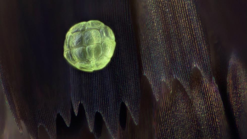
A pollen grain rests on a Protographium agesilaus butterfly wing. (Linden Gledhill)
The above image shows a magnified Salamis Parhassus wing. (Linden Gledhill)
The above image shows a Citharias aurorean wing. (Linden Gledhill)
More images:

Many photographers aim to catch the beauty of butterflies. But Linden Gledhill takes his photography a step further, showing us the gorgeous patterns at the cellular level.

Gledhill develops biopharmaceuticals to treat cancer and diabetes at an international pharmaceutical company. His photography blends his love of science into an artform. He spends nights and vacations working on his photography projects.

I love applying my knowledge and skills in science to achieve images which often people don’t see,” Gledhill told weather.com.

According to the Library of Congress, the powder seen on butterflies’ wings is similar to scales. Their wings are made of very thin layers of chitin, a derivative of glucose. Similar to solar panels, the wings soak up heat from the sun in order to keep the butterfly warm, The New York Times reported.

In the past, Gledhill has used a standard camera fitted with old microscope lenses on extension tubes. He now uses an Olympus BHT metrology microscope fitted with a StackShot drive to create the images. He also uses LED lighting and high-speed flash along with the microscope.

He usually uses damaged preserved specimens from farm-raised butterflies.

Gledhill’s images cover a range of species and they include moths as well. “I select those with interesting coloration or scale shapes… one of the most spectacular examples is the sunset moth, Urania ripheus,” said Gledhill. “People often think of moths as being drab. This is a day-flying species, hence the bright colors, most of which are achieved by physical structure and not pigments.

ORIGINAL: Weather Channel
By Nicole Bonaccorso weather.com

viernes, 27 de diciembre de 2013

Olympus BioScapes 2013 Winners Gallery

Thumbnail images of the Olympus BioScapes 2013 winners and honorable mentions are displayed in this gallery. In order to view a larger version of the images (or to play videos), please click on the individual thumbnails.

2013 Winning Entries
The Olympus BioScapes 2013 winners, honorable mentions, and technical merit awards are displayed in this gallery. In order to view the images, please click on the individual links.
Specimen: Carnivorous U. gibba plant
HHMI Janelia Farm Research Campus
Ashburn, Virginia, United States
Specimen: Open trap of aquatic carnivorous plant, humped bladderwort Utricularia gibba, with single-cell organisms inside.
Technique: Confocal imaging, 100x
2nd Prize - Miss Dorit Hockman Specimen: Molossus rufus embry
Miss Dorit Hockman
University of Oxford
Oxfordshire, United Kingdom
Specimen: Embryo of black mastiff bat Molossus rufus.
Technique: Stereo microscopy
3rd Prize - Dr. Igor Siwanowicz Specimen: Desmids
Dr. Igor Siwanowicz
HHMI Janelia Farm Research Campus
Ashburn, Virginia, United States
Specimen: Single-cell fresh water algae (desmids). Composite image including, concentric from the outside: Micrasterias rotata, Micrasterias sp., M. furcata, M. americana, 2x M. truncata, Euastrum sp. and Cosmarium sp.
Technique: Confocal imaging, 400x
4th Prize - Mr. Spike Walker Specimen: Lily flower bud
Staffordshire, United Kingdom
Specimen: Lily flower bud, transverse section.
Technique: Darkfield illumination, stitched images
5th Prize - Dr. Dylan Burnette Specimen: Mouse fibroblasts
Dr. Dylan Burnette
National Institutes of Health
Bethesda, Maryland, United States
Specimen: Mouse embryonic fibroblasts showing actin filaments (red), mitochondria (green) and DNA (blue).
Technique: Structured illumination microscopy (SIM) fluorescence, acquired with a 60x objective
6th Prize - Mr. Kurt Wirz Specimen: Gonocerus acuteangulatus
Mr. Kurt Wirz
Basel, Switzerland
Specimen: "Brother bugs." Gonocerus acuteangulatus, two hours old. Size 3mm.
7th Prize - Mr. Charles Krebs Specimen: Phantom Midge larva
Mr. Charles Krebs
Issaquah, Washington, United States
Specimen: Phantom midge larva (Chaoborus) "Glassworm." Birefringent musculature that is usually clear and colorless is made visible here by specialized illumination.
Technique: Polarized light, 100X

8th Prize - Dr. Yaron Fuchs Specimen: Mouse tail with stem cells
Dr. Yaron Fuchs
Howard Hughes Medical Institute/The Rockefeller University
New York, NY USA
Specimen: Mouse tail whole mounts showing hair follicle stem cells and proliferating cells.
Technique: Confocal imaging
9th Prize - Mr. Fabrice Parais Specimen: Sericostoma sp.
Mr. Fabrice Parais
DREAL (Regional Directorate of Environment, Planning and Housing) of Basse-Normandie
Caen, France
Specimen: Head and legs of a caddisfly larva: Sericostoma sp., a benthic macroinvertebrate that can be used for freshwater biomonitoring; because it is relatively sensitive to organic pollution and dies if water is dirty, it is a good indicator of water quality.
Technique: Stereo microscopy, 15x
10th Prize - Mr. Ralph Grimm Specimen: Video: Paramecium
Mr. Ralph Grimm
Jimboomba Queensland, Australia
Specimen: Paramecium, showing contractile vacuole and ciliary motion.
Technique: Differential interference contrast, 350x-1000x


2013 Honorable Mentions


C. Barros

M. Boyle

T. Burns

M. Clarke

M. Crutchley

N. Cuenca

S. Di Talia

J. Dolan

G. Drange

J. Ducharme

A. Dumitrache

A. Ertürk

A. Ferrand

M. Ghabril and C. Babbey

M. Gibson

M. Gibson

G. Günther

J. Hallfeldt

T. Hickman

P. Honkakoski

C. Jackson

M. Kandasamy

M. Khodaverdi

M. Klinghardt

L. Knight

L. Knight

A. Kobitski et al.

A. Kohn and J. Kubo

C. Krebs

C. Krebs

M. Lehnert and C. Mulvane

N. Lindström

X. Lu and C. Bolt

G. Luna

D. Maitland

J. Michels

J. Michels

D. Millard

M. Miś

D. Moore

R. Moreno Gill

S. Mouchet

J. Myslowski

W. Nell

J. Nicholson

S. Nishimura

A. Pan

J. Petersen

J. Petersen

A. Phillips-Yzaguirre

C. Pintér

C. Pintér

P. Ray

G. Rouse

A. Salehi

A. Singh

I. Siwanowicz

V. Sýkora

E. Tabdanov

R. Taiariol

V. Tobias Santos

M. Turzańska

M. Turzańska

W. van Egmond

P. Verrees

D. von Wangenheim

L. Windus

K. Wirz

A. Woolley and A. Gilmour

All image copyrights belong to the individual contestants.
For image use permissions, contact ilene@olympusbioscapes.com

ORIGINAL: Olympus Bioscapes