Mostrando entradas con la etiqueta Microfluídica. Mostrar todas las entradas

domingo, 2 de noviembre de 2014

Lego-like modular components make building 3-D 'labs-on-a-chip' a snap


Modular fluidic and instrumentation components developed by researchers at the University of Southern California Viterbi School of Engineering. Credit: USC Viterbi School of Engineering

Thanks to new LEGO-like components developed by researchers at the USC Viterbi School of Engineering, it is now possible to build a 3-D microfluidic system quickly and cheaply by simply snapping together small modules by hand.

Microfluidic systems are used in many fields including engineering, chemistry and biotechnology to precisely manipulate small volumes of fluids for use in applications such as 
  • enzymatic or DNA analysis, 
  • pathogen detection, 
  • clinical diagnostic testing, and 
  • synthetic chemistry. 
Traditionally, microfluidic devices are built in a cleanroom on a two-dimensional surface using the same technology developed to produce integrated circuits for the electronics industry.

Though tiny, designing, assembling and testing a new microfluidics system can take a lot of time and money. Building a single device can often require multiple iterations, each of which can take up to two weeks and several thousand dollars to manufacture. And the more complex the system, the higher the number of iterations needed.

"You test your device and it never works the first time," said Krisna Bhargava, materials science graduate student at the USC Viterbi School of Engineering. "If you've grown up to be an engineer or scientist, you've probably been influenced by LEGO at some point in your childhood. I think every scientist has a secret fantasy that whatever they're building will be as simple to assemble."

Frustrated that reproducing a simple microfluidic circuit could cost him so much time and money, Bhargava set out simplify the construction process. First, he identified the primitive elements commonly used in microfluidic systems, much like how circuitry is broken down in electrical engineering. Basic microfluidic functions would be separated into standardized modular components, not an entirely revolutionary concept. But then, he abandoned the two-dimensional method of building microfluidic devices altogether.

"The founders of the microfluidics field took the same approach as the semiconductor industry: to try to pack in as much integrated structure as possible into a single chip," explained Bhargava. "In electronics, this is important because a high density of transistors has many direct and indirect benefits for computation and signal processing. In microfluidics, our concerns are not with bits and symbolic representations, but rather with the way fluidics are routed, combined, mixed, and analyzed; there's no need to stick with continuing to integrate more and more complex devices."

Borrowing an approach from the electronics industry, which uses prototype boards to build circuits, Bhargava conceived of three-dimensional modular components that encapsulated the common elements of microfluidic systems, as well as a connector that could join the separate components together. Inspired by recent advancements in micron-scale 3D-printing, he and a USC Viterbi research team that included chemical engineering and materials science professor Noah Malmstadt and biomedical engineering graduate student Bryant Thompson, designed computer models for eight modular fluidic and instrumentation components (MFICs, pronounced "em-fix") that would each perform a simple operation. Examples are a "helix" component that can mix two fluid streams and a component that contains an integrated optical sensor for measuring the size of small droplets. The components constructed for this study are approximately 1 cm3, slightly smaller than a standard 6-sided die.

The team's development of these MFICs represents the first attempt to break a device into separate components that can be assembled, disassembled and re-assembled over and over.


lunes, 25 de agosto de 2014

Sorting cells with sound waves

Acoustic device that separates tumor cells from blood cells could help assess cancer’s spread.
Illustration: Christine Daniloff/MIT
Researchers from MIT, Pennsylvania State University, and Carnegie Mellon University have devised a new way to separate cells by exposing them to sound waves as they flow through a tiny channel. Their device, about the size of a dime, could be used to detect the extremely rare tumor cells that circulate in cancer patients’ blood, helping doctors predict whether a tumor is going to spread.
This microfluidic device uses sound waves to sorts cells as they flow through the channel, from left to right. Image courtesy of the researchers
Separating cells with sound offers a gentler alternative to existing cell-sorting technologies, which require tagging the cells with chemicals or exposing them to stronger mechanical forces that may damage them.

Acoustic pressure is very mild and much smaller in terms of forces and disturbance to the cell. This is a most gentle way to separate cells, and there’s no artificial labeling necessary,” says Ming Dao, a principal research scientist in MIT’s Department of Materials Science and Engineering and one of the senior authors of the paper, which appears this week in the Proceedings of the National Academy of Sciences.

Subra Suresh, president of Carnegie Mellon, the Vannevar Bush Professor of Engineering Emeritus, and a former dean of engineering at MIT, and Tony Jun Huang, a professor of engineering science and mechanics at Penn State, are also senior authors of the paper. Lead authors are MIT postdoc Xiaoyun Ding and Zhangli Peng, a former MIT postdoc who is now an assistant professor at the University of Notre Dame.

The researchers have filed for a patent on the device, the technology of which they have demonstrated can be used to separate rare circulating cancer cells from white blood cells.

To sort cells using sound waves, scientists have previously built microfluidic devices with two acoustic transducers, which produce sound waves on either side of a microchannel. When the two waves meet, they combine to form a standing wave (a wave that remains in constant position). This wave produces a pressure node, or line of low pressure, running parallel to the direction of cell flow. Cells that encounter this node are pushed to the side of the channel; the distance of cell movement depends on their size and other properties such as compressibility.

However, these existing devices are inefficient: Because there is only one pressure node, cells can be pushed aside only short distances.

The new device overcomes that obstacle by tilting the sound waves so they run across the microchannel at an angle — meaning that each cell encounters several pressure nodes as it flows through the channel. Each time it encounters a node, the pressure guides the cell a little further off center, making it easier to capture cells of different sizes by the time they reach the end of the channel.


ORIGINAL: MIT
Anne Trafton | MIT News Office 
August 25, 2014

viernes, 18 de abril de 2014

Neurology and microfluidic science kits take over for kids in the 21st century

Today kids are not happy enough to play around with the old chemistry kits from yesteryear, they want more, they want real science. Recently the people behind the ISEF science fair, The Society for Science & the Public, launched a competition for children with the aim of letting children make their own modern version of a science kit for kids in the 21st century. Of course in today’s world, children do have access to a great deal more modern technology than what was around in the 60s and 70s.

[Image Courtesy of George Korir]

The results of the competition are in and it was a handmade microfluidic kit that took first prize in the competition. This was made by a graduate student along with the help of his biochemistry professor. For those who don’t know, microfluidic tech is the stuff that is found in lab-on-a-chip devices which are used to analyse small amounts of liquid. These move around and mix chemicals through channels, which are of course very small, on platforms that are very small, such as the size of a computer chip. Scientists have been busy working on devising microfluidic devices that can mimic human organs. First they had to decide what chemical reactions to make in the kit for children.

Holes were then punched out of paper card and these holes corresponded to sequences and were then loaded with the correct chemicals. In order to make the reactions between the chemicals, kids had to then use the cards with a reader that worked with a hand crank. This then released the chemicals slowly, just one single drop for every hole that was punched in the card. Manu Prakash from the Stanford University, who was the professor who helped to make the system, made a statement saying that he could see kids in the future trading the reaction cards and could even become as popular as trading baseball cards were in the past.


In second place came a design of electrodes that when placed onto the body can sense electricity after it sent messages to the brain produced by the body whilst flexing muscles. To do this, electrodes were hooked up to amplifiers and then an electrical device such as a motor or light bulb. This allowed those using it to turn on something, for instance a propeller, simply by squeezing their hand or even just thinking about it.


[Image Courtesy of George Korir]


Chemistry sets were always on the Christmas list of some boys and girls during the 60s and 70s but today they are more than just attaching wires to a battery and small light bulb. The competition could have sparked off a whole new trend when it comes to science kits and they could become the in-thing for budding inventors and scientists for Christmas 2014 and beyond.

For a full list of all winners and their bios head over to the Gordon and Betty Moore Foundation.


April 15th, 2014 

miércoles, 26 de marzo de 2014

Biochip quickly tests results of cancer therapy


The inside structure of the biochip is comprised of 3 layers, each testing a key element of the therapy: drug, oxygen, and light. (Credit: Xia Lou/University of Michigan)


University of Michigan right Original Study ("A high-throughput photodynamic therapy screening platform with on-chip control of multiple microenvironmental factors")

A new type of lab-on-a-chip could make it easier to determine the effectiveness of a promising cancer treatment that combines photosensitive drugs, light, and oxygen.

The treatment, known as photodynamic therapy (PDT), uses a unique type of drug, called a photosensitizer, that generates high-energy oxygen when activated by light, in this case an LED laser.

The oxygen reaction destroys cells locally only in the immediate surrounding area, which in this case is the tumor, without damaging other healthy cells in the rest of the body. PDT may also prompt the patient’s immune system to attack the tumor, whereas without treatment it will likely ignore it.

PDT is a fairly complicated therapy because it requires light, oxygen, and the drug, all three of which need to be carefully controlled,” says Xia Lou, a postdoctoral fellow at the University of Michigan. “With chemotherapy, for example, you only control how much of the drug is used.”

The biochip can test the interaction of the drug, light, and oxygen simultaneously, generating results in a fraction of the time of current testing practices.

In cancer research doctors are always looking for better drugs,” adds Lou. “But there has always been a lack in the ability to efficiently test new drugs.

Researchers are also hoping to get more reliable test results than is the norm. We are providing more precise drug test conditions to insure that the results we are getting will more closely match the actual results from cancer treatment.
Test all three components There are two primary challenges in PDT today:
  • One is determining the best treatment plan for the patient, and 
  • the other is testing the efficacy of new and existing drugs.
For example, the location of a tumor in the body will determine the amount of light needed to excite the drug, and will impact the amount of drug that is administered. Also, when testing new drugs, it is again important to know the amount of light needed to excite the oxygen reaction, and the strength of the reaction.

This adds a high level of complexity compared to conventional drug testing that only tests one component, the drug itself.

Current PDT testing methods might take more than 24 hours to acquire just 10 data points.

With our device,” says Lou, “we can finish the testing in 1 hour and have 1,000 data points. The result is a very comprehensive understanding of how the 3 elements need to be manipulated for maximum effect in an individual patient.

The researchers reported the details of their findings in the journal Lab on a Chip. The NSF Engineering Research Center for Wireless Integrated Microsystems, Thermo Fisher Scientific, and the National Institute of Health supported the project.

Source: University of Michigan

ORIGINAL: Futurity
February 28, 2014 

domingo, 4 de agosto de 2013

Cell-targeting automata, mimicking QM, DNA analysis via Phone, brain-to-brain interface, nanoribbons, cell reprogramming, amazing microfluidics. Science Sunday. Digest 26 - 4th Aug 2013.

ORIGINAL: Mark Bruce

SciTech #ScienceSunday Digest 26 - 4th Aug 2013.

1. Molecular Automata Improve Cell Targeting.By linking specific antibodies to specific short DNA sequences termed “molecular automata” unique subpopulations of cells can be targeted and identified http://www.hss.edu/newsroom_molecular-robots-build-targeted-therapeutics.asp. In a proof-of-concept with human blood samples researchers were able to target specific subpopulations of white blood cells. The antibody component binds to the desired receptor expressed on the cell surface, while the DNA component is a linker molecule that can fluoresce. By adding a number of different “automata” you can progressively select only those cells that express those particular receptors on their surface and no others; the DNA strands from the different automata go through complementary binding due to their proximity and fluoresce, enabling them to be identified and separated. This is a great addition of modularity into cell and drug targeting technology - individual advances in drug carriers and cell targeting are now possible and complimentary.

2. Mimicking Quantum Mechanics with Fluid Dynamics.The fluidic analogue of a classic quantum experiment has been produced, but instead of electrons being confined by a circular ring of iron atoms, bouncing fluid droplets are confined in a dish http://web.mit.edu/newsoffice/2013/when-fluid-dynamics-mimic-quantum-mechanics-0729.html. In this deceptively simple experiment the fluid droplets were shown to mimic the statistical behaviour of electrons with remarkable accuracy; the same distribution appeared in both systems. This provides evidence for de Broglie’s Pilot Wave theory - at least for fluid droplets bouncing along vibrating fluid baths, driven by interfering waves produced by their own bounces. The video is worth a watch The pilot-wave dynamics of walking droplets.

3. DNA Analysis on Your Smartphone.A company called Biomeme is to launch a new smartphone docking station that plugs into your phone and makes use of its processing power to perform real-time DNA analysis http://www.medgadget.com/2013/07/mobile-real-time-dna-analysis-on-your-smartphone.html. The dock takes biological samples and allows your phone to conduct PCR on the sample and subsequently determine the presence of specific DNA sequences of interest that are linked to pathogens or other diseases. Cheap ultra-mobile and portable DNA analysis would be quite a game-changer for many applications. Detecting single DNA point-mutations is getting easier too http://www.washington.edu/news/2013/07/28/breakthrough-in-detecting-dna-mutations-could-help-treat-tuberculosis-cancer/.

4. Non-Invasive Brain-to-Brain Communication Allows Mind Control of Rat’s Tail.The first brain-to-brain interface between a human and a rat has been demonstrated and, in a proof-of-concept study allowed the human to mind-control the movement of the rat’s tail http://www.plosone.org/article/info:doi/10.1371/journal.pone.0060410. The article includes a nice embedded video showing the experimental setup of how an animal’s body was remote-controlled by someone else's brain - still very rudimentary but such capabilities will only get easier and better with time. A new technique can also measure neuronal activity at a faster rate http://blogs.princeton.edu/research/2013/07/25/a-faster-vessel-for-charting-the-brain-nature-communications/.

5. New Meniscus Lithography Creates Long Graphene Nanoribbons.Long graphene nanoribbons can now be created easily and cheaply using a new water-enabled lithographic technique that uses water or the meniscus of at a water interface as a mask http://news.rice.edu/2013/07/29/water-clears-path-for-nanoribbon-development/. The new technique allows sub-10nm nanoribbons to be constructed and built into field-effect transistors using much cheaper fabrication equipment. In future work the group aims to improve the technique to produce ribbons of uniform thickness and controllable edge state to better define the electronic properties. Graphene supercapacitors are moving along nicely http://monash.edu.au/news/show/soft-approach-leads-to-revolutionary-energy-storage.
A thin line of platinum sits atop a substrate. The metal nanowire was created with a new meniscus mask process discovered at Rice University. Image by Tour Group/Rice University

6. Computer Model Predicts How to Change Cell Differentiation.A new computer model can make accurate predictions concerning which differentiated cells can be efficiently changed into different cell types - from skin to nerve cells for example http://wwwen.uni.lu/universite/actualites/a_la_une/cells_reprogrammed_on_the_computer. This is a big step in computational biology, for producing computer-based instructions for reprogramming cells for various applications. Previously such differentiated cell conversions required a precisely delivered cocktail of factors that largely arose as a result of trial and error. Now, with the new model, this trial and error approach has been replaced by calculations that determine which genes, at what time, and in what order need to be switched on to achieve the desired results; these predictions were proved accurate in the lab.

7. Microfluidic Breakthrough: Merging, Transporting, Splitting Addressable Microdroplets.In a big step for microfluidics a new microfluidic system is able to merge, transport, and split microdroplets on the chip over long timescales and involving potentially thousands of operations on each of the hundreds of addressable microdroplets http://phys.org/news/2013-07-microfluidic-breakthrough-biotechnology.html. The droplets are addressable by each having a unique optoelectronic identifier that allows the user to monitor, at any time, what chemical operations have been performed on each droplet. The prototype system successfully manipulated and analysed microdroplets that were each loaded with thousands of bacteria - they showed that bacteria were confined to their original droplet and could not move to others.

8. Controllably Binding Together Atoms that Repel.By adding a controlled amount of energetic noise into a system atoms that normally repel one another can be made to form a strong and stable bond http://phys.org/news/2013-07-repelling-atoms.html. The addition of the controlled noise into the quantum system of the atoms, in this case from vacuum fluctuations from the electromagnetic field, induces an interference phenomenon that traps the atoms and forces them into a robust bond. The group quotes possible applications in cooling atomic quantum gases but this is tapping into a new natural phenomenon - if we can understand it more completely and achieve mastery it will open the way to interesting new technologies.

9. Guided Self-Assembling, Self-Integrating Nanowire Circuits.A new technique for growing nanowires not only allows nanowires to be grown in ordered horizontal arrays, but to be guided to automatically assemble into transistors and then subsequently “self-integrate” to form logic circuits http://phys.org/news/2013-07-growth-nanowires-self-integrated-circuits.html. The advance makes it possible to determine the arrangement of nanowires in advance to suit the desired electronic circuit that the user wishes to make. This is a great example of controlled bottom-up assembly of complex structures.

10. A Cheap Billion-Pixel Microscope.A new computational image-processing method allows a standard microscope to be fitted-out with $200 worth of add-ons and become a billion-pixel imaging system that can outperform the best standard microscopes http://www.caltech.edu/content/pushing-microscopy-beyond-standard-limits. The system allows both large fields of view and very high resolution - basic tests showed it could produce 100 times more information than the unmodified systems, and improve the performance of a 2x objective lens to the level of a 20x objective lens. The system actually measures both light intensity and phase from different angles . . . and because it measures the phase I wonder if this might find application in holography? 
The Yang lab's new microscope setup (left). A raw image taken with a 2X objective lens is shown (top right) along with the reconstructed image produced by the new microscope setup (bottom right).
Credit: Guoan Zheng. CalTech


The weekly SciTech Digests are also available as a Google Currents Edition here:
https://www.google.com/producer/editions/CAow4-hB/scitech_digest

+ScienceSunday, with your hosts +Buddhini Samarasinghe, +Rajini Rao, +Chad Haney, +Allison Sekuler
2013-08-04
2 photos

jueves, 1 de agosto de 2013

Lifelike cooling for sunbaked windows

ORIGINAL: Wyss Institute - Harvard
Jul 30, 201

Adaptable microfluidic circulatory system could cut air-conditioning costs

Microfluidic window
A specially fabricated sheet of silicone rubber (PDMS) creates a network of channels that function as an artificial circulatory system. Water flows through those channels on hot, sunny days, which should help keep windows -- and the air inside buildings -- cool. [Credit: Wyss Institute]
Boston, Mass. -- Sun-drenched rooms make for happy residents, but large glass windows also bring higher air-conditioning bills. Now a bioinspired microfluidic circulatory system for windows developed by researchers at the Wyss Institute for Biologically Inspired Engineering at Harvard University could save energy and cut cooling costs dramatically -- while letting in just as much sunlight.

The same circulatory system could also cool rooftop solar panels, allowing them to generate electricity more efficiently, the researchers report in the July 29 online edition of Solar Energy Materials and Solar Cells.

The circulatory system functions like those of living animals, including humans, which contain an extensive network of tiny blood vessels near the surface of the skin that dilate when we are hot. This allows more blood to circulate, which promotes heat transfer through our skin to the surrounding air.

The artificial circulatory system can cool a glass window pane significantly -- enough, if used throughout a building, to save significant amounts of energy and chop cooling costs. [Credit: Wyss Institute]

Similarly, the new window-cooling system contains an extensive network of ultrathin channels near the "skin" of the window -- the pane -- through which water can be pumped when the window is hot. The channels consist of long, narrow troughs that are molded into a thin sheet of clear silicone rubber that, when stretched over a flat pane of glass, create sealed channels.

"The water comes in at a low temperature, runs next to a hot window, and carries that thermal energy away," said Benjamin Hatton, Ph.D., lead author of the study. Hatton, who is now an assistant professor of materials science and engineering at the University of Toronto, was a member of the Advanced Technology Team at the Wyss Institute. He worked on the Adaptive Material Technologies platform led by Joanna Aizenberg, Ph.D., who is a Core Faculty member of the Wyss Institute and the Amy Smith Berylson Professor of Materials Science at Harvard School of Engineering and Applied Sciences.

Today's insulation and construction methods do a good job keeping heat from leaking through walls, but heat transfer through glass windows remains one of the major stumbling blocks to energy-efficient buildings. In large part, that is because the molecules in glass absorb the sun's infrared light, heating the window, which heats the air inside the building significantly.

The idea to cool glass windows when they get hot emerged from work on microfluidics by Don Ingber, M.D., Ph.D., the Wyss Institute's Founding Director, and his team working on biomimetic microsystems. Ingber is also the Judah Folkman Professor of Vascular Biology at Harvard Medical School and Boston Children's Hospital, and Professor of Bioengineering at Harvard School of Engineering and Applied Sciences.

Microfluidic devices circulate fluids through tiny, ultrathin channels and are typically used to build small devices for laboratory research and clinical diagnosis. In contrast, Ingber's team developed an innovative method to build large-scale microfluidic devices for organ-on-chip applications. They first use a vinyl cutter -- a computer-controlled device that cuts intricate patterns on large vinyl sheets -- to create a plastic mold. Then they pour liquid silicone rubber into the mold, let it solidify, and remove it, which creates the thin sheet imbued with long, narrow troughs.


The channels that make up the artificial circulatory system are visible when they're empty (left), but transparent when they're filled with water (right). Windows with this system installed would remain transparent. [Credit: Wyss Institute]

When Ingber's microfluidics team met with Aizenberg's adaptive materials team in cross-platform meetings, the idea emerged that this microfluidics technology could be applied to building materials to control heat transfer, much like capillary blood flow warms the feet of Antarctic penguins as they wait for their mates near the South Pole.

Hatton and the Wyss Institute team then created and tested a four-inch-square microfluidic windowpane. They found that when these channels were filled with water, they were also transparent to the eye -- which is just what people want in a window, Hatton said.

They then used a heat lamp to heat a pane with this vasculature to 100 F -- as hot as a window might get on a sunny summer day. Using a special infrared camera, they showed that the circulatory system could readily cool the pane.

The Wyss Institute team then worked with Matthew Hancock, an applied mathematician at the Broad Institute in Cambridge, Mass., who developed a mathematical model that predicts how the circulatory system would perform on normal-size windows. Pumping just half a soda can's worth of water through the window's circulatory system would cool a full-size window pane by a full 8 C (14 F), they calculated. The energy needed to pump water would be far less than the heat energy the water absorbed. This suggested that installing the cooled windows throughout a building would generate a big net win.

"The idea of using nature's lesson to create kind of a living skin on a building is a very important and promising direction for how buildings should and will be constructed in the future," said Chuck Hoberman, an award-winning U.S. designer, expert in adaptive architecture, and Wyss Institute Visiting Scholar.

"Our new window technology marries advances in microfluidics with creative thinking about adaptive architecture, and it's the sort of cross-disciplinary research that the Wyss Institute was designed to foster," Ingber said. "We are optimistic that microfluidic windows will go a long way toward helping us cool our homes and commercial buildings more efficiently."

Next, the researchers plan to team up with architecture researchers to meld their mathematical model with existing architectural energy-modeling software to see how much energy microfluidic windows would save if installed over an entire building.

This work was funded by the Wyss Institute. In addition to Hatton, Aizenberg, Ingber and Hancock, the research team included: Ian Wheeldon, Ph.D., a former Wyss postdoctoral researcher who's currently an assistant professor in the department of chemical and environmental engineering at the University of California, Riverside, and Matthias Kolle, Ph.D., a postdoctoral fellow on Aizenberg's team.

PRESS CONTACT

Dan Ferber

dan.ferber@wyss.harvard.edu

+1 617-432-1547

IMAGES AND VIDEO AVAILABLE

###

About the Wyss Institute for Biologically Inspired Engineering at Harvard University

The Wyss Institute for Biologically Inspired Engineering at Harvard University (http://wyss.harvard.edu) uses Nature's design principles to develop bioinspired materials and devices that will transform medicine and create a more sustainable world. Working as an alliance among Harvard's Schools of Medicine, Engineering, and Arts & Sciences, and in partnership with Beth Israel Deaconess Medical Center, Brigham and Women's Hospital, Boston Children's Hospital, Dana Farber Cancer Institute, Massachusetts General Hospital, the University of Massachusetts Medical School, Spaulding Rehabilitation Hospital, Boston University and Tufts University, the Institute crosses disciplinary and institutional barriers to engage in high-risk research that leads to transformative technological breakthroughs. By emulating Nature's principles, Wyss researchers are developing innovative new engineering solutions for healthcare, energy, architecture, robotics, and manufacturing. These technologies are translated into commercial products and therapies through collaborations with clinical investigators, corporate alliances, and new start-ups. The Wyss Institute recently won the prestigious World Technology Network award for innovation in biotechnology.

lunes, 8 de julio de 2013

Chemists Work to Desalt the Ocean for Drinking Water, One Nanoliter at a Time

ORIGINAL: U of Texas
June 27, 2013

AUSTIN, Texas —

??By creating a small electrical field that removes salts from seawater, chemists at The University of Texas at Austin and the University of Marburg in Germany have introduced a new method for the desalination of seawater that consumes less energy and is dramatically simpler than conventional techniques. The new method requires so little energy that it can run on a store-bought battery. The process evades the problems confronting current desalination methods by eliminating the need for a membrane and by separating salt from water at a microscale.

The technique, called electrochemically mediated seawater desalination, ?was ? described last week in the journal?? Angewandte Chemie. The research team was led by?? Richard Crooks of The University of Texas at Austin and Ulrich Tallarek of the University of Marburg. It’s patent-pending and is in commercial development by startup company Okeanos Technologies. “The availability of water for drinking and crop irrigation is one of the most basic requirements for maintaining and improving human health,” said Crooks, the Robert A. Welch Chair in Chemistry in the College of Natural Sciences. “Seawater desalination is one way to address this need, but most current methods for desalinating water rely on expensive and easily contaminated membranes. The membrane-free method we’ve developed still needs to be refined and scaled up, but if we can succeed at that, then one day it might be possible to provide fresh water on a massive scale using a simple, even portable, system.

This new method holds particular promise for the water-stressed areas in which about a third of the planet’s inhabitants live. Many of these regions have access to abundant seawater but not to the energy infrastructure or money necessary to desalt water using conventional technology. As a result, millions of deaths per year in these regions are attributed to water-related causes.

People are dying because of a lack of freshwater,” said Tony Frudakis, founder and CEO of Okeanos Technologies. “And they’ll continue to do so until there is some kind of breakthrough, and that is what we are hoping our technology will represent.
The left panel shows the salt (which is tagged with a fluorescent tracer) flowing upward after a voltage is applied by an electrode (the dark rectangle) jutting into the channel at just the point where it branches. In the right panel no voltage is being applied.

To achieve desalination, the researchers apply a small voltage (3.0 volts) to a plastic chip filled with seawater. The chip contains a microchannel with two branches. At the junction of the channel an embedded electrode neutralizes some of the chloride ions in seawater to create an “ion depletion zone” that increases the local electric field compared with the rest of the channel. This change in the electric field is sufficient to redirect salts into one branch, allowing desalinated water to pass through the other branch.

The neutralization reaction occurring at the electrode is key to removing the salts in seawater,” said Kyle Knust, a graduate student in Crooks’ lab and first author on the paper.

Like a troll at the foot of the bridge, the ion depletion zone prevents salt from passing through, resulting in the production of freshwater.

Thus far Crooks and his colleagues have achieved 25 percent desalination. Although drinking water requires 99 percent desalination, they are confident that goal can be achieved.

This was a proof of principle,” said Knust. “We’ve made comparable performance improvements while developing other applications based on the formation of an ion depletion zone. That suggests that 99 percent desalination is not beyond our reach.


The other major challenge is to scale up the process. Right now the microchannels, about the size of a human hair, produce about 40 nanoliters of desalted water per minute. To make this technique practical for individual or communal use, a device would have to produce liters of water per day. The authors are confident that this can be achieved as well.

If these engineering challenges are surmounted, they foresee a future in which the technology is deployed at different scales to meet different needs.

You could build a disaster relief array or a municipal-scale unit,” said Frudakis. “Okeanos has even contemplated building a small system that would look like a Coke machine and would operate in a standalone fashion to produce enough water for a small village.


The fundamental scientific breakthroughs that led to this advance were primarily supported by the Office of Basic Energy Sciences in the U.S. Department of Energy. Okeanos Technologies is funded by venture capital and grants from the U.S. Environmental Protection Agency. The intellectual property is owned by The University of Texas at Austin through the Office of Technology Commercialization (OTC). In the event of eventual profits, patent holders, including Crooks and Knust, will be paid according to the OTC’s standard licensing agreement. Okeanos Technologies is also currently supporting Knust’s stipend and tuition via a gift to UT.

For more information, contact: Daniel Oppenheimer, College of Natural Sciences, 512 745 3353; Richard Crooks, 512-475-8639, 

lunes, 3 de junio de 2013

Popular Scientist. Aydogan Ozcan (LUCAS Inventor)

ORIGINAL: UCLA MAGAZINE
By Robin Keats
Apr 1, 2013


UCLA Associate Professor of Electrical and Bioengineering Aydogan Ozcan is one of the world's "Brilliant 10" scientists, as proclaimed by Popular Science magazine. Ozcan is the director of the Bio-and Nano-Photonics Laboratory at the UCLA Henry Samueli School of Engineering and Applied Science, where he leads a team of students in creating breakthrough—and inexpensive—technological devices that bring the frontiers of medicine to ordinary individuals.
Photo by: Clara Richmond.
We could begin with any one of more than a dozen startlingly original inventions to frame the contributions of Aydogan Ozcan, but let's start with the crowd-sourced bio-game he and his team of researchers created to diagnose malaria. One thousand online gamers from around the world were directed to identify, then "kill" or "bank," infected red-blood cells that appeared on their screens as electronic images. Their success rate at identifying the disease was very comparable to that of medical experts. This decidedly non-expert methodology is designed for use in developing nations where hugely expensive diagnostic equipment is unavailable.

And then there is BigFoot, the name Ozcan gave to the monitoring software he pioneered that allows diabetes patients and others with chronic foot ailments to track their conditions at home using a conventional flatbed scanner and a PC.

These are just two of an apparently endless lineup of incredible ideas that spring from the mind of the 34-year-old scientist who already holds 22 patents, with more than 15 pending, for inventions in wide-field and lens-less imaging, nonlinear and fiber optics, critical coherence tomography and nanoscopy.

Ozcan's self-described "prize child," though, is LUCAS, an acronym for "Lens-less, Ultra-wide-field blood Cell monitoring Array platform based on Shadow imaging." It turns a cell phone into a diagnostic device by clipping on a gadget that combines an LED light, a spatial filter and a slot for a medical slide. Information is gathered by the device, which sends it off to diagnosticians anywhere in the world. Bring LUCAS into the jungle where people get little or no medical attention, and voila! Expert diagnosis.

Through the device, millions of people in such remote areas as sub-Saharan Africa who would otherwise go undiagnosed will be screened or monitored for malaria, and eventually for tuberculosis and HIV. It will also help prevent waterborne illnesses that kill about 5 million people a year. And the cost to clip LUCAS onto your cell phone? Five to 10 bucks.

No 'Aha' Moment
Ozcan was born in Istanbul, Turkey, in 1978. His father was a government worker, his mother a housewife. His education began in the small, Black Sea city of Sinope, which also gave the world Diogenes, founder of Cynic philosophy. "My family moved around a lot," he says. "Five different schools for the five years of elementary school we have in Turkey ... I think those moves caused me to develop skills that have helped me quickly adapt to things."

Ozcan went to Bilkent University in Ankara for his bachelor's degree. He earned a master's degree and Ph.D. at Stanford University, followed by two years in Boston on the research faculty of Harvard Medical School's Wellman Center for Photomedicine. Ozcan joined UCLA as an assistant professor in 2007.

What turned him onto scientific innovation? "There was," he says, "no 'aha' moment." But as early as his freshman year in high school, "the idea of proving something and learning the framework of proofs was so unique," he recalls. "That's when I said 'I love it' [and] thought I'd be in a profession that allows for the creation of useful things.
Add caption
For his breakthrough medical diagnostic inventions, Ozcan has been honored by NASA, the U.S. Department of State, the Gates Foundation, National Geographic and Popular Mechanics, among other institutions and publications. In the photo above, he is holding a prototype of his LUCAS device in his lab.

Teaching Invention "Being a scientist and going to the frontier of knowledge requires a lot of dedication, a lot of small things put together," he says. "I insist that my students know that those who are successful in this profession are the ones who learn how to cope with failures. You improve from rejection; you write a new manuscript; you try again. Some people would say, 'It's enough.' I won't play that game."

It has to be that way, Ozcan says of his role as an educator. "Within the school of engineering, the function of a professor is to create new information and new technologies. You also have to train the next generation of engineers and scientists, so that new know-how is continually created to make life simpler."

Ozcan adds, "The definition of engineering, for me, is to make life simpler and better. That's the most important thing that differentiates an engineer's mind from that of a physicist. A physicist is looking at interesting questions, but not how a combination of disciplines could be applied to create things that will enhance life. That's the intersection of science and engineering and I want to be, professionally, at that juncture. Engineers need to be very multidisciplinary. That's what I stress in my lab."

Creating Invention

The man whose world involves complex technologies like optics, photonics and imaging has a simple vision of his own future as an innovator. "It's like a single picture," he says. "You can give yourself 10 or 15 years to paint it, but then it's going to be something you can admire. That's how I take my career. I'm in the middle of that picture now [with] the expansion of the cell phone into an array of telemedicine tools."

Cell phones, he points out, have better graphics processors than IBM supercomputers had in the late 1980s. "There are already 5 billion cell phones in use," he explains. "We manufacture 2 billion of them every year … There will be more of them everywhere on the planet."

With this phenomenal global network of smart gadgets, he points out, computation is almost free.

"Because cell-phone use is so ubiquitous and inexpensive," he adds, "I can rely on it … to design new microscopes and micro-analysis tools that look at bodily fluids using technology attached to the cell phone or running on it."

The "Nano Internet" and Beyond
The young scientist's next challenge is already obvious to him. He foresees spending the next decade or so building the "Nano Internet," which he describes as "the development of these personal micro-analysis tools [that] give us something more valuable than the tools themselves."

What he envisions are millions of his tiny devices interconnecting and streaming personal health information in real time, from brainwaves to blood-pressure counts.

"If my lab and others like it do our work well," he predicts, "then we'll have this kind of Nano Internet. The opportunities that lie ahead of us, with our eyes, with our information, channeled into the micro and Nano worlds."

These and other trails yet unimagined will be blazed from Westwood. "UCLA maintains and extends such vision," says Ozcan. "It's got everything I need to fulfill whatever I dream."

martes, 2 de abril de 2013

Luke Bawazer on Genetically Evolving Technology

ORIGINAL: 33rd Square
April 1, 2013 


Luke Bawazer's research in synthetic biology aims to utilize droplet microfluidics to advance studies of solution-based mineralization and to develop new biomimetic materials engineering strategies. Recently he spoke at TEDxWarwick about the promise and potential of this work. 
Luke Bawazer works in Fiona Meldrum's laboratory at University of Leeds, where he conducts research at the interface of chemistry, materials science, and synthetic biology.

Bawazer's research aims to utilize droplet microfluidics to advance studies of solution-based mineralization and to develop new biomimetic materials engineering strategies. A major anticipated outcome of our research is the ability probe interesting materials chemistry and biochemistry questions that are important to biomineralization, using novel low-cost and high-throughput platforms.

His work is inspired by living organisms, such as bones, teeth, and seashells. These biominerals will help us create new materials technologies using genetic engineering. With synthetic biology we can now make and evolve synthetic genes that encode new inorganic composite materials with useful electronic functions.

"There are many examples in nature of really amazing materials from the perspective of a materials scientist or a materials engineer, they are really amazing materials," Bawazer told Materials Today late last year. "Some examples are abalone shell, which is made of calcium carbonate, or it’s 95% calcium carbonate, and less than 5% organic matter, and yet that composite has a 3,000 times higher fracture toughness than calcium carbonate alone, which is of course just chalk, and it’s due to a unique micro-architecture. So if you examine and look at abalone shell in a microscope, large sections of it look like a brick-and-mortar structure, with micro-bricks in micro-mortar being biomolecules."


Bawazer has recently been highlighted for his work towards genetically evolving semiconductors. He has received his PhD in Biochemistry and Molecular Biology from Dan Morse's laboratory at the University of California focusing on the directed laboratory evolution of biomineralizing enzymes.

According to Bawazwer, "a very important part of this evolution approach is the capacity of these artificial cells, and that’s something I’m very interested in, and that I’d like to develop further. Currently, I’m working with new microfluidic-based techniques to create artificial cells that are more robust, probed in different ways. So, for example, if we’d like to look for materials that are electronically active, then we want to be able to probe these cells electronically, and that requires certain properties of the cell membrane. The main things I’m working on is developing new sets of artificial cells that are more robust and amenable to directed laboratory evolution."

SOURCE TEDx Talks

miércoles, 20 de marzo de 2013

Harvard's Wyss Institute and Sony DADC Announce Collaboration on Organs-on-Chips

ORIGINAL: Wyss Institute
Date: Mar 18, 2013

Boston, MA -- Today the Wyss Institute for Biologically Inspired Engineering at Harvard University and Sony DADC announced a collaboration that will harness Sony DADC's global manufacturing expertise to further advance the Institute's Organs-on-Chips technologies. 
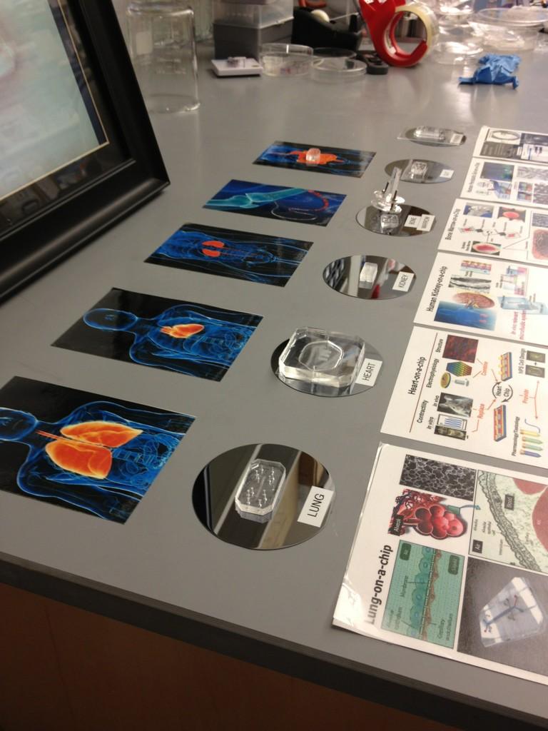
Human Organs-on-Chips are composed of a clear, flexible polymer about the size of a computer memory stick, and contain hollow microfluidic channels lined by living human cells -- allowing researchers to recapitulate the physiological and mechanical functions of the organs, and to observe what happens in real time. The goal is to provide more predictive and useful measures of the efficacy and safety of new drugs in humans -- and at a fraction of the time and costs associated with traditional animal testing.

"We are excited to apply Sony DADC's deep manufacturing expertise to confront one of the major challenges in the life sciences by helping to accelerate the translation of the Wyss Institute's Organ-on-Chips from the benchtop to the marketplace," said Christoph Mauracher, Senior Vice President of the BioSciences division of Sony DADC. "The Organs-on-Chips have the potential to revolutionize testing of drugs, chemicals, toxins and cosmetics."


This collaboration builds on the momentum the Wyss Institute team has gained recently on its Organs-on-Chips research program. With support from Defense Advanced Research Projects Agency (DARPA)*, National Institutes of Health (NIH), Food and Drug Administration (FDA), and pharmaceutical partners, more than ten Organs-on-Chips are currently under development at the Wyss Institute, including a lung, heart, liver, kidney, bone marrow, and gut-on-a-chip; there is also a major effort to integrate these organ chips into "human body on-chips" that mimic whole body physiology.

In February, Wyss Founding Director Don Ingber, M.D., Ph.D., who leads the Organs-on-Chips research program, received the prestigious 3Rs Prize from the UK's National Centre for the Replacement, Refinement and Reduction of Animals in Research for the lung-on-a-chip. This month, the Society of Toxicology awarded him the Leading Edge in Basic Science Award for his "seminal scientific contributions and advances to understanding fundamental mechanisms of toxicity."

"Our work with Sony is a wonderful example of the Wyss Institute model in action," said Ingber. "We collaborate with industry to help de-risk the technologies we develop, both technically and commercially, and therefore expedite their translation into real world applications."

###

*Part of this research was sponsored by the U.S. Army Research Office (ARO) and DARPA; the views and conclusions contained in this document are those of the authors and should not be interpreted as representing the official policies, either expressed or implied, of ARO, DARPA or the U.S. Government.

Contacts
Wyss Institute for Biologically Inspired Engineering 
Kristen M. Kusek
+1 617-432-8266
Kristen.kusek@wyss.harvard.edu 

Sony DADC
Manfred Koranda
+43 6246 880 8143
manfred.koranda@sonydadc.com

lunes, 7 de enero de 2013

Building a body, one organ chip at a time

ORIGINAL: Vector
by TOM ULRICH
JANUARY 4, 2013

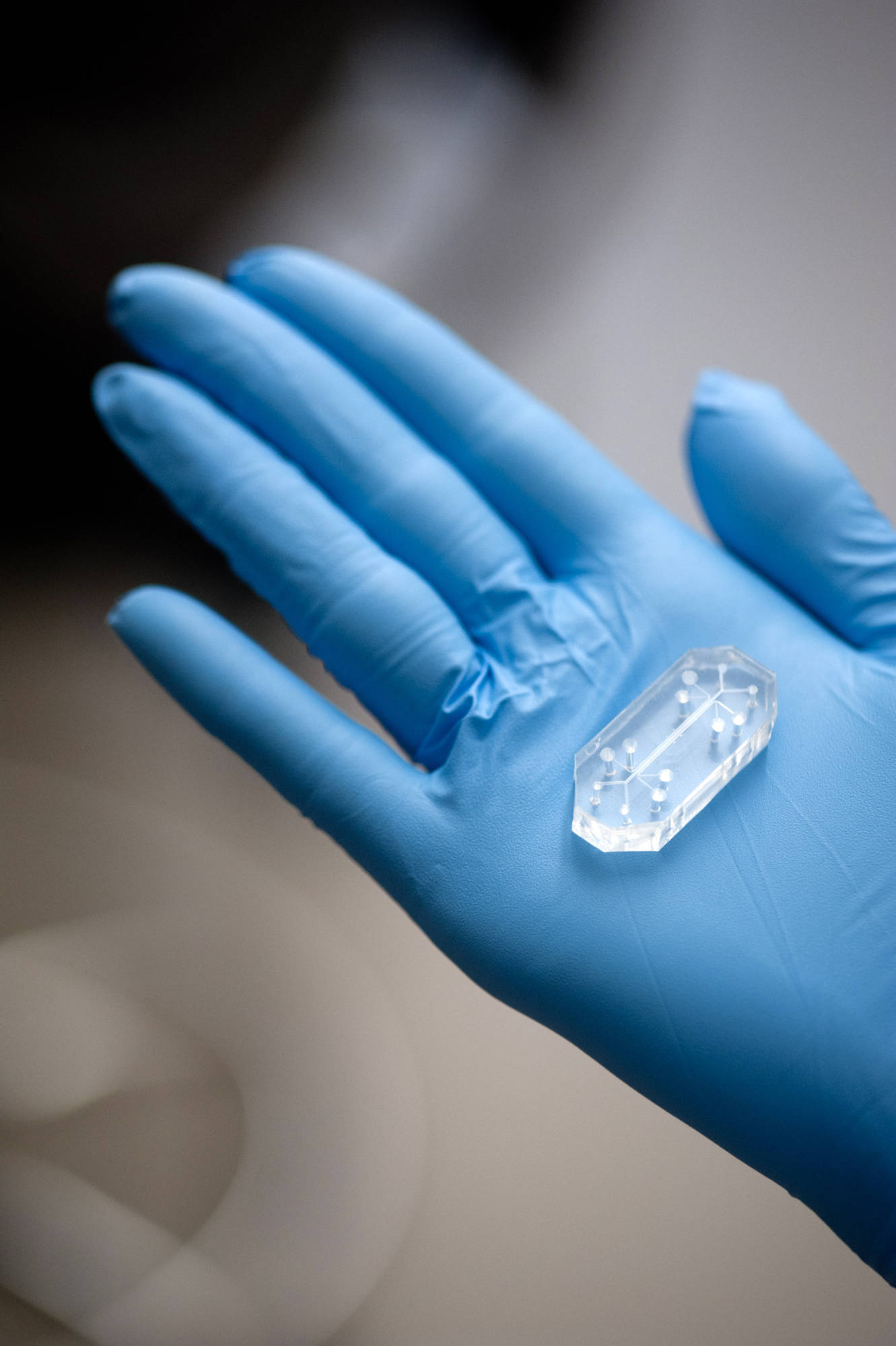
It may not look like it, but it's a lung, just in chip form
They don’t look like much sitting in your hand. A few pieces of clear plastic, each smaller than an Altoids tin, with channels visible inside and holes for plugging tubing into them.

But fill them with cells and treat those cells the right way, and they turn into something amazing: tiny hearts, lungs, guts, kidneys.

They’re “organs on chips,” and they represent what’s probably the most comprehensive effort to date to physically model the functions of whole organs for drug development and disease research.

Developed by a team of biologists and engineers led by Donald Ingber, MD, PhD, a member of Boston Children’s Hospital’s Vascular Biology Program and director of the Wyss Institute for Biologically Inspired Engineering at Harvard, they’re the building blocks for an ambitious project to create an artificial multi-organ system—essentially, a whole body on a chip.

Each of the chips—Ingber’s team is currently developing 10 different organs—is built using microfabrication techniques like those common in the semiconductor industry.This allows us to create features and structures that we can control at the size scale in which cells live, and also apply physiological fluid flows and mechanical forces,” Ingber explains. “We have precise control over where cells live in the device and what they experience.

Going through the motions
The two features Ingber mentioned, flow and force, are instrumental in faithfully mimicking organ function. His lung on a chip, for instance, has a central microfluidic channel that is split into two parallel channels by porous flexible membrane. One side of the membrane is coated with human lung “air sac” cells (over which air can pass), the other with human lung capillary blood vessel cells. A combination of pumps subject the cells to the sensations of continuous blood flow and rhythmic breathing by moving culture medium through the blood vessel channel and applying suction that deforms the cell-coated membrane.

This video from the Wyss Institute explains in more detail:


Those forces incite dramatic responses from the cells in the chips. “You can put endothelial [blood vessel] and epithelial [air sac] cell cultures together, but they won’t reflect the range of functions you see in a real lung,” Ingber says. “This is because respiratory physiology relies on the mechanics of breathing and blood flow.

For instance, once we added breathing motions to the chip,” he adds, “the epithelial cells started producing surfactant, just like what happens in the lining of a normal lung when a baby takes its first breath.
“[O]nce we added breathing motions to the [lung] chip, the epithelial cells started producing surfactant, just like what happens…when a baby takes its first breath.”
Similarly, Ingber’s lab recently revealed that breathing motions were essential for using the lung chip to mimic the pulmonary edema (fluid leakage from blood vessels into the lungs) that occurs in some cancer patients treated with the drug interleukin-2—the first demonstration that chips could model a complex human disease, as well as a drug toxicity.

Ingber’s team has also developed a chip that models the complex environment of the human gut—including its microbial inhabitants. “Once we added a trickling flow of medium and peristaltic motions, the cells in the chip started forming structures similar to intestinal villi,” he says. “Now we’ve added bacteria, and can start to study the relationship between the microbiome and human intestinal disease processes.

Ingber's expanding repertoire of organs-on-chips could soon be linked together to create a whole body on a chip.
With a host of chips now in hand, Ingber and his team are now working with the federal Defense Advanced Research Project Agency to create a system for linking chips representing different organs together. The system will allow broader study of organ physiology and also how drugs affect multiple organs—the first step toward simulating a complete living body.

We want to be able to administer a drug via the ‘gut’ or ‘lungs,’ see how it is metabolized by the ‘liver,’ excreted by the ‘kidney,’ and whether it causes toxicity in the ‘heart,’” says Ingber. “We also want to model a broad range of disease states, like asthma, Crohn’s, radiation exposure and so on.

He’s also talking to the Food and Drug Administration about potentially accepting organ-on-chip data as part of the drug approval process in addition to or, in the future, in lieu of animal data.We’ve only just started the discussion, but we hope they’ll one day accept human organ chip data instead of certain animal studies, just as they’ve begun to accept biomarker data.


jueves, 15 de noviembre de 2012

Organ Printing from Stem Cells


Researchers Have Figured Out How to Use Modified Inkjet Technology to Print Cells

Tissue engineering is bringing together advances from stem cell biology, microfluidics, robotics, and 3-D cell culture to develop novel products for the drug development and toxicity testing sectors. The ability to create miniature tissue or even human organs-on-a-chip is valuable for a number of reasons.

First, it has been shown that cells growing in more physiological 3-D cultures behave differently to the same cells when grown in the type of 2-D cultures currently being used in the drug development sector. Second, when analyzing the potential toxic effects that drug metabolites may have on other cell types, it would be useful to have an in vitro system that links the human liver to a chamber containing the test cell type.

Third, current analysis of potential metabolite toxicity involves the use of a large number of experimental animals (mostly rodents). This is not ideal as animal models are both costly to run and less likely to give an accurate representation of metabolism and toxicity in the human organ. Ongoing model development also seeks to reduce the number of animals used in such work for ethical reasons.

Researchers at Edinburgh’s Heriot-Watt University have developed valve-based cell-printing processes that are able to deliver cells in specific patterns in volumes as low as 2 nL or less than 5 cells per droplet. Using cells derived by Roslin Cells, sister company Roslin Cellab has demonstrated the first example of printing human embryonic stem cells using this valve-based printing approach. The printed cells are to be subjected to a directed differentiation protocol to produce human hepatocyte-like cells. In order to analyze the printed and differentiated cells, a development product from Reinnervate Limited will be used to enable us to maintain printed 3-D stem cells on the upper surface.

The Cell Printer

Figure 1. Schematic drawing of the cell printer system. Three days after printing, hES cells remained positive for the Oct4 pluripotency marker (right).

In recent years, the use of a simple inkjet technology for cell printing has triggered tremendous interest and established the field of biofabrication. In laboratories, we have seen exciting demonstrations of printing 2-D tissue like skins and 3-D structures such as artery and kidney; however, there are still many challenges to overcome before the technology can be used clinically to generate transplantable organs.

One of the key challenges has been the development of printing nozzles that are more controllable and gentle on the cells to preserve cell and tissue viability. We recently developed a valve-based dual-nozzle printer (Figure 1) that has been validated to print highly viable cells including the first example of printing human embryonic stem cells for tissue regeneration.

The Cells

Figure 2. 2-D culture of RC10 derived hepatocyte-like cells (HLCs). Day 17 HLCs are highly positive (green) for albumin and hepatocyte nuclear factor 4a expression and also show good tight junction formation (ZO-1).
Human embryonic stem cells (hESC) are pluripotent and can thus be used to generate many cell types present in the human body. In addition, the cells are highly expandable, which enables large numbers to be produced prior to differentiation into the cell type required.

A panel of 5 of the 20 hESC lines derived by Roslin Cells were used to make embryoid bodies, which are cell aggregates that are allowed to differentiate in an undirected manner. Our special interest is in the production of hepatocytes, so we selected the hESC line that showed the strongest natural tendency to make these cells (RC10).

After 17 days of directed differentiation, key markers of mature hepatocytes were expressed by RC10 derived hepatocyte-like cells (Figure 2).

In addition, the hepatocyte-like cells were shown to be metabolically active, demonstrating basal CYP3A activity, albumin secretion, urea genesis, and testosterone metabolism.

A feature of hESC lines that has made them more difficult to work with is that, when dissociated into a single-cell suspension and re-seeded into fresh plates, they have a tendency to differentiate spontaneously. For this reason, many laboratories prefer to passage the cells physically without using trypsin; however, this introduces great variance in cell number between wells after re-plating and should be avoided.

Interestingly, the RC10 hESC line is also more resistant to the deleterious effects of single-cell passaging, making it a good choice for producing hepatocytes for cell-based assays. This feature also means that RC10 can be used effectively in cell printing processes to establish artificial stem cell colonies of specific sizes and shapes and to create cell spheroids.


Figure 3. Production of uniform-sized stem cell spheroids for subsequent differentiation.
For some applications it is useful to deliver cell aggregates rather than a cell suspension. For example, when differentiating stem cells into blood cells such as macrophages, it is necessary to proceed via an embryoid body (EB) intermediate. The size of these EBs partly determines the efficiency with which the macrophages can be produced and is controlled by the number of cells used to make the initial pre-EB spheroids.

Printing of pre-formed stem cell spheroids may also enable 3-D tissue to be built up more quickly. Figure 3 shows how stem cell spheroids of varying size can be created reproducibly using a dual printing head that delivers cells in medium or medium alone. After printing, the well plate is inverted to allow cells to gravity aggregate and begin dividing.

Once spheroid growth has reached the required level, the spheroids can be transferred to the surface upon which the new tissue is to be created. Establishing spheroids with between 5 and 140 dissociated cells resulted in spheroids of 0.25–0.6 mm diameter.

Discussion and Perspectives

This work demonstrates that the valve-based printing process is gentle enough to maintain stem cell viability, accurate enough to produce spheroids of uniform size, and that printed cells maintain their pluripotency.

In subsequent experiments, we will print and differentiate cells into hepatocytes and test for metabolic activity and other markers of mature hepatocyte phenotype. Following on from this we will look to establish 3-D stem cell cultures that will be differentiated and analyzed for an expected improvement in metabolic activity and functional life span. These will be made either by delivering the cells to a surface in a hydrogel/medium mixture, or printing them on top of a matrix and building up cell layers with incorporation of additional matrix compounds.

We see these products as being potentially valuable to the in vitro drug development and toxicity-testing sectors, though clearly by demonstrating improved hepatocyte function and longevity in 3-D cultures we will also be paving the way for cells to be incorporated into clinical protocols either for patient implantation or inclusion in a bio-artificial liver device.


Will Wenmiao Shu, Ph.D. (W.Shu@hw.ac.uk), is a lecturer at Heriot-Watt University, Jason King, Ph.D. (Jason.King@RoslinCellab.com), is business development manager at Roslin Cellab. Stem cell printing and analyses were carried out by Seb Greenhough and Alan Faulkner.