Mostrando entradas con la etiqueta enzima. Mostrar todas las entradas

miércoles, 24 de septiembre de 2014

Hacked photosynthesis could boost crop yields

Algal enzyme can speed up rate at which plants make food.

chain45154/Moment/Getty

Rice is one of the crops that could increase their yields by converting solar energy more efficiently.

It is difficult to find fault with a process that can create food from sunlight, water and air, but for many plants, there is room for improvement. Researchers have taken an important step towards enhancing photosynthesis by engineering plants with enzymes from blue-green algae that speed up the process of converting carbon dioxide into sugars.

The results, published today in Nature1, surmount a daunting hurdle on the path to boosting plant yields — a goal that is taking on increasing importance as the world’s population grows.

“With the limited ability to increase land use for agriculture, there’s a huge interest in trying to improve yield across all the major crops,” says Steven Gutteridge, a research fellow at chemical firm DuPont’s crop-protection division in Newark, Delaware.

Researchers have long wanted to increase yields by targeting Rubisco, the enzyme responsible for converting carbon dioxide into sugar. Rubisco is possibly the most abundant protein on Earth, and can account for up to half of all the soluble protein found in a leaf.

But one reason for its abundance is its inefficiency: plants produce so much Rubisco in part to compensate for its slow catalysis. Some have estimated that tinkering with Rubisco and ways to boost the concentration of carbon dioxide around it could generate up to a 60% increase2 in the yields of crops such as rice and wheat.

Light speed
Plant geneticist Maureen Hanson of Cornell University in Ithaca, New York, and her colleagues decided to borrow a faster Rubisco from the cyanobacterium Synechococcus elongatus.

Related stories
Solar energy: Springtime for the artificial leaf
Experiment aims to steep rainforest in carbon dioxide
Life found deep under the sea

More related stories

A team including Hanson and plant physiologist Martin Parry of Rothamsted Research in Harpenden, UK, shuttled bacterial Rubisco genes into the genome of the chloroplast — the cellular organelle where photosynthesis takes place — in the tobacco plant (Nicotiana tabacum), a common model organism for genetic-engineering research. In some of the plants the researchers also added a bacterial protein that is thought to help Rubisco to fold properly. In others, they added a bacterial protein that structurally supports Rubisco.

Both lines of tobacco were able to use the bacterial Rubisco for photosynthesis, and both converted CO2 to sugar faster than normal tobacco1.

The work provides an important foundation for testing the hypothesis that a faster Rubisco can yield a more productive plant, says Donald Ort, a plant biologist at the University of Illinois at Urbana–Champaign. But Hanson is quick to note that her team will need to do more before that hypothesis has been proven.

Although the bacterial Rubisco works faster than the tobacco enzyme, it is also more prone to wasting energy by reacting with oxygen rather than CO2. Photosynthetic bacteria overcome this problem by creating specialized structures called carboxysomes, which enclose the enzyme and create a CO2-rich environment, discouraging wasteful reactions.

Without carboxysomes, Hanson’s engineered plants — which also express much less Rubisco than normal plants — must be grown in chambers that can maintain artificially high CO2 concentrations.

There is hope, however, that they may soon be weaned off this requirement. In June, Hanson’s team reported3 the creation of tobacco that could generate structures resembling bacterial carboxysomes. The next step, says Hanson, will be to try this experiment in plants that express the turbocharged bacterial Rubisco.

Ort says that it may be possible to generate tobacco plants with functional carboxysomes in the next five years. Naturedoi:10.1038/nature.2014.15949
Read the related Editorial and News & Views.

References

Lin, M. T., Occhialini, A., Andralojc, P. J., Parry, M. A. J. & Hanson, M. R. Nature http://dx.doi.org/10.1038/nature13776 (2014).
PubMed Show context

McGrath, J. M. & Long, S. P. Plant Physiol. 164, 2247–2261 (2014).
Article
PubMed
ISI
ChemPort Show context

Lin, M. T. et al. Plant J. 79, 1–12 (2014).
Article
PubMed
ISI
ChemPort Show context


ORIGINAL: Nature
By Heidi Ledford
17 September 2014

domingo, 7 de abril de 2013

Laura Deming: “Mi meta es extender la esperanza de vida”

ORIGINAL: Prensa Libre
POR ROBERTO VILLALOBOS VIATOD REVISTA D
07/04/13

Es una chica sonriente y espontánea. Delgada y alta. Conversadora. Muy inteligente. Laura Deming nació hace 18 años en Nueva Zelanda, un país insular de Oceanía localizado en el suroeste del océano Pacífico, formado por dos grandes islas y muchas otras más pequeñas.

Laura Deming. Una fundación le dio US$100 mil para que abandonara sus estudios y se dedicara a la investigación. La joven lo hizo y le va bastante bien. Esta es su historia.
Laura, aún bastante joven, ya es una destacada bióloga. Apenas tenía 12 años cuando, con su familia, se mudó a San Francisco, Estados Unidos, para trabajar con Cynthia Kenyon, una prestigiosa bióloga molecular que estudia el proceso del envejecimiento. A los 14 se mudó a Boston para ingresar en el Instituto Tecnológico de Massachusetts (MIT, en inglés), una auténtica “fábrica de cerebros”.

A los 17, la Fundación Thiel puso en sus manos US$100 mil a cambio de que abandonara sus estudios —eso fue en el 2011—. Claro, el trato no era “así por así”. La brillante Laura, además, debía implementar tecnologías de punta con teorías innovadoras en el campo en el que se desenvolvía. En su caso, se enfoca en crear una terapia para incrementar la esperanza de vida del ser humano; es decir, obtener una fórmula para detener el envejecimiento. Fue así que cofundó la compañía Floreat Capital, con la cual lucha para alcanzar esa meta.

La joven Deming estuvo hace unos días en Guatemala, invitada por la Universidad Francisco Marroquín, donde impartió varias conferencias, todas en inglés, donde expuso las investigaciones y resultados que ha efectuado en su ya vasta y fructífera carrera científica.

¿Cómo te vino la idea de que el envejecimiento puede ser tratado?
Sucedió cuando tenía 8 años. Mi mamá me habló acerca de la muerte y no podía parar de llorar. Eso me parecía una tragedia. La vida es increíble, pero la muerte es inevitable. Para entonces ya me había gustado la biología. Desde entonces, mi concepción de ciencia cambió, pues la empecé a ver como un factor que puede salvar vidas.

Pese a esa corta edad, decidiste ponerte manos a la obra.
(Ríe). Sí. Cuando tenía 12 años —vivía en Nueva Zelanda— le escribí un correo electrónico a Cinthia Kenyon —biogerontóloga y bióloga molecular—, quien para mí es la mejor científica que estudia el envejecimiento en Estados Unidos. No tenía nada qué perder. Ella, sorprendentemente, me contestó y me invitó a trabajar en su laboratorio en la Universidad de California, en San Francisco (UCSF). Tomó el riesgo de permitir que yo, entonces una niña, empezara a investigar sobre ese tema. Fue así que Kenyon se convirtió en mi mentora.

Siendo tan pequeña, ¿entendías lo que ella te enseñaba?
Kenyon tenía una manera adecuada para describir las cosas. Por ejemplo, me decía que los científicos son como detectives, que tratan de resolver misterios y descubrir a los “culpables” genéticos del envejecimiento.

¿Qué fue lo que hiciste en la UCSF?
Juguetear con pequeños gusanos —C. elegans— en el laboratorio de biología, tratando de que estos vivieran más de lo esperado, lo cual logré. También tomé clases de genética y bioquímica, entre otras materias.

Luego vinieron tus estudios en el prestigioso Instituto Tecnológico de Massachusetts (MIT, en inglés).
Sí. Ingresé cuando tenía 14 años.

***

En el laboratorio Guarente, del MIT, Deming investigó el potencial terapéutico de la enzima SIRT1 en la osteoporosis y en el envejecimiento de mamíferos. Efectuó, asimismo, ensayos bioquímicos para estudiar la función proteica y trabajó con factores mutantes en ratones para establecer ensayos sobre osteoporosis. También efectuó estudios en el iGEM (International Genetically Engineered Machine Competition) y el Laboratorio Weiss, ambos del MIT.

***

Luego te ofrecieron US$100 mil para que dejaras tus estudios.
(Ríe). Así es. Es la Beca 20 under 20 proporcionada por la Fundación Thiel.

***

Dicha beca es facilitada por el magnate Peter Thiel, cofundador de la empresa de pagos en línea PayPal y uno de los primeros que apostó por Facebook.

Thiel incentiva con US$100 mil a jóvenes con mentes brillantes por debajo de los 20 años para que abandonen sus estudios y pasen los próximos dos años en Silicon Valley, San Francisco, para desarrollar proyectos innovadores.

Este empresario, quien tiene dos títulos por la Universidad de Stanford, considera que la educación superior actual está en una “burbuja loca”, como la ha denominado, pues hay miles de estudiantes que se endeudan para pagar una cuantiosa matrícula, la cual deben pagar al graduarse, consiguiendo, además, un trabajo que solo produce un pequeño retorno a la inversión.

Jim O’Neill, director de la Fundación Thiel, expresó en una entrevista con la BBC de Londres: “Hay obsesión por los títulos y las credenciales y no se presta suficiente atención a las habilidades y el talento (...) Hay muchos ejemplos de empresarios exitosos que crean un negocio rentable y no vuelven a la universidad, tales como Bill Gates, de Microsoft, o Steve Jobs, de Apple”.

Jim Danielson, otro de los becarios, también expresó en esa ocasión: “A veces el sistema universitario frena la innovación”. De momento, Danielson desarrolla un motor eléctrico para un automóvil Porsche 924S con energía electrónica que él mismo inventó.

Para Deming, el objetivo es diferente.

***

¿Cuál es tu meta con esa beca?
Conseguir la primera terapia que permita incrementar la esperanza de vida del ser humano. Hasta ahora hemos tenido resultados alentadores. Con ratones de laboratorio, con la misma edad y características, pero algunos con modificaciones respecto de otros, se ha logrado incrementar su esperanza de vida. ¿Funcionará en humanos? Es posible, pero se necesitan más estudios. Para eso, quizás hagan falta otros 20 años.

¿Qué causa el envejecimiento?
Es causado por una complicada mezcla de factores genéticos y estocásticos.

¿Por qué para ti es tan importante vivir más?
Porque si no se encuentra algo para vivir más, el ser humano seguirá muriendo en torno a los cien años. Además, en el camino, es posible que se encuentren formas para detener o curar enfermedades propias del envejecimiento, como el alzhéimer, párkinson o Huntington.

Aparte de las enfermedades que me mencionaste, ¿crees posible curar el cáncer?
Quizás en unos 20 años se pueda conseguir una cura para el alzhéimer, pero el cáncer es muy complicado, ya que es muy diferente a otras enfermedades.

¿Temes envejecer?
No, no le temo a ese proceso, pero sí creo que es necesario encontrar una terapia que lo retrase.

Me has hablado de crear una terapia antienvejecimiento. ¿Consideras que tal proceso es una enfermedad?
La Administración de Alimentos y Medicamentos (FDA, en inglés) no lo cataloga como tal.

Entonces, ¿cómo es que has podido crear un negocio alrededor de eso?

No es una hazaña. Mi compañía —Floreat Capital, de la cual es cofundadora y en la que trabaja con Corey Goodman, una destacada biotecnóloga—, procuramos crear una estructura rentable y autosostenible que permita tener varios proyectos antienvejecimiento, y luego comercializar las investigaciones. Es importante, además, que los científicos tengan fuentes estables para financiar proyectos de este tipo.

La Iglesia Católica no tiene una postura oficial en cuanto a la investigación genética. Ahora se ha elegido a un nuevo papa —Francisco—. ¿Esperas que esa institución se pronuncie al respecto?
Ese es un punto interesante. En realidad no sé qué decir. No soy católica, pero lo que esperaría es que la Iglesia esté abierta a la ciencia, pues las investigaciones que se llevan a cabo son para mejorar la salud y la calidad de vida en general. Sería terrible si hubiera algo que detuviera el desarrollo de este tipo de estudios.

¿Cuando termine tu beca —al final de este año—, planeas regresar a la Universidad?
Extraño estudiar para los exámenes, pero creo que podría aprender mucho más acerca de biotecnología —tanto en el aspecto científico como en lo empresarial— sumergiéndome en ella.

Al hablarte se nota tu entusiasmo por lo que haces. ¿Cómo pueden los jóvenes encontrar su aptitud?
Es una pregunta bastante difícil, pero creo que todo se dirige a encontrar aquello que a cada quien le apasiona. ¿Cómo se halla? Pues hay que hacer muchas cosas; experimentar. En ese proceso, esa persona se dará cuenta de cuáles son las cosas que le gustan hacer.

¿Es posible que todos logremos nuestros sueños?
Es posible que no sea posible lograr todo lo que quieras, pero si no lo intentas nunca lo sabrás. Los sueños grandes, lógicamente, son los más difíciles de alcanzar. Son realmente duros. Pero vale la pena intentarlo. No importa la edad. El solo hecho de estar vivo quiere decir que tienes la oportunidad de luchar por eso que quieres.

PERFIL
Laura Deming, neozelandesa de 18 años, estudió con la bióloga molecular Cinthia Kenyon, de junio del 2006 a mayo del 2009.

Del 2009 al 2011 ingresó en el Instituto Tecnológico de Massachusetts (MIT, en inglés), para estudiar Física y Biología.

En el 2011 fue seleccionada para la Beca Thiel, que proporciona US$100 mil como incentivo para dejar los estudios, a cambio de desarrollar negocios y tecnologíasinnovadoras. Laura Deming se enfoca en crear una terapia efectiva para incrementar la esperanza de vida del ser humano.

jueves, 7 de marzo de 2013

Breakthrough could lead to drugs that better combat 'superbugs'

February 28, 2013

NDM-1, present in a number of pathogenic bacteria, including Klebsiella pneumonia and Escherichia coli, is able to defeat many of the world’s most widely used antibiotics, including penicillin derivatives, cephalosporins, monobactams and carbapenems.
LEMONT, Ill. – In the never-ending battle between antibiotic developers and the bacteria they fight, scientists at the U.S. Department of Energy’s (DOE) Argonne National Laboratory have made a key breakthrough that could allow for the development of new drugs to more effectively combat antibiotic-resistant “superbugs.”

An Argonne team led by Youngchang Kim of the Structural Biology Center, in collaboration with researchers from the Midwest Center for Structural Genomics, the University of Texas-Pan American and Texas A&M University, recently determined the structure of NDM-1, a harmful enzyme able to overcome several antibiotics. The team used a combination of X-ray crystallography at Argonne’s Advanced Photon Source (APS), biochemical assays, and computational modeling using resources at two Texas universities.

Structure of NDM-1 in complex with two cadmium ions and ampicillin. The intermediate (int-amp) is superposed with the modeled unhydrolyzed ampicillin (yellow).
NDM-1, present in a number of pathogenic bacteria, including Klebsiella pneumonia and Escherichia coli, is able to defeat many of the world’s most widely used antibiotics, including penicillin derivatives, cephalosporins, monobactams and carbapenems. The enzyme works by effectively binding to and breaking – in a process known as hydrolysis – a structure called a β-lactam ring, which is necessary for antibiotics to function.

The traditional view of enzymes, particularly those most essential for the bacteria to survive, is that they have a very specific substrate (or target – in this case the antibiotic) to bind and act on, and they interact with this target directly by forming a well-fitted lock-and-key-like complex. However NDM-1, and others like it, can act on a broad range of substrates.

“These kinds of enzymes can recognize many different targets,” said Andrzej Joachimiak, head of Argonne’s Structural Biology Center and the Midwest Center for Structural Genomics.

“The appearance of NDM-1 among pathogenic bacteria represents a major concern because the enzyme can inactivate so many of the antibiotics that we use to treat infections,” said Charles Edmonds of the National Institutes of Health’s National Institute of General Medical Sciences, which partially supported the study. “This work, by providing a detailed understanding of the structure of the enzyme and its mechanism of action, brings an invaluable tool to the design of new drugs to combat this significant threat to public health.”

The act of NDM-1 binding to the antibiotic, however, does not represent the entire story, because for the enzyme to effectively overcome the antibiotic, it must also cleave the β-lactam ring. The researchers found that certain metals, including zinc, manganese and cadmium, can bind to the NDM-1 active site – and to the β-lactam ring. While zinc provides the most favorable environment for cleavage to occur, cadmium tends to inhibit the enzyme’s ability to cleave the ring.

“The next step in the research is to look for inhibitors that we can create that would block the functioning of the enzyme,” Joachimiak said. “If we can stop the enzyme from cutting the ring, the antibiotics stand a much better chance of staying effective.”

The results of the research were recently published in The FASEB Journal in an article titled “NDM-1, the ultimate promiscuous enzyme: substrate recognition and catalytic mechanism.”

The research was supported by the National Institutes of Health and the National Science Foundation. The APS and the Argonne Structural Biology Center are supported by DOE’s Office of Science, while the Argonne Midwest Center for Structural Genomics is supported by the National Institutes of Health. The researchers also used computational resources at the University of Texas at Austin and the University of Texas-Pan American.

Argonne National Laboratory seeks solutions to pressing national problems in science and technology. The nation's first national laboratory, Argonne conducts leading-edge basic and applied scientific research in virtually every scientific discipline. Argonne researchers work closely with researchers from hundreds of companies, universities, and federal, state and municipal agencies to help them solve their specific problems, advance America's scientific leadership and prepare the nation for a better future. With employees from more than 60 nations, Argonne is managed by UChicago Argonne, LLC for the U.S. Department of Energy's Office of Science.- See more at: http://www.anl.gov/articles/breakthrough-could-lead-drugs-better-combat-superbugs#sthash.exRUc0SD.dpuf

viernes, 18 de enero de 2013

Descrita una nueva ruta de señalización clave en la transferencia de genes virulentos entre bacterias

ORIGINAL: DCYT

El trabajo, publicado en la revista Molecular Cell, asigna a una enzima una nueva función señalizadora
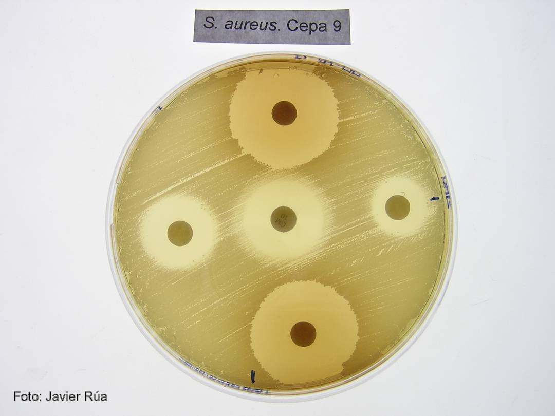
Cultivo de 'Staphylococcus aureus' tratado con los fenoles BHA (derecha) e hidroquinona (arriba).

CSIC/DICYT Un trabajo con participación del Consejo Superior de Investigaciones Científicas (CSIC) ha asignado a una enzima presente en todos los organismos vivos una nueva función en la transferencia de genes virulentos entre bacterias, un proceso que acaba provocando una infección. Los resultados, que aparecen publicados en el último número de Molecular Cell, y que han empleado como modelo la bacteria Staphylococcus aureus, la más frecuente en las infecciones adquiridas en hospitales, establecen el mecanismo de actuación de estas moléculas.

Las bacterias son capaces de transferir material genético entre sí mediante mecanismos de transferencia horizontal de genes. Cuando estos genes son virulentos, las bacterias que los reciben adquieren la capacidad de provocar enfermedades. “Algunos de los genes que codifican para toxinas y otros factores de virulencia están presentes en unas regiones denominadas islas de patogenicidad. Estas islas se transfieren de unas bacterias a otras utilizando virus que infectan bacterias, los llamados bacteriófagos”, explica el investigador del CSIC José Rafael Penadés, que trabaja en el Instituto de Biomedicina de Valencia.

El equipo de investigadores, formado también por científicos del Centro de Investigación y Tecnología Animal y la Universidad CEU Cardenal Herrera, en Valencia, han descubierto que las enzimas dUTPasas son capaces de despertar a las islas de patogenicidad para que detecten que la bacteria está siendo atacada por un virus. Antes de que la bacteria muera infectada, las islas inician su replicación y se transfieren a otras bacterias inocuas, a las que convierten en virulentas.

“El proceso evolutivo ha hecho que las islas detecten que un virus está infectando a las bacterias, lo que producirá su muerte, y utilicen la presencia del bacteriófago para activarse e iniciar su ciclo. Esto ocurre porque algunas proteínas del fago se unen a un represor que bloquea la isla”, explica el investigador del CSIC.

Proteínas G protooncogénicas
Los resultados confirman que las dUTPasas son moléculas señalizadoras que emplean un mecanismo similar al descrito para una familia de proteínas presentes en células eucariotas: las proteínas G protooncogénicas. “Las dUTPasas son activas como señalizadoras cuando se unen a un nucleótido dUTP. Es entonces cuando cambian su conformación y, una vez cumplida su función, degradan el nucleótido y pasan a estar apagadas. Este mecanismo de encendido y apagado es el mismo que el empleado por los protooncogenes”, destaca Penadés.

Según el investigador del CSIC Alberto Marina, el estudio sugiere por primera vez que las dUTPasas cumplen una función señalizadora no sólo en la mayoría de los virus, sino además en organismos vivos complejos como los eucariotas superiores. “Nuestros resultados aportan una visión completamente nueva del mecanismo de actuación de estas enzimas, que depende de una serie de características presentes en las enzimas de los bacteriófagos de Staphylococcus aureus y que están también presentes en otras muchas dUTPasas de un gran número de organismos vivos. Todo ello sugiere que el mecanismo descrito es universal”, concluye el investigador del CSIC.

Referencia bibliográfica 
María Ángeles Tormo-Más, Jorge Donderis, María García-Caballer, Aaron Alt, Ignacio Mir-Sanchís, Alberto Marina y José R. Penadés. Phage dUTPases Control Transfer of Virulence Genes by a Proto-Oncogenic G Protein-like Mechanism. Molecular Cell. DOI: 10.1016.

domingo, 22 de julio de 2012

MinION - $900 usb-powered DNA sequencer on sale this year

ORIGINAL: GizMag
February 19, 2012

The MinION is the size of a USB memory stick, and obtains both power and computer analysis from a normal laptop computer (Image: Oxford Nanopore)

The MinION is the size of a USB memory stick, and obtains both power and computer analysis from a normal laptop computer (Image: Oxford Nanopore)
Oxford Nanopore (ON) has been developing a disruptive nanopore-based technology for sequencing DNA, RNA, proteins, and other long-chain molecules since its birth in 2005. The company has just announced that within the next 6-9 months it will bring to market a fast, portable, and disposable protein sequencer that will democratize sequencing by eliminating large capital costs associated with equipment required to enter the field.

The MinION is the size of a USB memory stick, and obtains both power and computer analysis from a normal laptop computer. No polymerase chain reaction (PCR) or other DNA amplification technique is needed for optimum sensitivity. The MinION is capable of sequencing 100 million base pairs within its six hour working life. Samples of whole blood, plasma, and serum are accepted by the device for immediate analysis and the reagents needed for the analytic process come ready to use with each MinION.

What are nanopores?

A nanopore is an organic molecule penetrated by a very small (~1-2 nanometer) hole (Image: Oxford Nanopore)
A nanopore is an organic molecule penetrated by a very small (~1-2 nanometer) hole. These nanopores are embedded in a synthetic polymer membrane which is only two molecular layers in thickness. Initially bilayer lipid membranes, similar to those appearing throughout the body, were used, but proved far too fragile to use in a production sequencer. Development of such a thin membrane that has extremely large electrical resistance and is sufficiently rugged for use in the field was a major (and successful) ON project.


A nanopore embedded in a synthetic polymer membrane collects small molecules for identification (Image: Oxford Nanopore)
A nanopore is mounted upon such a membrane so that the hole in the nanopore forms a path from one side of the membrane to the other. The membrane is then positioned so that it divides a volume of an electrophysiological fluid in half.


This diagram shows a protein nanopore set in an electrically resistant membrane bilayer (Image: Oxford Nanopore)

The image above shows a nanopore embedded in a membrane. A voltage difference is placed between the two halves of the fluid. The fluid contains mobile ions, so an ion current then passes through the center of the nanopore. This ionic current acts to sweep nearby molecules, proteins, and even DNA or RNA into the mouth of the nanopore.

If a target molecule is drawn into or upon the nanopore, it disrupts the flow of ions in a characteristic manner which can be detected and interpreted.

Although in the image it appears that the detection current is only due to physical obstruction of the nanopore, one must remember that the chemical bonds of the nanopore and the target molecules have unique distributions of electronic charge. The target molecule will attempt to lodge in the nanopore so that the potential energy of the system is lowest. This effect, together with the possibility of modifying the structure of the nanopore to increase the difference in the ionic current for a set of target molecules implies that selectivity can be adjusted to detect small differences in molecular structure.

A wide range of molecules can be detected and identified using the nanopore ionic current measurements, ranging from small physiological or pharmaceutical molecules to proteins. However, the MinION is directed toward DNA sequencing, so the nanopore and enzymes in the electrophysiological fluid are optimized to differentiate the four amino acids (adenine, cytosine, guanine, and thymine) which encode genes in DNA. In the final result, three amino acids are read simultaneously by the nanopore, which possesses the delicacy to differentiate between the 64 possible combinations.

Strand sequencing
The MinION uses a strand sequencing technique for DNA analysis. Strand sequencing is shown in the following excellent video from Oxford Nanopore.


Here's a quick overview: The DNA strand is attracted to an enzyme that is part of the electrophysiological fluid. In turn, the enzyme is attracted to a nanopore, in which it lodges. The enzyme is engineered to unzip the double-helix structure of the DNA strand so that one strand is directed back into the fluid, while the other enters the nanopore for sequencing. More amazing is that the enzyme incorporates a ratcheting structure, so the single DNA strand does not pass quickly through the nanopore, but rather advances one base pair at a time. The rate at which the DNA strand passes through the nanohole is controlled by the magnitude of the unobstructed ionic current, and can be as large as 15 base pairs per second. Better accuracy is associated with slower rates.


If a target molecule is drawn into or upon the nanopore, it disrupts the flow of ions in a characteristic manner which can be detected and interpreted (Image: Oxford Nanopore)

The sensing electronics for the ionic current is fabricated on a sample chip, which comprises 512 nanopores, giving a total strand reading rate of about 7500 bases/second. The MinION has a limited operational life of about 6 hours, but in that time can read over 150 million bases.

Sample chips for the MinION incorporate 512 electrically distinct nanopores, each with its own sensing electronics (Image: Oxford Nanopore)
For comparison, the largest human chromosome, chromosome 1, contains about 250 million base pairs. Attempting to read a smaller complete chromosome (say, number 22, which has about 50 million base pairs) would require that the chromosome be split into a large number of pieces which would be analyzed concurrently, and the pieces reassembled. This is not the primary purpose of MinION, but it does provide data which would make possible such a feat.

Bringing genetic analysis out of the research lab
The MinION has successfully been used to read the genome of the lambda bacteriophage, about 48500 base pairs in length, twice in a single pass. This was accomplished by closing the end of the DNA with a hairpin closure, so that both strands in the genome would pass through the same nanopore. Reading roughly 100,000 base pairs in a single DNA capture is unheard of using more traditional sequencing techniques.

The MinION has successfully been used to read the genome of the lambda bacteriophage, about 48500 base pairs in length (Image: Oxford Nanopore)
During interviews just proceeding the product announcement at AGBT 2012, Clive Brown, the Chief Technology Officer of Oxford Nanopores, revealed that the expected $900 price tag for the MinION has a good bit of margin built in. We can thus expect prices to fall quickly as production becomes routine in its challenges.

The science and engineering research which has gone into making a device like the MinION possible is a remarkable feat for the ages. It is difficult to point to any portion of the development which was straightforward. It is to be hoped that the impact of MinION and future developments thereof is to truly begin to bring genetic analysis out of the research lab as a vital tool for physicians' office.

Source: Oxford Nanopore

About the Author
From an early age Brian wanted to become a scientist. He did, earning a Ph.D. in physics and embarking on an R&D career which has recently broken the 40th anniversary. What he didn't expect was that along the way he would become a patent agent, a gourmet cook, a biotech entrepreneur, an opera tenor and a science writer.

viernes, 25 de mayo de 2012

Telomerase Gene Therapy Extends Lives Of Mice By Up To 24 Percent

ORIGINAL: SingularityHub
by Peter Murray
May 24th, 2012

After being injected with the telomerase gene, adult and old mice lived 24 percent and 13 percent longer, respectively.
Scientists are doing their best to give us the gift of immortality. The latest in the fight against ever dying is a gene therapy that gives mice a healthy dose of telomerase, the enzyme that keeps our chromosomes – and thus our cells and bodies – “young.” The therapy extended the lifespans of mice by 24 percent and, at least so far, the therapy appears to be completely safe.

As we age the dying cells in our body are replenished through cell division. But with each cell division the bits of DNA at the ends of chromosomes – the telomeres – deteriorate. At some point the shortened telomeres signal to the cell that it’s time to stop dividing, leading to tissue degradation – one of the hard facts of life for the now aged cells. But now scientists have given cells a kind of molecular fountain of youth – at least in mice. They injected the mice with the telomerase gene which then slowed the cellular aging process by extending the dwindling telomere ends. They gave the gene therapy to one year old mice, considered adults, and two year old mice, considered old. The lifespan of the one year olds were extended by 24 percent, the two year olds by 13 percent. Not only did the mice live longer, but they reaped beneficial effects across a range of conditions associated with aging including insulin sensitivity, osteoporosis, and physical coordination.

An inactive form of telomerase had no effect on lifespan, confirming that its telomere-lengthening enzymatic activity was crucial. The study was led by María Blasco at the Spanish National Cancer Research Centre and published in EMBO Molecular Medicine.

Dr. María Blasco, Spanish National Cancer Research Centre 
The treatment involved replacing the genes of a virus with the gene for telomerase. This viral vector had several advantages. First, viruses are good at getting into the body and infecting a large number of cells. Inserting telomerase into a small handful of cells won’t have much impact on an organism’s lifespan. Second, the gene remains active for years. And lastly, the viral DNA did not insert itself into the DNA of the mouse cells. Past attempts at gene therapy that work this way run the risk of insertion errors that turn the cell into a tumor, as was the case in the trial which caused leukemia in two of nine participants testing a gene therapy for “bubble boy disease.”

Longevity through telomerase is nothing new. Adding telomerase to human cells in culture allowed them to extend their lifespans by at least an extra 20 divisions. And mice genetically engineered to make telomerase lived 40 percent longer and showed improved glucose tolerance, coordination, and less inflammation compared to normal mice. But genetically engineering people isn’t an option (yet), so a treatment form of telomerase such as the injectable virus in the current study – extending the lifespans of adult and old mice – is a much more conceivable approach.

Aging is a complex process with lots of components, many of which we might not even be aware. But if telomere shortening is really so powerfully rate-limiting to our lifespans, then it could turn out to be as close to a silver bullet for longevity as we’re likely to find. Maybe telomerase treatments could buy us those extra years crucial to reaching Aubrey de Grey’s “longevity escape velocity” beyond which new treatments will save us from the disease of death – indefinitely.

[image credits: Science Daily, publico.es, and Science Creative Daily]

miércoles, 23 de mayo de 2012

DNA used as rewritable data storage in cells

ORIGINAL: ScienceNews
Monday, May 21st, 2012

Genetically encoded memory could track cell division inside the body

They aren’t yet competition for Intel, but bioengineers have created a one-bit “memory” made of DNA that can record, erase and rewrite data within living cells.

One day, doctors might be able to insert such devices into a cancer patient to tally how many times a cell divides and flag when to shut the cancer down. Or researchers might track exactly what happens inside cells as they age.

The work is a step forward in synthetic biology, a new field in which scientists create tools to control life’s basics from the cell on up.

“We can write and erase DNA in a living cell,” says Jerome Bonnet, a bioengineer at Stanford University. “Now we can bring logic and computation inside a cell itself.”

Bonnet and his colleagues, led by Stanford’s Drew Endy, describe the feat in a paper published online May 21 in the Proceedings of the National Academy of Sciences.

Figure S2: Alternate architecture for a reset circuit. A, Schematic diagram of the decoupled reset circuit where integrase is expressed from a low-copy plasmid while excisionase is expressed from a medium-copy plasmid. B, Cells bearing the chromosomal LR DNA register were transformed with both plasmids encoding integrase and excisionase, pulsed with arabinose and analyzed by flow cytometry. Cells relaxed to the BP state after induction with approximatively 85% efficiency.
Scientists have long dreamed of putting tiny computers inside the body to monitor and perhaps even control what’s going on. But nobody has yet made a silicon-based computer chip small enough to embark on a fantastic computing voyage inside a cell.

So researchers are turning instead to biological tools, such as enzymes and DNA. Some biologists have devised DNA switches that can be turned on and off within a cell. And in 2009, bioengineers reported making a genetic “counter” that could tally the number of times a particular event, like a cell dividing, took place (SN: 6/20/09, p. 5).

But these previous efforts made systems that could write a piece of information only once. Truly useful digital data storage allows the information to be erased and rewritten over and over again, like burning new information onto a CD with each pass. “What we didn’t have is some kind of logic that also has memory,” says Pakpoom Subsoontorn, a graduate student on the team.

The researchers chose DNA as the stuff of memory and used enzymes called recombinases as the tools to flip it on and off. Those enzymes came from bacteriophages, which are viruses that infect bacteria. These viruses use one enzyme to integrate into the genome of the bacterium they’re infecting.

In the experiment, the enzyme traveled to a particular place on the sequence of DNA that contains genetic information and flipped a small section so that it read backward. Sending a second signal then flipped the sequence back to its original state. The flipped and unflipped versions thus represent the “0” and “1” states of a computer bit, says Bonnet.

Working in the bacterium Escherichia coli, the team also tweaked the DNA so that it would fluoresce in different colors depending on the orientation of the strand in question. By watching the cells’ glow change between red and green and then back again, the scientists could tell when the DNA strand had been flipped.

So far, Endy’s team has just one bit of memory. Next they hope to scale up to eight bits, or a byte — a goal that could take many more years, Bonnet says. The scientists are also working on speeding up the flips; it currently takes about an hour to invert a DNA segment.

But the team has gotten the flips to hold for more than 100 generations within a living cell, a laboratory first. “This was an important proof of concept that it was doable,” says Bonnet. “Now we want to build a more complex system, something other people can use.”

Though interesting, it’s not yet clear whether DNA-based memory will ever replace silicon-based memory for certain applications, says Roger Brent, a researcher at the Fred Hutchinson Cancer Research Center and director of the Center for Biological Futures in Seattle. “It will need to prove itself in the marketplace of ideas.”

sábado, 3 de marzo de 2012

Immortal worms defy aging

ORIGINAL: KurzweilAI
February 29, 2012

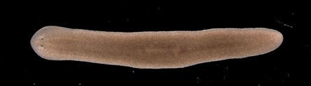
Planarian flatworm (credit: The University of Nottingham)
Researchers from The University of Nottingham have discovered how planarian flatworms overcome the aging process to be potentially immortal: they can rejuvenate their telomeres.

The discovery, funded by the Biotechnology and Biological Sciences Research Council (BBSRC) and Medical Research Council (MRC), may eventually lead to alleviating aging and age-related characteristics in human cells.

Planarian worms have amazed scientists with their apparently limitless ability to regenerate. Researchers have been studying their ability to replace aged or damaged tissues and cells in a bid to understand the mechanisms underlying their longevity.

“We’ve been studying two types of planarian worms; those that reproduce sexually, like us, and those that reproduce asexually, simply dividing in two,” said Dr. Aziz Aboobaker from the University’s School of Biology.

“Both appear to regenerate indefinitely by growing new muscles, skin, guts and even entire brains over and over again.

“Usually when stem cells divide — to heal wounds, or during reproduction or for growth — they start to show signs of aging. This means that the stem cells are no longer able to divide and so become less able to replace exhausted specialized cells in the tissues of our bodies.

“Our aging skin is perhaps the most visible example of this effect. Planarian worms and their stem cells are somehow able to avoid the aging process and to keep their cells dividing.”

Each time an animal cell divides, the protective telomere “cap” gets shorter. When they get too short, the cell loses its ability to renew and divide. In an immortal animal, we would therefore expect cells to be able to maintain telomere length indefinitely so that they can continue to replicate. Aboobaker predicted that planarian worms actively maintain the ends of their chromosomes in adult stem cells, leading to theoretical immortality.

Previous work, leading to the award of the 2009 Nobel Prize for Physiology or Medicine, had shown that telomeres could be maintained by the activity of an enzyme called telomerase. In most sexually reproducing organisms, the enzyme is most active only during early development. So as we age, telomeres start to reduce in length.

This project identified a possible planarian version of the gene coding for this enzyme and turned down its activity. This resulted in reduced telomere length and proved it was the right gene. They were then able to confidently measure its activity and resulting telomere length and found that asexual worms dramatically increase the activity of this gene when they regenerate, allowing stem cells to maintain their telomeres as they divide to replace missing tissues.

“The next goals for us are to understand the mechanisms in more detail and to understand more about how you evolve an immortal animal,” said Aboobaker.

“The worms are a model system in which we can ask questions, like is it possible for a multicellular animals to be immortal and avoid the effects of aging?,” he told KurzweilAI.

“If so, how does this animal do this in comparison to animals that don’t? Of course we hope that this impacts humans, that’s why we do it. But we aren’t planning on making any drugs or medicines… other people are, I’m sure.”

Ref.: Thomas C. J. Tan et al., , Telomere maintenance and telomerase activity are differentially regulated in asexual and sexual worms, Proceedings of the National Academy of Sciences, 2012 [DOI: 10.1073/pnas.1118885109] (open access)