Mostrando entradas con la etiqueta NIH. Mostrar todas las entradas

martes, 24 de enero de 2017

TSRI Scientists Create First Stable Semisynthetic Organism


LA JOLLA, CA – January 23, 2017 – Life’s genetic code has only ever contained four natural bases. These bases pair up to form two “base pairs”—the rungs of the DNA ladder—and they have simply been rearranged to create bacteria and butterflies, penguins and people. Four bases make up all life as we know it.

Until now. Scientists at The Scripps Research Institute (TSRI) have announced the development of the first stable semisynthetic organism. Building on their 2014 study in which they synthesized a DNA base pair, the researchers created a new bacterium that uses the four natural bases (called A, T, C and G), which every living organism possesses, but that also holds as a pair two synthetic bases called X and Y in its genetic code.

TSRI Professor Floyd Romesberg and his colleagues have now shown that their single-celled organism can hold on indefinitely to the synthetic base pair as it divides. Their research was published January 23, 2017, online ahead of print in the journal Proceedings of the National Academy of Sciences.

“We’ve made this semisynthetic organism more life-like,” said Romesberg, senior author of the new study.

While applications for this kind of organism are still far in the future, the researchers say the work could be used to create new functions for single-celled organisms that play important roles in drug discovery and much more.

Building a Unique Organism
When Romesberg and his colleagues announced the development of X and Y in 2014, they also showed that modified E. coli bacteria could hold this synthetic base pair in their genetic code. What these E. coli couldn’t do, however, was keep the base pair in their code indefinitely as they divided. The X and Y base pair was dropped over time, limiting the ways the organism could use the additional information possessed in their DNA.

“Your genome isn’t just stable for a day,” said Romesberg. “Your genome has to be stable for the scale of your lifetime. If the semisynthetic organism is going to really be an organism, it has to be able to stably maintain that information.”

Romesberg compared this flawed organism to an infant. It had some learning to do before it was ready for real life.

In stepped TSRI Graduate Student Yorke Zhang and Brian Lamb, an American Cancer Society postdoctoral fellow in the Romesberg lab at the time of the study. Together, they helped develop the means for the single-celled organism to retain the artificial base pair.

First, Zhang and Lamb, co-first authors of the study, optimized a tool called a nucleotide transporter, which brings the materials necessary for the unnatural base pair to be copied across the cell membrane. “The transporter was used in the 2014 study, but it made the semisynthetic organism very sick,” Zhang explained. The researchers discovered a modification to the transporter that alleviated this problem, making it much easier for the organism to grow and divide while holding on to X and Y.

Next, the researchers optimized their previous version of Y. The new Y was a chemically different molecule that could be better recognized by the enzymes that synthesize DNA molecules during DNA replication. This made it easier for cells to copy the synthetic base pair.

A New Use for CRISPR-Cas9
Finally, the researchers set up a “spell check” system for the organism using CRISPR-Cas9, an increasingly popular tool in human genome editing experiments. But instead of editing a genome, the researchers took advantage of CRISPR-Cas9’s original role in bacteria.

The genetic tools in CRISPR-Cas9 (a DNA segment and an enzyme) originated in bacteria as a kind of immune response. When a bacterium encounters a threat, like a virus, it takes fragments of the invader genome and pastes them into its own genome—a bit like posting a “wanted” poster on the off chance it sees the invader again. Later, it can use those pasted genes to direct an enzyme to attack if the invader returns.

Knowing this, the researchers designed their organism to see a genetic sequence without X and Y as a foreign invader. A cell that dropped X and Y would be marked for destruction, leaving the scientists with an organism that could hold on to the new bases. It was like the organism was immune to unnatural base pair loss.

“We were able to address the problem at a fundamental level,” said Lamb, who now serves as a research scientist at Vertex Pharmaceuticals.

Their semisynthetic organism was thus able to keep X and Y in its genome after dividing 60 times, leading the researchers to believe it can hold on to the base pair indefinitely.

“We can now get the light of life to stay on,” said Romesberg. “That suggests that all of life’s processes can be subject to manipulation.”

A Foundation for Future Research
Romesberg emphasized that this work is only in single cells and is not meant to be used in more complex organisms. He added that the actual applications for this semisynthetic organism are “zero” at this point. So far, scientists can only get the organism to store genetic information.

Next, the researchers plan to study how their new genetic code can be transcribed into RNA, the molecule in cells needed to translate DNA into proteins. “This study lays the foundation for what we want to do going forward,” said Zhang.
Professor Floyd Romesberg (right) and Graduate Student Yorke Zhang led the new study at The Scripps Research Institute, along with Brian Lamb (not pictured).(Photo by Madeline McCurry-Schmidt.)

Additional authors of the study, “A semisynthetic organism engineered for the stable expansion of the genetic alphabet,” were Aaron W. Feldman and Anne Xiaozhou Zhou of TSRI; Thomas Lavergne of the University of Grenoble; and Lingjun Li of Henan Normal University.

The study was supported by the National Institutes of Health (grant GM060005), a National Science Foundation Graduate Research Fellowship (grant DGE-1346837), the National Natural Science Foundation of China (grant 21472036), a Labex ARCANE grant (ANR-11-LABX-0003-01), NanoBio-ICMG platforms (FR 2607) and a postdoctoral fellowship from the American Cancer Society, Illinois Division.

About The Scripps Research Institute
The Scripps Research Institute (TSRI) is one of the world's largest independent, not-for-profit organizations focusing on research in the biomedical sciences. TSRI is internationally recognized for its contributions to science and health, including its role in laying the foundation for new treatments for cancer, rheumatoid arthritis, hemophilia, and other diseases. An institution that evolved from the Scripps Metabolic Clinic founded by philanthropist Ellen Browning Scripps in 1924, the institute now employs more than 2,500 people on its campuses in La Jolla, CA, and Jupiter, FL, where its renowned scientists—including two Nobel laureates and 20 members of the National Academies of Science, Engineering or Medicine—work toward their next discoveries. The institute's graduate program, which awards PhD degrees in biology and chemistry, ranks among the top ten of its kind in the nation. In October 2016, TSRI announced a strategic affiliation with the California Institute for Biomedical Research (Calibr), representing a renewed commitment to the discovery and development of new medicines to address unmet medical needs. For more information, see www.scripps.edu.

# # #

For information:
Office of Communications 
Tel: 858-784-2666 
Fax: 858-784-8118 

jueves, 14 de enero de 2016

ORNL cell-free protein synthesis is potential lifesaver

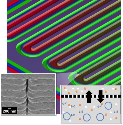
This section of a serpentine channel reactor shows the parallel reactor and feeder channels separated by a nanoporous membrane. At left is a single nanopore viewed from the side; at right is a diagram of metabolite exchange across the membrane.
OAK RIDGE, Tenn., Dec. 29, 2015 – Lives of soldiers and others injured in remote locations could be saved with a cell-free protein synthesis system developed at the Department of Energy’s Oak Ridge National Laboratory.

The device, a creation of a team led by Andrea Timm and Scott Retterer of the lab’s Biosciences Division, uses microfabricated bioreactors to facilitate the on-demand production of therapeutic proteins for medicines and biopharmaceuticals. Making these miniature factories cell-free, which eliminates the maintenance of a living system, simplifies the process and lowers cost.

“With this approach, we can produce more protein faster, making our technology ideal for point-of-care use,” Retterer said. “The fact it’s cell-free reduces the infrastructure needed to produce the protein and opens the possibility of creating proteins when and where you need them, bypassing the challenge of keeping the proteins cold during shipment and storage.”

ORNL’s bioreactor features elegance through a permeable nanoporous membrane and serpentine design fabricated using a combination of electron beam and photolithography and advanced material deposition processes. This design enables prolonged cell-free reactions for efficient production of proteins, making it easily adaptable for use in isolated locations and at disaster sites.

From a functional perspective, the design uses long serpentine channels integrated in a way to allow the exchange of materials between parallel reactor and feeder channels. With this approach, the team can control the exchange of metabolites, energy and species that inhibit production of the desired protein. Through other design features, researchers extend reaction times and improve yields.

“We show that the microscale bioreactor design produces higher protein yields than conventional tube-based batch formats and that product yields can be dramatically improved by facilitating small molecule exchange with the dual-channel bioreactor,” the authors wrote in their paper, published in the journal Small.

The researchers also note that on-demand biologic synthesis would aid the production of drugs that are costly to mass-produce, including orphan drugs and personalized medicines.

Other authors of the paper, titled “Towards Microfluidic Reactors for Cell-Free Protein Synthesis at the Point-of-Care,” are ORNL’s Peter Shankles, Carmen Foster and Mitchel Doktycz.

Funding for this project was provided by the Defense Advanced Research Projects Agency through a collaboration with researchers from Leidos (https://www.leidos.com) and Northwestern University. This accomplishment represents the culmination of research led by Doktycz and Retterer funded by multiple grants from the National Institutes of Health and DOE over the last decade. A portion of the work was performed at the Center for Nanophase Materials Sciences, a DOE Office of Science User Facility.

UT-Battelle manages ORNL for the DOE's Office of Science. The Office of Science is the single largest supporter of basic research in the physical sciences in the United States, and is working to address some of the most pressing challenges of our time. For more information, please visit http://science.energy.gov/.

ORIGINAL: ORNL
Ron Walli, Communications. wallira@ornl.gov, 865.576.0226
December 29, 2015

martes, 31 de marzo de 2015

NCATS Support Leads to Clinical Trial to Test Repurposed Cancer Treatment as Alzheimer's Therapy

In a mouse model of Alzheimer's disease, amyloid beta clusters (red) build up among neurons (green) in a memory-related area of the brain. (Strittmatter Laboratory, Yale University Photo/Adam Kaufman)
As Baby Boomers get older, the number of people with age-related conditions such as cancer and Alzheimer's disease continues to grow. Alzheimer's disease is the most common form of dementia, a group of disorders that cause progressive loss of memory and other mental processes. About 5 million Americans have Alzheimer's disease, and current drug therapies can only ease symptoms of the disease without stopping its progression. New treatments — so-called disease-modifying therapies — are needed to halt Alzheimer's by targeting its underlying mechanisms.

Blocking that path to therapeutic success is the costly, complex process of drug development. The average length of time from discovery of a therapeutic target to approval of a new drug is about 14 years. The failure rate during this process exceeds 95 percent.

NCATS is addressing these translational bottlenecks through programs such as the Discovering New Therapeutic Uses for Existing Molecules (New Therapeutic Uses) program. Launched in 2012, this initiative matches academic researchers with pharmaceutical industry assets that have undergone significant research and development to accelerate the process of finding new therapies.

Now, NCATS is celebrating one of the first promising results from the New Therapeutic Uses program: Center-supported scientists at Yale University School of Medicine have found that an experimental compound originally developed as a cancer therapy potentially could be used to treat Alzheimer's disease. The compound successfully reversed brain problems in mouse models of the condition, and now the researchers are testing it in humans. The results of the animal study were published for early view on March 21, 2015, in the Annals of Neurology. Read the NIH news release.

A NEW THERAPEUTIC TARGET
Back in 2012, Yale neurobiology researcher, neurologist and senior author of the animal study Stephen Strittmatter, M.D., Ph.D., and his colleagues found an important, missing piece of the Alzheimer's puzzle. They wanted to understand more completely the molecular events that occur in the brain to produce symptoms.
Two types of brain cells, microglia (red, left) and astrocytes (red, right), surround amyloid beta clusters (green) located in the brain of a mouse that exhibits Alzheimer's-like symptoms. Strittmatter Laboratory, Yale University Photo/Adam Kaufman
In Alzheimer's disease, abnormal clumps of amyloid beta protein build up in the brain. These protein clusters damage brain cells (neurons), eventually killing them. However, howamyloid beta harms cells has been unclear.

The Yale team discovered that aggregated amyloid beta activates a series of signals within neurons that leads to abnormal functioning and loss of synapses, which are the spaces between neurons that enable the cells to "talk" to each other and form memories. Central to this process is the activation of a protein called Fyn kinase through another molecule, cellular prion protein. These results suggested that a compound that blocks Fyn activity might represent a potential disease-modifying therapy for Alzheimer's.

THE POWER OF CROWDSOURCING
Around the time Strittmatter's Fyn finding emerged, NCATS launched the New Therapeutic Uses program and released a list of pharmaceutical industry assets, inviting scientists to pitch new ideas for diseases that might be treated with those assets. That 2012 list included a Fyn kinase inhibitor, saracatinib (AZD0530), developed by biopharmaceutical company AstraZeneca. Strittmatter, along with co-principal investigators Haakon Nygaard, M.D., Ph.D., and Christopher van Dyck, M.D., submitted a proposal to test the hypothesis that saracatinib could improve Alzheimer's-related brain abnormalities. The team received one of the first New Therapeutic Uses awards in June 2013.

"AstraZeneca developed saracatinib to treat cancer outside the brain, so nobody had thought to link it to Alzheimer's disease," Strittmatter said. "This connection — between our knowledge of Fyn kinase in the brain and AstraZeneca's information on this compound — would never have happened without the New Therapeutic Uses program."

A PROMISING EFFECT IN MICE
AstraZeneca scientists teamed with the Yale group, providing saracatinib for the mouse study and sharing knowledge and data gathered from previous studies.

"No one individual or group has complete knowledge of disease pathways and treatment targets," said Craig Wegner, Ph.D., Head, Boston Emerging Innovations Unit, Scientific Partnering & Alliances within AstraZeneca's Innovative Medicines and Early Development Biotech Unit. "This program successfully unites scientists from government, academia and industry and is a great example of how we are working together to push the boundaries of science."

The Yale team gave the experimental drug to mice with Alzheimer's-like symptoms, such as memory problems and age-related buildup of abnormal amyloid beta clusters, modeling the development of the disease in humans. After four weeks, the Alzheimer's mice showed complete reversal of spatial learning and memory loss. When the scientists examined the brains of the mice, they found that the characteristic synapse loss had been fully restored, providing a biological explanation for the memory improvement.

The treatment also reduced several other Alzheimer's-related biochemical changes in the mice and did not appear to be toxic. Although many experimental treatments have aimed to reduce abnormal amyloid beta buildup in the brain, this one is unique in that it targets the toxic effects of the protein clusters within a cell, appearing to protect it from damage.

The Yale research team also has completed a successful Phase 1b safety study of saracatinib in humans with Alzheimer's disease, showing that the compound reaches the brains of patients at levels similar to those beneficial in mice. The study results have been accepted for publication later in 2015. The data are encouraging, but the researchers cautioned that more human studies are needed to determine if saracatinib is an effective treatment for Alzheimer's.

AN ACCELERATED PATH TO THE CLINIC
Saracatinib's prior development and the Yale team's successful completion of animal and human studies enabled the compound to advance rapidly into a larger, multisite Phase 2a trial in Alzheimer's patients now ongoing. "The speed and efficiency with which this research has advanced has set new standards of excellence, enabling us to jointly push the boundaries of medical science," Wegner said.

Using the pre-negotiated NCATS template agreements, which were designed to streamline the legal and administrative process for research collaboration by multiple organizations, "really facilitated our collaboration with Yale, so scientists could be scientists," he added.

"Through this project, NCATS and AstraZeneca have provided us with an incredible shortcut in the drug development process and have accelerated the path to finding a more effective Alzheimer's disease treatment," Strittmatter said.

In the Phase 2a trial, 152 participants will receive saracatinib or placebo for one year. Researchers will assess safety, tolerability and effectiveness of the experimental drug, and they will use brain imaging to visualize the effect of saracatinib on synapse function — the same feature improved by the drug in mice. Study investigators currently are enrolling older adults with Alzheimer's disease to participate in the trial and expect to have results in about two years. Learn more about the trial via ClinicalTrials.gov or the study website .

Both human trials were funded by the New Therapeutic Uses program. The Phase 2a study will take place at multiple clinical sites as part of the Alzheimer's Disease Cooperative Study , an initiative for multisite studies sponsored by the National Institute on Aging (NIA) to facilitate the development and testing of new therapeutics for the condition. In addition to funding from NCATS, the NIH Common Fund, NIA, BrightFocus Foundation, Alzheimer's Association and Falk Medical Research Trust provided support for the animal study.

"The Yale team's awareness of this new Alzheimer's drug target, combined with AstraZeneca's drug development resources, allowed the rapid advancement of saracatinib to clinical testing, demonstrating the power of NCATS' New Therapeutics Uses crowdsourcing approach," said NCATS Director Christopher P. Austin, M.D. "By reengineering the drug development pipeline through projects like this, we can more quickly deliver new and better treatments to patients."

ORIGINAL: NCATS-NIH
March 2015

lunes, 20 de enero de 2014

How EnChroma’s smart sunglasses can help solve color blindness

ABC 7. The EnChroma glasses

 Wearable technology is proving to be hugely beneficial to people with a variety of disabilities and health conditions.

In Berkeley, a group of engineers is developing smart sunglasses that can help color-blind people identify and better discriminate between colors. The startup, called EnChroma, initially received funding for its research from the National Institutes of Health (NIH).

It’s a huge potential market. According to the website We Are Colorblind, around 8 percent of the male population of the planet is color blind.

For some color-blind people, driving is challenging, as it can be difficult to discern between a red and green traffic light. Former New York Times writer David Pogue is color blind and gave the glasses a try. Pogue doesn’t have any trouble driving, but doesn’t see deep greens and reds.
EnChroma

The Explorer Glasses will set you back $600

“Yards full of leafy trees and plants suddenly had different shades of green. Everywhere I looked, desaturated or barely discernible red things were popping,” wrote Pogue. “There was a weird sensation of seeing red and green areas in the periphery of my vision.”


According to a company spokesperson, color-blind wearers of Enchroma’s smart “Cx Explorer” glasses experience up to a 30 percent improvement in ability to identify colors and a 70 percent improvement in color discrimination.

The sunglasses are designed to be worn outdoors in bright light — not artificial light. The company wouldn’t advise wearing them at work, to read, or watch videos on a computer screen.

In an email interview, cofounders Don McPherson and Tony Dykes detailed how EnChroma’s technology rivals other products. According to the founders, most products on the market will use strongly tinted red and magenta lenses.

“A side effect is that these aids distort your overall perception of color, completely eliminating some colors and flattening out your depth perception,” the company wrote to me. “They also look rather silly.”

Meanwhile, EnChroma uses a mathematical formula to directly communicate with the brain’s visual system. This system is calculated to help a color-blind person observe the correct ratios the brain needs for normal color vision. The company uses over 100 reflective coatings at different opacities rather than a single tinted lens.
McPherson, a material sciences expert, initially had the idea for the technology when making very expensive glass laser-protective eyewear. These glasses would sell to hospitals for $1,600 a pop. McPherson consistently received reorders, as the glasses would be stolen by the surgeons to wear as normal sunglasses. He began wearing them too and noticed at a frisbee game that he could see orange cones for the first time.

After noticing he was color blind, McPherson began working on a prototype for others like him. EnChroma’s Explorer glasses are available for a more affordable (but still heart-skipping) $600.

EnChroma has raised $1.3 million from angel investors and the NIH to date. The company plans to raise about $2 to $4 million in February from venture capitalists. The goal is to use the same technology to benefit people with a variety of vision problems.

To see the glasses in action, check out the video from ABC’s San Francisco affiliate. You can also order a pair to try — there’s a 30 day money-back guarantee.


ORIGINAL: Venture Beat
January 20, 2014

viernes, 3 de enero de 2014

Getting CLARITY: Hydrogel process developed at Stanford creates transparent brain

Stanford bioengineers have transformed an intact, post-mortem mouse brain into a transparent three-dimensional structure that keeps all the fine wiring and molecular structures in place. Known as CLARITY, the technique stands to transform our understanding of the brain and indeed of any biological tissue.
Combining neuroscience and chemical engineering, researchers at Stanford University have developed a process that renders a mouse brain transparent. The postmortem brain remains whole — not sliced or sectioned in any way — with its three-dimensional complexity of fine wiring and molecular structures completely intact and able to be measured and probed with visible light and chemicals.

The process, called CLARITY, ushers in an entirely new era of whole-organ imaging that stands to fundamentally change our scientific understanding of the most important, but least understood of organs, the brain, and potentially other organs, as well.

The process is described in a paper to be published online April 10 in Nature by bioengineer and psychiatrist Karl Deisseroth leading a multidisciplinary team, including postdoctoral scholar Kwanghun Chung.

“Studying intact systems with this sort of molecular resolution and global scope — to be able to see the fine detail and the big picture at the same time — has been a major unmet goal in biology, and a goal that CLARITY begins to address,” Deisseroth said.


CLARITY provides the ability to do a fly-through of an intact mouse brain using a fluorescent imaging technique on a complete brain that previously could only be performed on a brain sectioned into thin slices. (Video: Karl Deisseroth and Kwanghun Chung, Stanford University)

“This feat of chemical engineering promises to transform the way we study the brain’s anatomy and how disease changes it,” said Thomas Insel, MD, director of the National Institute of Mental Health. “No longer will the in-depth study of our most important three-dimensional organ be constrained by two-dimensional methods.”

The research in this study was performed primarily on a mouse brain, but the researchers have used CLARITY on zebrafish and on preserved human brain samples with similar results, establishing a path for future studies of human samples and other organisms.

“CLARITY promises to revolutionize our understanding of how local and global changes in brain structure and activity translate into behavior,” said Paul Frankland, PhD, a senior scientist in neurosciences and mental health at the Hospital for Sick Children Research Institute in Toronto, who was not involved in the research. Frankland’s colleague, senior scientist Sheena Josselyn, PhD, added that the process could turn the brain from “a mysterious black box” into something essentially transparent. 

An inscrutable place

The mound of convoluted grey matter and wiring that is the brain is a complex and inscrutable place. Neuroscientists have struggled to fully understand its circuitry in their quest to comprehend how the brain works, and why, sometimes, it doesn’t.

CLARITY is the result of a research effort in Deisseroth’s lab to extract the opaque elements — in particular the lipids — from a brain and yet keep the important features fully intact. Lipids are fatty molecules found throughout the brain and body. In the brain, especially, they help form cell membranes and give the brain much of its structure. Lipids pose a double challenge for biological study, however, because they make the brain largely impermeable both to chemicals and to light.

Neuroscientists would have liked to extract the lipids to reveal the brain’s fine structure without slicing or sectioning, but for one major hitch: removing these structurally important molecules causes the remaining tissue to fall apart.

Prior investigations have focused instead on automating the slicing/sectioning approach, or in treating the brain with organic molecules that facilitate the penetration of light only, but not macromolecular probes. With CLARITY, Deisseroth’s team has taken a fundamentally different approach.

“We drew upon chemical engineering to transform biological tissue into a new state that is intact but optically transparent and permeable to macromolecules,” said Chung, the paper’s first author.

This new form is created by replacing the brain’s lipids with a hydrogel. The hydrogel is built from within the brain itself in a process conceptually similar to petrification, using what is initially a watery suspension of short, individual molecules known as hydrogel monomers. The intact, postmortem brain is immersed in the hydrogel solution and the monomers infuse the tissue. Then, when “thermally triggered,” or heated slightly to about body temperature, the monomers begin to congeal into long molecular chains known as polymers, forming a mesh throughout the brain. This mesh holds everything together, but, importantly, it does not bind to the lipids.

With the tissue shored up in this way, the team is able to vigorously and rapidly extract lipids through a process called electrophoresis. What remains is a 3-D, transparent brain with all of its important structures — neurons, axons, dendrites, synapses, proteins, nucleic acids and so forth — intact and in place.

Going things one better

CLARITY then goes one better. In preserving the full continuity of neuronal structures, CLARITY not only allows tracing of individual neural connections over long distances through the brain, but also provides a way to gather rich, molecular information describing a cell’s function is that is not possible with other methods.

“We thought that if we could remove the lipids nondestructively, we might be able to get both light and macromolecules to penetrate deep into tissue, allowing not only 3-D imaging, but also 3-D molecular analysis of the intact brain,” said Deisseroth, who holds the D.H. Chen Professorship.

Using fluorescent antibodies that are known to seek out and attach themselves only to specific proteins, Deisseroth’s team showed that 
  • it can target specific structures within the CLARITY-modified — or “clarified” — mouse brain and 
  • make those structures and only those structures light up under illumination. The researchers 
  • can trace neural circuits through the entire brain or 
  • explore deeply into the nuances of local circuit wiring. They 
  • can see the relationships between cells and 
  • investigate subcellular structures. They
  • can even look at chemical relationships of protein complexes, nucleic acids and neurotransmitters.
“Being able to determine the molecular structure of various cells and their contacts through antibody staining is a core capability of CLARITY, separate from the optical transparency, which enables us to visualize relationships among brain components in fundamentally new ways,” said Deisseroth, who is one of 15 experts on the “dream team” that will map out goals for the $100 million brain research initiative announced April 2 by President Obama.

A three-dimensional rendering of clarified brain imaged from below (ventral half). (Image: Courtesy of the Deisseroth lab)

And in yet another significant capability from a research standpoint, researchers are now able to destain the clarified brain, flushing out the fluorescent antibodies and repeating the staining process anew using different antibodies to explore different molecular targets in the same brain. This staining/destaining process can be repeated multiple times, the authors showed, and the different data sets aligned with one another.

Opening the door

CLARITY has accordingly made it possible to perform highly detailed, fine-structural analysis on intact brains — even human tissues that have been preserved for many years, the team showed. Transforming human brains into transparent-but-stable specimens with accessible wiring and molecular detail may yield improved understanding of the structural underpinnings of brain function and disease.

Beyond the immediate and apparent benefit to neuroscience, Deisseroth cautioned that CLARITY has leapfrogged our ability to deal with the data. “Turning massive amounts of data into useful insight poses immense computational challenges that will have to be addressed. We will have to develop improved computational approaches to image segmentation, 3-D image registration, automated tracing and image acquisition,” he said.

Indeed, such pressures will increase as CLARITY could begin to support a deeper understanding of large-scale intact biological systems and organs, perhaps even entire organisms.

“Of particular interest for future study are intrasystem relationships, not only in the mammalian brain but also in other tissues or diseases for which full understanding is only possible when thorough analysis of single, intact systems can be conducted,” Deisseroth said. “CLARITY may be applicable to any biological system, and it will be interesting to see how other branches of biology may put it to use.”

Other co-authors include undergraduate student Jenelle Wallace; graduate students Sung-Yon Kim, Kelly Zalocusky, Joanna Mattis, Aleksandra Denisin and Logan Grosenick; research assistants Sandhiya Kalyanasundaram, Julie Mirzabekov, Sally Pak and Charu Ramakrishnan; postdoctoral scholars Aaron Andalman, PhD, and Tom Davidson, PhD; former undergraduate student Hannah Bernstein; and former staff scientist Viviana Gradinaru.

The research is supported by the National Institute of Mental Health (grant MH099647); the National Science Foundation; the Simons Foundation; the President and Provost of Stanford University; the Wiegers, Snyder, Reeves, Gatsby and Yu foundations; the DARPA REPAIR program; and the Burroughs Wellcome Fund.

Information about Stanford’s Department of Bioengineering, which also supported the work, is available at http://bioengineering.stanford.edu. The department is jointly operated by the School of Engineering and the School of Medicine.

Andrew Myers is associate director of communications for the Stanford University School of Engineering.


ORIGINAL: Stanford U
Andrew Myers | Stanford Engineering 

 Tom Abate
tabate@stanford.edu
Jamie Beckett
jbeckett@stanford.edu
April 10, 2013

miércoles, 22 de mayo de 2013

Extracting human DNA with full genetic data in minutes

ORIGINAL: KurzweilAI
May 13, 2013

Hand-held device for extracting DNA (credit: UW/NanoFacture/KNR)
University of Washington engineers and NanoFacture, a Bellevue, Wash., company, have created a device that can extract human DNA from fluid samples in a simpler, more efficient and environmentally friendly way than conventional methods.

The device will give hospitals and research labs a much easier way to separate DNA from human fluid samples, which will help with genome sequencing, disease diagnosis and forensic investigations.

Separating DNA from bodily fluids is a cumbersome process that’s become a bottleneck as scientists make advances in genome sequencing, particularly for disease prevention and treatment. The market for DNA preparation alone is about $3 billion each year.

Conventional methods use a centrifuge to spin and separate DNA molecules or strain them from a fluid sample with a micro-filter, but these processes take 20 to 30 minutes to complete and can require excessive toxic chemicals.

A close-up view of the portable device (credit: UW/NanoFacture/KNR)

UW engineers designed microscopic probes that dip into a fluid sample – saliva, sputum or blood – and apply an electric field within the liquid. That draws particles to concentrate around the surface of the tiny probe. Larger particles hit the tip and swerve away, but DNA-sized molecules stick to the probe and are trapped on the surface. It takes two or three minutes to separate and purify DNA using this technology.

“This simple process removes all the steps of conventional methods,” said Jae-Hyun Chung, a UW associate professor of mechanical engineering who led the research.

The hand-held device can clean four separate human fluid samples at once, but the technology can be scaled up to prepare 96 samples at a time, which is standard for large-scale handling.

The tiny probes, called microtips and nanotips, were designed and built at the UW in a micro-fabrication facility where a technician can make up to 1 million tips in a year, which is key in proving that large-scale production is feasible, Chung said.

Engineers in Chung’s lab also have designed a pencil-sized device using the same probe technology that could be sent home with patients or distributed to those serving in the military overseas. Patients could swab their cheeks, collect a saliva sample, then process their DNA on the spot to send back to hospitals and labs for analysis.

This could be useful as efforts ramp up toward sequencing each person’s genome for disease prevention and treatment, Chung said.

The market for this device isn’t developed yet, but Chung’s team will be ready when it is. Meanwhile, the larger device is ready for commercialization, and its creators have started working with distributors.

A UW Center for Commercialization grant of $50,000 seeded initial research in 2008, and since then researchers have received about $2 million in funding from the National Science Foundation and the National Institutes of Health.


lunes, 29 de abril de 2013

Scripps Research Institute Scientists Discover How a Protein Finds Its Way


Katrin Karbstein.
Photo: TSRI
JUPITER, FL, April 29, 2013 – Proteins, the workhorses of the body, can have more than one function, but they often need to be very specific in their action or they create cellular havoc, possibly leading to disease.

Scientists from the Florida campus of The Scripps Research Institute (TSRI) have uncovered how an enzyme co-factor can bestow specificity on a class of proteins with otherwise nonspecific biochemical activity.

The protein in question helps in the assembly of ribosomes, large macromolecular machines that are critical to protein production and cell growth. This new discovery expands scientists’ view of the role of co-factors and suggests such co-factors could be used to modify the activity of related proteins and their role in disease.

“In ribosome production, you need to do things very specifically,” said TSRI Associate Professor Katrin Karbstein, who led the study. “Adding a co-factor like Rrp5 forces these enzymes to be specific in their actions. The obvious possibility is that if you could manipulate the co-factor, you could alter protein activity, which could prove to be tremendously important.”

The new study, which is being published the week of April 29, 2013, in the online Early Edition of the Proceedings of the National Academy of Science, sheds light on proteins called DEAD-box proteins, a provocative title actually derived from their amino acid sequence. These proteins regulate all aspects of gene expression and RNA metabolism, particularly in the production of ribosomes, and are involved in cell metabolism. The link between defects in ribosome assembly and cancer and between DEAD-box proteins and cancer is well documented.

The findings show that the DEAD-box protein Rok1, needed in the production of a small ribosomal subunit, recognizes the RNA backbone, the basic structural framework of nucleic acids. The co-factor Rrp5 then gives Rok1 the ability to target a specific RNA sequence by modulating the structure of Rok1.

“Despite extensive efforts, the roles of these DEAD-box proteins in the assembly of the two ribosomal subunits remain largely unknown,” Karbstein said. “Our study suggests that the solution may be to identify their cofactors first.”

The first author of the study, “Cofactor-Dependent Specificity of a DEAD-box Protein,” is Crystal L. Young. Also a co-author of the paper is Sohail Khoshnevis.

The study was supported by National Institutes of Health Grant R01-GM086451 and the American Heart Association.

About The Scripps Research Institute
The Scripps Research Institute (TSRI) is one of the world's largest independent, not-for-profit organizations focusing on research in the biomedical sciences. TSRI is internationally recognized for its contributions to science and health, including its role in laying the foundation for new treatments for cancer, rheumatoid arthritis, hemophilia, and other diseases. An institution that evolved from the Scripps Metabolic Clinic founded by philanthropist Ellen Browning Scripps in 1924, the institute now employs about 3,000 people on its campuses in La Jolla, CA, and Jupiter, FL, where its renowned scientists—including three Nobel laureates—work toward their next discoveries. The institute's graduate program, which awards PhD degrees in biology and chemistry, ranks among the top ten of its kind in the nation. For more information, see www.scripps.edu.

# # #

For information:
Office of Communications 
Tel: 858-784-2666
Fax: 858-784-8136

miércoles, 20 de marzo de 2013

Harvard's Wyss Institute and Sony DADC Announce Collaboration on Organs-on-Chips

ORIGINAL: Wyss Institute
Date: Mar 18, 2013

Boston, MA -- Today the Wyss Institute for Biologically Inspired Engineering at Harvard University and Sony DADC announced a collaboration that will harness Sony DADC's global manufacturing expertise to further advance the Institute's Organs-on-Chips technologies. 
Human Organs-on-Chips are composed of a clear, flexible polymer about the size of a computer memory stick, and contain hollow microfluidic channels lined by living human cells -- allowing researchers to recapitulate the physiological and mechanical functions of the organs, and to observe what happens in real time. The goal is to provide more predictive and useful measures of the efficacy and safety of new drugs in humans -- and at a fraction of the time and costs associated with traditional animal testing.

"We are excited to apply Sony DADC's deep manufacturing expertise to confront one of the major challenges in the life sciences by helping to accelerate the translation of the Wyss Institute's Organ-on-Chips from the benchtop to the marketplace," said Christoph Mauracher, Senior Vice President of the BioSciences division of Sony DADC. "The Organs-on-Chips have the potential to revolutionize testing of drugs, chemicals, toxins and cosmetics."


This collaboration builds on the momentum the Wyss Institute team has gained recently on its Organs-on-Chips research program. With support from Defense Advanced Research Projects Agency (DARPA)*, National Institutes of Health (NIH), Food and Drug Administration (FDA), and pharmaceutical partners, more than ten Organs-on-Chips are currently under development at the Wyss Institute, including a lung, heart, liver, kidney, bone marrow, and gut-on-a-chip; there is also a major effort to integrate these organ chips into "human body on-chips" that mimic whole body physiology.

In February, Wyss Founding Director Don Ingber, M.D., Ph.D., who leads the Organs-on-Chips research program, received the prestigious 3Rs Prize from the UK's National Centre for the Replacement, Refinement and Reduction of Animals in Research for the lung-on-a-chip. This month, the Society of Toxicology awarded him the Leading Edge in Basic Science Award for his "seminal scientific contributions and advances to understanding fundamental mechanisms of toxicity."

"Our work with Sony is a wonderful example of the Wyss Institute model in action," said Ingber. "We collaborate with industry to help de-risk the technologies we develop, both technically and commercially, and therefore expedite their translation into real world applications."

###

*Part of this research was sponsored by the U.S. Army Research Office (ARO) and DARPA; the views and conclusions contained in this document are those of the authors and should not be interpreted as representing the official policies, either expressed or implied, of ARO, DARPA or the U.S. Government.

Contacts
Wyss Institute for Biologically Inspired Engineering 
Kristen M. Kusek
+1 617-432-8266
Kristen.kusek@wyss.harvard.edu 

Sony DADC
Manfred Koranda
+43 6246 880 8143
manfred.koranda@sonydadc.com

jueves, 7 de marzo de 2013

Breakthrough could lead to drugs that better combat 'superbugs'

February 28, 2013

NDM-1, present in a number of pathogenic bacteria, including Klebsiella pneumonia and Escherichia coli, is able to defeat many of the world’s most widely used antibiotics, including penicillin derivatives, cephalosporins, monobactams and carbapenems.
LEMONT, Ill. – In the never-ending battle between antibiotic developers and the bacteria they fight, scientists at the U.S. Department of Energy’s (DOE) Argonne National Laboratory have made a key breakthrough that could allow for the development of new drugs to more effectively combat antibiotic-resistant “superbugs.”

An Argonne team led by Youngchang Kim of the Structural Biology Center, in collaboration with researchers from the Midwest Center for Structural Genomics, the University of Texas-Pan American and Texas A&M University, recently determined the structure of NDM-1, a harmful enzyme able to overcome several antibiotics. The team used a combination of X-ray crystallography at Argonne’s Advanced Photon Source (APS), biochemical assays, and computational modeling using resources at two Texas universities.

Structure of NDM-1 in complex with two cadmium ions and ampicillin. The intermediate (int-amp) is superposed with the modeled unhydrolyzed ampicillin (yellow).
NDM-1, present in a number of pathogenic bacteria, including Klebsiella pneumonia and Escherichia coli, is able to defeat many of the world’s most widely used antibiotics, including penicillin derivatives, cephalosporins, monobactams and carbapenems. The enzyme works by effectively binding to and breaking – in a process known as hydrolysis – a structure called a β-lactam ring, which is necessary for antibiotics to function.

The traditional view of enzymes, particularly those most essential for the bacteria to survive, is that they have a very specific substrate (or target – in this case the antibiotic) to bind and act on, and they interact with this target directly by forming a well-fitted lock-and-key-like complex. However NDM-1, and others like it, can act on a broad range of substrates.

“These kinds of enzymes can recognize many different targets,” said Andrzej Joachimiak, head of Argonne’s Structural Biology Center and the Midwest Center for Structural Genomics.

“The appearance of NDM-1 among pathogenic bacteria represents a major concern because the enzyme can inactivate so many of the antibiotics that we use to treat infections,” said Charles Edmonds of the National Institutes of Health’s National Institute of General Medical Sciences, which partially supported the study. “This work, by providing a detailed understanding of the structure of the enzyme and its mechanism of action, brings an invaluable tool to the design of new drugs to combat this significant threat to public health.”

The act of NDM-1 binding to the antibiotic, however, does not represent the entire story, because for the enzyme to effectively overcome the antibiotic, it must also cleave the β-lactam ring. The researchers found that certain metals, including zinc, manganese and cadmium, can bind to the NDM-1 active site – and to the β-lactam ring. While zinc provides the most favorable environment for cleavage to occur, cadmium tends to inhibit the enzyme’s ability to cleave the ring.

“The next step in the research is to look for inhibitors that we can create that would block the functioning of the enzyme,” Joachimiak said. “If we can stop the enzyme from cutting the ring, the antibiotics stand a much better chance of staying effective.”

The results of the research were recently published in The FASEB Journal in an article titled “NDM-1, the ultimate promiscuous enzyme: substrate recognition and catalytic mechanism.”

The research was supported by the National Institutes of Health and the National Science Foundation. The APS and the Argonne Structural Biology Center are supported by DOE’s Office of Science, while the Argonne Midwest Center for Structural Genomics is supported by the National Institutes of Health. The researchers also used computational resources at the University of Texas at Austin and the University of Texas-Pan American.

Argonne National Laboratory seeks solutions to pressing national problems in science and technology. The nation's first national laboratory, Argonne conducts leading-edge basic and applied scientific research in virtually every scientific discipline. Argonne researchers work closely with researchers from hundreds of companies, universities, and federal, state and municipal agencies to help them solve their specific problems, advance America's scientific leadership and prepare the nation for a better future. With employees from more than 60 nations, Argonne is managed by UChicago Argonne, LLC for the U.S. Department of Energy's Office of Science.- See more at: http://www.anl.gov/articles/breakthrough-could-lead-drugs-better-combat-superbugs#sthash.exRUc0SD.dpuf

lunes, 25 de febrero de 2013

piRNAs Key Role in Coordinating Biological Activity

ORIGINAL: SciTechDaily
by Staff
February 25, 2013 

Image: DNA from Shutterstock

A team of Yale researchers discovered that specialized RNAs called piRNAs guide epigenetic factors to numerous sites throughout the genome of the fruit fly Drosophila, where these switches work to turn genes on or off.

If a genome is the blueprint for life, then the chief architects are tiny slices of genetic material that orchestrate how we are assembled and function, Yale School of Medicine researchers report February 21 in the journal Developmental Cell.

The study pinpoints the molecular regulators of epigenetics — the process by which unchanging genes along our DNA are switched on and off at precisely right time and place.

“Our genome is like a landscape with lakes, mountains, and rivers, but it is not yet a community or a city full of buildings,” said Haifan Lin, director of the Yale Stem Cell Center and senior author of the study. “What this system does is decide where and when to send out the masons, carpenters, and electricians to build a city or a community.”

In the past 20 years, scientists have discovered that some proteins, called epigenetic factors, traverse the static genome and turn the genes on or off. The staggering number of potential combinations of active and inactive genes explains why a relatively small number of genes can carry out such a wide range of functions. But what guides these epigenetic factors to their target? The answer, the Yale team has found, is specialized RNAs called piRNAs.

In the latest study, the Yale team discovered that piRNAs guide epigenetic factors to numerous sites throughout the genome of the fruit fly Drosophila, where these switches work to turn genes on or off. The dramatic change in gene expression patterns found illustrated piRNAs key role in coordinating biological activity.

“This is the first major mechanism discovered that controls where epigenetic factors —the gene switches — are to be placed in the genome,” Lin said.

Several types of cancers appeared to be triggered when the wrong kinds of piRNAs guide epigenetic factors to activate the wrong genes. Blocking the action of these piRNAs should become a new opportunity to treat cancers, Lin said.

Xiao A. Huang and Hang Yin of Yale are co-lead authors of the paper.

The research was funded by a National Institutes of Health Pioneer Award to Haifan Lin and a grant from Connecticut Stem Cell Research Fund to Lin and former Yale professor and co-author Michael Snyder, now of Stanford University.

Publication: Haifan Lin, et al., “A Major Epigenetic Programming Mechanism Guided by piRNAs,” Developmental Cell, 21 February 2013; DOI: 10.1016/j.devcel.2013.01.023

domingo, 24 de febrero de 2013

Obama Seeking to Boost Study of Human Brain

ORIGINAL: NYTimes
Published: February 17, 2013

The Obama administration is planning a decade-long scientific effort to examine the workings of the human brain and build a comprehensive map of its activity, seeking to do for the brain what the Human Genome Project did for genetics.


Francis S. Collins, the director of the National Institutes of Health, one of the federal agencies involved in the project. Danny Moloshok/Reuters. 
George M. Church, a molecular biologist at Harvard, said he was helping to plan the project, the Brain Activity Map. Jessica Rinaldi/Reuters.
The project, which the administration has been looking to unveil as early as March, will include federal agencies, private foundations and teams of neuroscientists and nanoscientists in a concerted effort to advance the knowledge of the brain’s billions of neurons and gain greater insights into perception, actions and, ultimately, consciousness.

Scientists with the highest hopes for the project also see it as a way to develop the technology essential to understanding diseases like Alzheimer’s and Parkinson’s, as well as to find new therapies for a variety of mental illnesses.

Moreover, the project holds the potential of paving the way for advances in artificial intelligence.

The project, which could ultimately cost billions of dollars, is expected to be part of the president’s budget proposal next month. And, four scientists and representatives of research institutions said they had participated in planning for what is being called the Brain Activity Map project.

The details are not final, and it is not clear how much federal money would be proposed or approved for the project in a time of fiscal constraint or how far the research would be able to get without significant federal financing.

In his State of the Union address, President Obama cited brain research as an example of how the government should “invest in the best ideas.”

“Every dollar we invested to map the human genome returned $140 to our economy — every dollar,” he said. “Today our scientists are mapping the human brain to unlock the answers to Alzheimer’s. They’re developing drugs to regenerate damaged organs, devising new materials to make batteries 10 times more powerful. Now is not the time to gut these job-creating investments in science and innovation.”

Story C. Landis, the director of the National Institute of Neurological Disorders and Stroke, said that when she heard Mr. Obama’s speech, she thought he was referring to an existing National Institutes of Health project to map the static human brain. “But he wasn’t,” she said. “He was referring to a new project to map the active human brain that the N.I.H. hopes to fund next year.”

Indeed, after the speech, Francis S. Collins, the director of the National Institutes of Health, may have inadvertently confirmed the plan when he wrote in a Twitter message: “Obama mentions the #NIH Brain Activity Map in #SOTU.”

A spokesman for the White House Office of Science and Technology Policy declined to comment about the project.

The initiative, if successful, could provide a lift for the economy. “The Human Genome Project was on the order of about $300 million a year for a decade,” said George M. Church, a Harvard University molecular biologist who helped create that project and said he was helping to plan the Brain Activity Map project. “If you look at the total spending in neuroscience and nanoscience that might be relative to this today, we are already spending more than that. We probably won’t spend less money, but we will probably get a lot more bang for the buck.”

Scientists involved in the planning said they hoped that federal financing for the project would be more than $300 million a year, which if approved by Congress would amount to at least $3 billion over the 10 years.