Mostrando entradas con la etiqueta NSF. Mostrar todas las entradas

martes, 24 de enero de 2017

TSRI Scientists Create First Stable Semisynthetic Organism


LA JOLLA, CA – January 23, 2017 – Life’s genetic code has only ever contained four natural bases. These bases pair up to form two “base pairs”—the rungs of the DNA ladder—and they have simply been rearranged to create bacteria and butterflies, penguins and people. Four bases make up all life as we know it.

Until now. Scientists at The Scripps Research Institute (TSRI) have announced the development of the first stable semisynthetic organism. Building on their 2014 study in which they synthesized a DNA base pair, the researchers created a new bacterium that uses the four natural bases (called A, T, C and G), which every living organism possesses, but that also holds as a pair two synthetic bases called X and Y in its genetic code.

TSRI Professor Floyd Romesberg and his colleagues have now shown that their single-celled organism can hold on indefinitely to the synthetic base pair as it divides. Their research was published January 23, 2017, online ahead of print in the journal Proceedings of the National Academy of Sciences.

We’ve made this semisynthetic organism more life-like,” said Romesberg, senior author of the new study.

While applications for this kind of organism are still far in the future, the researchers say the work could be used to create new functions for single-celled organisms that play important roles in drug discovery and much more.

Building a Unique Organism
When Romesberg and his colleagues announced the development of X and Y in 2014, they also showed that modified E. coli bacteria could hold this synthetic base pair in their genetic code. What these E. coli couldn’t do, however, was keep the base pair in their code indefinitely as they divided. The X and Y base pair was dropped over time, limiting the ways the organism could use the additional information possessed in their DNA.

Your genome isn’t just stable for a day,” said Romesberg. “Your genome has to be stable for the scale of your lifetime. If the semisynthetic organism is going to really be an organism, it has to be able to stably maintain that information.

Romesberg compared this flawed organism to an infant. It had some learning to do before it was ready for real life.

In stepped TSRI Graduate Student Yorke Zhang and Brian Lamb, an American Cancer Society postdoctoral fellow in the Romesberg lab at the time of the study. Together, they helped develop the means for the single-celled organism to retain the artificial base pair.

First, Zhang and Lamb, co-first authors of the study, optimized a tool called a nucleotide transporter, which brings the materials necessary for the unnatural base pair to be copied across the cell membrane. The transporter was used in the 2014 study, but it made the semisynthetic organism very sick,” Zhang explained. The researchers discovered a modification to the transporter that alleviated this problem, making it much easier for the organism to grow and divide while holding on to X and Y.

Next, the researchers optimized their previous version of Y. The new Y was a chemically different molecule that could be better recognized by the enzymes that synthesize DNA molecules during DNA replication. This made it easier for cells to copy the synthetic base pair.

A New Use for CRISPR-Cas9
Finally, the researchers set up a “spell check” system for the organism using CRISPR-Cas9, an increasingly popular tool in human genome editing experiments. But instead of editing a genome, the researchers took advantage of CRISPR-Cas9’s original role in bacteria.

The genetic tools in CRISPR-Cas9 (a DNA segment and an enzyme) originated in bacteria as a kind of immune response. When a bacterium encounters a threat, like a virus, it takes fragments of the invader genome and pastes them into its own genome—a bit like posting a “wanted” poster on the off chance it sees the invader again. Later, it can use those pasted genes to direct an enzyme to attack if the invader returns.

Knowing this, the researchers designed their organism to see a genetic sequence without X and Y as a foreign invader. A cell that dropped X and Y would be marked for destruction, leaving the scientists with an organism that could hold on to the new bases. It was like the organism was immune to unnatural base pair loss.

We were able to address the problem at a fundamental level,” said Lamb, who now serves as a research scientist at Vertex Pharmaceuticals.

Their semisynthetic organism was thus able to keep X and Y in its genome after dividing 60 times, leading the researchers to believe it can hold on to the base pair indefinitely.

We can now get the light of life to stay on,” said Romesberg. “That suggests that all of life’s processes can be subject to manipulation.

A Foundation for Future Research
Romesberg emphasized that this work is only in single cells and is not meant to be used in more complex organisms. He added that the actual applications for this semisynthetic organism are “zero” at this point. So far, scientists can only get the organism to store genetic information.

Next, the researchers plan to study how their new genetic code can be transcribed into RNA, the molecule in cells needed to translate DNA into proteins. “This study lays the foundation for what we want to do going forward,” said Zhang.
Professor Floyd Romesberg (right) and Graduate Student Yorke Zhang led the new study at The Scripps Research Institute, along with Brian Lamb (not pictured).(Photo by Madeline McCurry-Schmidt.)

Additional authors of the study, “A semisynthetic organism engineered for the stable expansion of the genetic alphabet,” were Aaron W. Feldman and Anne Xiaozhou Zhou of TSRI; Thomas Lavergne of the University of Grenoble; and Lingjun Li of Henan Normal University.

The study was supported by the National Institutes of Health (grant GM060005), a National Science Foundation Graduate Research Fellowship (grant DGE-1346837), the National Natural Science Foundation of China (grant 21472036), a Labex ARCANE grant (ANR-11-LABX-0003-01), NanoBio-ICMG platforms (FR 2607) and a postdoctoral fellowship from the American Cancer Society, Illinois Division.

About The Scripps Research Institute
The Scripps Research Institute (TSRI) is one of the world's largest independent, not-for-profit organizations focusing on research in the biomedical sciences. TSRI is internationally recognized for its contributions to science and health, including its role in laying the foundation for new treatments for cancer, rheumatoid arthritis, hemophilia, and other diseases. An institution that evolved from the Scripps Metabolic Clinic founded by philanthropist Ellen Browning Scripps in 1924, the institute now employs more than 2,500 people on its campuses in La Jolla, CA, and Jupiter, FL, where its renowned scientists—including two Nobel laureates and 20 members of the National Academies of Science, Engineering or Medicine—work toward their next discoveries. The institute's graduate program, which awards PhD degrees in biology and chemistry, ranks among the top ten of its kind in the nation. In October 2016, TSRI announced a strategic affiliation with the California Institute for Biomedical Research (Calibr), representing a renewed commitment to the discovery and development of new medicines to address unmet medical needs. For more information, see www.scripps.edu.

# # #

For information:
Office of Communications 
Tel: 858-784-2666 
Fax: 858-784-8118 

miércoles, 7 de septiembre de 2016

New genus of bacteria found living inside hydraulic fracturing wells

'Frackibacter' one of dozens of microbes forming sustainable ecosystems there, study finds

OHIO STATE UNIVERSITY

Ohio State University researchers and their colleagues have identified a new genus of bacteria living inside hydraulic fracturing wells. These jars contain samples of "produced water fluids" -- the fluid that is collected at the surface of a hydraulic fracturing well after fracturing -- from wells in Marcellus and Utica shale formations. The fluids are orange because they contain large amounts of iron that oxidizes when the fluids are brought to the surface. By analyzing the genomes of microbes in the water, the researchers are piecing together the existence of microbial communities inside the wells. 
CREDIT: Photo by Rebecca Daly, courtesy of The Ohio State University.


COLUMBUS, Ohio--Researchers analyzing the genomes of microorganisms living in shale oil and gas wells have found evidence of sustainable ecosystems taking hold there--populated in part by a never-before-seen genus of bacteria they have dubbed "Frackibacter."

The new genus is one of the 31 microbial members found living inside two separate fracturing wells, Ohio State University researchers and their colleagues report in the Sept. 5 online edition of the journal Nature Microbiology.

Even though the wells were hundreds of miles apart and drilled in different kinds of shale formations, the microbial communities inside them were nearly identical, the researchers discovered.

Almost all the microbes they found had been seen elsewhere before, and many likely came from the surface ponds that energy companies draw on to fill the wells. But that's not the case with the newly identified Candidatus Frackibacter, which may be unique to hydraulic fracturing sites, said Kelly Wrighton, assistant professor of microbiology and biophysics at Ohio State.

In biological nomenclature, "Candidatus" indicates that a new organism is being studied for the first time using a genomic approach, not an isolated organism in a lab culture. The researchers chose to name the genus "Frackibacter" as a play on the word "fracking," shorthand for "hydraulic fracturing."

Candidatus Frackibacter prospered alongside the microbes that came from the surface, forming communities in both wells which so far have lasted for nearly a year.

"We think that the microbes in each well may form a self-sustaining ecosystem where they provide their own food sources," Wrighton explained. "Drilling the well and pumping in fracturing fluid creates the ecosystem, but the microbes adapt to their new environment in a way to sustain the system over long periods."

By sampling fluids taken from the two wells over 328 days, the researchers reconstructed the genomes of bacteria and archaea living in the shale. To the researchers' surprise, both wells--one drilled in Utica shale and the other drilled in Marcellus shale--developed nearly identical microbial communities.

In addition, the two wells are each owned by different energy companies that utilized different fracturing techniques. The two types of shale exist more than a mile and a half below ground, were formed millions of years apart, and contained different forms of fossil fuel. Yet one bacterium, Halanaerobium, emerged to dominate communities in both wells.

"We thought we might get some of the same types of bacteria, but the level of similarity was so high it was striking. That suggests that whatever's happening in these ecosystems is more influenced by the fracturing than the inherent differences in the shale," Wrighton said.

Wrighton and her team are still not 100 percent sure of the microbes' origins. Some almost undoubtedly came from the ponds that provide water to the wells, she said. But other bacteria and archaea could have been living in the rock before drilling began, Candidatus Frackibacter among them.

Shale energy companies typically formulate their own proprietary recipes for the fluid they pump into wells to break up the rock and release oil or gas, explained Rebecca Daly, research associate in microbiology at Ohio State and lead author of the Nature Microbiology paper. They all start with water and add other chemicals. Once the fluid is inside a well, salt within the shale leaches into it, making it briny.

The microorganisms living in the shale must tolerate high temperature, pressure and salinity, but this study suggests that salinity is likely the most important stressor on the microbes' survival. Salinity forces the microbes to synthesize organic compounds called osmoprotectants to keep themselves from bursting. When the cells die, the osmoprotectants are released into the water, where other microbes can use them for protection themselves or eat them as food. In that way, salinity forced the microbes to generate a sustainable food source.

In addition to the physical constraints in the environment, the microbes also must protect themselves from viruses. The researchers reconstructed the genomes of viruses living inside the wells, and found genetic evidence that some bacteria were indeed falling prey to viruses, dying, and releasing osmoprotectants into the water.

By examining the genomes of the different microbes, the researchers found that the osmoprotectants were being eaten by Halanaerobium and Candidatus Frackibacter. In turn, these bacteria provided food for other microbes called methanogens, which ultimately produced methane.

To validate their findings from the field, the researchers grew the same microbes in the lab under similar conditions. The lab-grown microbes also produced osmoprotectants that were converted into methane--a confirmation that the researchers are on the right track to understanding what's happening inside the wells.

One implication of the study is that methane produced by microbes living in shale wells could possibly supplement the wells' energy output.

Wrighton and Daly described the amount of methane produced by the microbes as likely minuscule compared to the amount of oil and gas harvested from the shale even a year after initial fracturing. But, they point out, there is a precedent in a related industry, that of coal-bed methane, to use microbes to greater advantage.

"In coal-bed systems they've shown that they can facilitate microbial life and increase methane yields," Wrighton said. "As the system shifts over time to being less productive, the contribution of biogenic methane could become significantly higher in shale wells. We haven't gotten to that point yet, but it's a possibility."

In the meantime, research led by co-author Michael Wilkins, assistant professor of earth sciences and microbiology, has used genomics information to grow Candidatus Frackibacter in the lab and is further testing its ability to handle high pressure and salinity.

###

This work is funded by the National Science Foundation's Dimensions of Biodiversity program, the Department of Energy and the Deep Carbon Observatory.

Among the study's co-authors from Ohio State is Paula Mouser, principal investigator on the Dimensions of Biodiversity grant. Other co-principal investigators and co-authors include Wrighton; Michael Wilkins, assistant professor of earth sciences and microbiology; and David Cole, professor of earth sciences and Ohio Research Scholar. Co-author David Hoyt of the Environmental Molecular Sciences Laboratory at the Pacific Northwest National Laboratory analyzed the compounds in the fluids that provided evidence of microbial metabolism.

Contact: Kelly Wrighton, 614-688-2189; Wrighton.1@osu.edu

Written by Pam Frost Gorder, 614-292-9475; Gorder.1@osu.edu

Disclaimer: AAAS and EurekAlert! are not responsible for the accuracy of news releases posted to EurekAlert! by contributing institutions or for the use of any information through the EurekAlert system.

ORIGINAL: EurekAlert
by Pam Frost Gorder
 5-SEP-2016

miércoles, 23 de marzo de 2016

Creating 3-D tissue and its potential for regeneration

Bioprinting technique may provide potential for tissue repair and regenerative medicine


Researchers are one step closer to embedding vascular networks into thick human tissues, which could result in tissue repair and regeneration — and ultimately even replacement of whole organs.

A team at the Wyss Institute for Biologically Inspired Engineering at Harvard University and the Harvard John A. Paulson School for Engineering and Applied Sciences (SEAS) has invented a method for 3-D bioprinting thick vascularized tissue constructs. The vasculature network enables fluids, nutrients, and cell growth factors to be perfused uniformly throughout the tissue.

The advance was reported Monday in the journal Proceedings of the National Academy of Sciences.

This latest work extends the capabilities of our multi-material bioprinting platform to thick human tissues, bringing us one step closer to creating architectures for tissue repair and regeneration,” says the study’s senior author, Jennifer A. Lewis, who is a Wyss core faculty member and the Hansjörg Wyss Professor of Biologically Inspired Engineering at SEAS.

Printing Vascular Tissue


Printing vessel vasculature is essential for sustaining functional living tissues. Until now, bioengineers have had difficulty building thick tissues, lacking a method to embed vascular networks. Credit: Lewis Lab/ Wyss Institute at Harvard University

In the study, Lewis and her team showed that their 3-D printed, vascularized tissues could thrive and function as living tissue architectures for upwards of six weeks.

To date, scaling up human tissues built of a variety of cell types has been limited by an inability to embed life-sustaining vascular networks. Building on their earlier work, Lewis and her team have now increased the tissue thickness threshold nearly tenfold, setting the stage for future advances in tissue engineering and repair. The method combines vascular plumbing with living cells and an extracellular matrix, enabling the structures to function as living tissues.

As an example of what can be done with the technology, Lewis’ team printed 1-centimeter-thick tissue containing human bone marrow stem cells surrounded by connective tissue. By pumping bone growth factors through supporting vasculature lined with the same endothelial cells found in human blood vessels, the scientists induced the cells to develop into bone cells over the course of one month, according to the study.

This research will help to establish the fundamental scientific understanding required for bioprinting of vascularized living tissues,” said Zhijian Pei, National Science Foundation program director for the Directorate for Engineering Division of Civil, Mechanical, and Manufacturing Innovation, which funded the project. “Research such as this enables broader use of 3-D human tissues for drug safety and toxicity screening and, ultimately, for tissue repair and regeneration.

Lewis’ novel 3-D bioprinting method uses a customizable, printed silicone mold to house the printed tissue structure. Inside this mold, layers of vascular channels made of pluronic (a material that liquefies at refrigerator temperature) and living stem cells are interdigitated like locking fingers. A cellular matrix is poured around this structure, and solidifies. The entire device is then refrigerated until the pluronic turns to liquid and is sucked out by a vacuum. This creates channels through which liquid containing endothelial cells, oxygen, nutrients, and growth factors — basically, simulated blood — can flow.

The bioprinted material can be used to create living tissue cultures as well as to drive directed tissue growth such as differentiating stem cells. To achieve a variety of tissue shapes, thicknesses, and composition, the shape of the printed silicone chip can be customized and the printable cellular material can be tuned to include a wide variety of cell types. In other words, this new method creates a fully controllable, living 3-D tissue environment, researchers say.

Having the vasculature prefabricated within the tissue allows enhanced cell functionality at the deep core of the tissue, and gives us the ability to modulate those cell functions through the use of perfusable substances such as growth factors,” said David Kolesky, a graduate researcher at the Wyss Institute and SEAS and one of the study’s first authors.

Jennifer and her team are shifting the paradigm in the field of tissue engineering based on their unique bioprinting approach,” said Wyss Institute Director Donald Ingber. “Their ability to build living 3-D vascularized tissues from the bottom up provides a potential way to form macroscale functional tissue replacements that can be surgically connected to the body’s own blood vessels to provide immediate perfusion of these artificial tissues, and thus, greatly increase their likelihood of survival. This would overcome many of the problems that held back tissue engineering from clinical success in the past.

Ingber is also the Judah Folkman Professor of Vascular Biology at Harvard Medical School and the vascular biology program at Boston Children’s Hospital, and professor of bioengineering at SEAS. In addition to Lewis and Kolesky, other team members on the new study include co-first authors Kimberly Homan, research associate at the Wyss Institute, and Mark Skylar-Scott, postdoctoral fellow at the Wyss Institute.

The work was supported by the National Science Foundation and the Wyss Institute for Biologically Inspired Engineering at Harvard University.

Adapted from a Wyss Institute press release written by Kat J. McAlpine, Wyss Institute Communications.

ORIGINAL: Harvard Gazzette
March 8, 2016

jueves, 28 de enero de 2016

Scientists Demonstrate Basics of Nucleic Acid Computing Inside Cells

.
DETAILS: Using strands of nucleic acid, scientists have demonstrated basic computing operations inside a living mammalian cell. Shown examining a cellular “AND” gate are associate professor Philip Santangelo and research scientist Chiara Zurla. (Credit: Rob Felt, Georgia Tech)
Using strands of nucleic acid, scientists have demonstrated basic computing operations inside a living mammalian cell. The research could lead to an artificial sensing system that could control a cell’s behavior in response to such stimuli as the presence of toxins or the development of cancer.

The research uses DNA strand displacement, a technology that has been widely used outside of cells for the design of molecular circuits, motors and sensors. Researchers modified the process to provide both “AND” and “OR” logic gates able to operate inside the living cells and interact with native messenger RNA (mRNA).

The tools they developed could provide a foundation for bio-computers able to sense, analyze and modulate molecular information at the cellular level. Supported by the Defense Advanced Research Projects Agency (DARPA) and the National Science Foundation (NSF), the research was reported December 21 in the journal Nature Nanotechnology.

The whole idea is to be able to take the logic that is used in computers and port that logic into cells themselves,” said .Philip Santangelo, an associate professor in the .Wallace H. Coulter Department of Biomedical Engineering at Georgia Tech and Emory University. “These devices could sense an aberrant RNA, for instance, and then shut down cellular translation or induce cell death.

Strand displacement reactions are the biological equivalent of the switches or gates that form the foundation for silicon-based computing. They can be programmed to turn on or off in response to an external stimuli such as a molecule. An “AND” gate, for example, would switch when both conditions were met, while an “OR” gate would switch when either condition was met.

In the switches the researchers used, a fluorophore reporter molecule and its complementary quenching molecule were placed side-by-side to create an “off” mode. Binding of RNA in one of the strands then displaced a portion of nucleic acid, separating the molecules and allowing generation of a signal that created an “on” mode. Two “on” modes on adjacent nucleic acid strands created an “AND” gate.

Demonstrating individual logic gates is only a first step,” said Georg Seelig, assistant professor of computer science and engineering and electrical engineering at the University of Washington. “In the longer term, we want to expand this technology to create circuits with many inputs, such as those we have constructed in cell-free settings.

The researchers used ligands designed to bind to specific portions of the nucleic acid strands, which can be created as desired and produced by commercial suppliers.

We sensed molecules and showed that we could respond to them,” said Santangelo. “We showed that we could utilize native molecules in the cell as part of the circuit, though we haven’t been able to control a cell yet.

Getting basic computing operations to function inside cells was no easy task, and the research required a number of years to accomplish. Among the challenges were getting the devices into the cells without triggering the switches, providing operation rapid enough to be useful, and not killing the human cell lines that researchers used in the lab.

We had to chemically change the probes to get them to work inside the cell and to make them stable enough inside the cells,” said Santangelo. “We found that these strand displacement reactions can be slow within the cytosol, so to get them to work faster, we built scaffolding onto the messenger RNA that allowed us to amplify the effects.”

The nucleic acid computers ultimately operated as desired, and the next step is to use their switching to trigger the production of signaling chemicals that would prompt the desired reaction from the cells. Cellular activity is normally controlled by the production of proteins, so the nucleic acid switches will have to be given the ability to produce enough signaling molecules to induce a change.

“We need to generate enough of whatever final signal is needed to get the cell to react,” Santangelo explained. “There are amplification methods used in strand displacement technology, but none of them have been used so far in living cells.”

Even without that final step, the researchers feel they’ve built a foundation that can be used to attain the goal.

We were able to design some of the basic logical constructs that could be used as building blocks for future work,” Santangelo said. “We know the concentrations of chemicals and the design requirements for individual components, so we can now start putting together a more complicated set of circuits and components.

Cells, of course, already know how to sense toxic molecules and the development malignant tendencies, and to then take action. But those safeguards can be turned off by viruses or cancer cells that know how to circumvent natural cellular processes.

Our mechanism would just give cells a hand at doing this,” Santangelo said. “The idea is to add to the existing machinery to give the cells enhanced capabilities.”

Applying an engineering approach to the biological world sets this example apart from other efforts to control cellular machinery.

What makes DNA strand displacement circuits unique is that all components are fully rationally designed at the level of the DNA sequence,” said Seelig. “This really makes this technology ideal for an engineering approach. In contrast, many other approaches to controlling the cellular machinery rely on components that are borrowed from biology and are not fully understood.

Beyond those already mentioned, the research team included Benjamin Groves, Yuan-Jyue Chen and Sergii Pochekailov from the University of Washington and Chiara Zurla and Jonathan Kirschman from Georgia Tech and Emory University.

This material is based on work supported by the Defense Advanced Research Projects Agency (DARPA) under contract W911NF-11-2-0068 and by National Science Foundation CAREER award 1253691. The content is solely the responsibility of the authors and does not necessarily represent the official views of DARPA or the NSF.

.
Image shows activation of “AND” gates in cells as observed by fluorescence microscopy.
(Credit: Chiara Zurla, Georgia Tech)
CITATION: Benjamin Groves, et al., “Computing in mammalian cells with nucleic acid strand exchange,” (Nature Nanotechnology, 2015)..http://dx.doi.org/10.1038/nnano.2015.278

Research News
Georgia Institute of Technology
177 North Avenue
Atlanta, Georgia 30332-0181 USA

Media Relations Contact: John Toon (404-894-6986) (.joon@gatech.edu).
Writer: John Toon


ORIGINAL: .Geogia Tech
January 19, 2016

miércoles, 26 de marzo de 2014

Biochip quickly tests results of cancer therapy


The inside structure of the biochip is comprised of 3 layers, each testing a key element of the therapy: drug, oxygen, and light. (Credit: Xia Lou/University of Michigan)


University of Michigan right Original Study ("A high-throughput photodynamic therapy screening platform with on-chip control of multiple microenvironmental factors")

A new type of lab-on-a-chip could make it easier to determine the effectiveness of a promising cancer treatment that combines photosensitive drugs, light, and oxygen.

The treatment, known as photodynamic therapy (PDT), uses a unique type of drug, called a photosensitizer, that generates high-energy oxygen when activated by light, in this case an LED laser.

The oxygen reaction destroys cells locally only in the immediate surrounding area, which in this case is the tumor, without damaging other healthy cells in the rest of the body. PDT may also prompt the patient’s immune system to attack the tumor, whereas without treatment it will likely ignore it.

PDT is a fairly complicated therapy because it requires light, oxygen, and the drug, all three of which need to be carefully controlled,” says Xia Lou, a postdoctoral fellow at the University of Michigan. “With chemotherapy, for example, you only control how much of the drug is used.”

The biochip can test the interaction of the drug, light, and oxygen simultaneously, generating results in a fraction of the time of current testing practices.

In cancer research doctors are always looking for better drugs,” adds Lou. “But there has always been a lack in the ability to efficiently test new drugs.

Researchers are also hoping to get more reliable test results than is the norm. We are providing more precise drug test conditions to insure that the results we are getting will more closely match the actual results from cancer treatment.
Test all three components There are two primary challenges in PDT today:
  • One is determining the best treatment plan for the patient, and 
  • the other is testing the efficacy of new and existing drugs.
For example, the location of a tumor in the body will determine the amount of light needed to excite the drug, and will impact the amount of drug that is administered. Also, when testing new drugs, it is again important to know the amount of light needed to excite the oxygen reaction, and the strength of the reaction.

This adds a high level of complexity compared to conventional drug testing that only tests one component, the drug itself.

Current PDT testing methods might take more than 24 hours to acquire just 10 data points.

With our device,” says Lou, “we can finish the testing in 1 hour and have 1,000 data points. The result is a very comprehensive understanding of how the 3 elements need to be manipulated for maximum effect in an individual patient.

The researchers reported the details of their findings in the journal Lab on a Chip. The NSF Engineering Research Center for Wireless Integrated Microsystems, Thermo Fisher Scientific, and the National Institute of Health supported the project.

Source: University of Michigan

ORIGINAL: Futurity
February 28, 2014 

viernes, 3 de enero de 2014

Getting CLARITY: Hydrogel process developed at Stanford creates transparent brain

Stanford bioengineers have transformed an intact, post-mortem mouse brain into a transparent three-dimensional structure that keeps all the fine wiring and molecular structures in place. Known as CLARITY, the technique stands to transform our understanding of the brain and indeed of any biological tissue.
Combining neuroscience and chemical engineering, researchers at Stanford University have developed a process that renders a mouse brain transparent. The postmortem brain remains whole — not sliced or sectioned in any way — with its three-dimensional complexity of fine wiring and molecular structures completely intact and able to be measured and probed with visible light and chemicals.

The process, called CLARITY, ushers in an entirely new era of whole-organ imaging that stands to fundamentally change our scientific understanding of the most important, but least understood of organs, the brain, and potentially other organs, as well.

The process is described in a paper to be published online April 10 in Nature by bioengineer and psychiatrist Karl Deisseroth leading a multidisciplinary team, including postdoctoral scholar Kwanghun Chung.

Studying intact systems with this sort of molecular resolution and global scope — to be able to see the fine detail and the big picture at the same time — has been a major unmet goal in biology, and a goal that CLARITY begins to address,” Deisseroth said.


CLARITY provides the ability to do a fly-through of an intact mouse brain using a fluorescent imaging technique on a complete brain that previously could only be performed on a brain sectioned into thin slices. (Video: Karl Deisseroth and Kwanghun Chung, Stanford University)

This feat of chemical engineering promises to transform the way we study the brain’s anatomy and how disease changes it,” said Thomas Insel, MD, director of the National Institute of Mental Health. “No longer will the in-depth study of our most important three-dimensional organ be constrained by two-dimensional methods.

The research in this study was performed primarily on a mouse brain, but the researchers have used CLARITY on zebrafish and on preserved human brain samples with similar results, establishing a path for future studies of human samples and other organisms.

CLARITY promises to revolutionize our understanding of how local and global changes in brain structure and activity translate into behavior,” said Paul Frankland, PhD, a senior scientist in neurosciences and mental health at the Hospital for Sick Children Research Institute in Toronto, who was not involved in the research. Frankland’s colleague, senior scientist Sheena Josselyn, PhD, added that the process could turn the brain from “a mysterious black box” into something essentially transparent. 

An inscrutable place

The mound of convoluted grey matter and wiring that is the brain is a complex and inscrutable place. Neuroscientists have struggled to fully understand its circuitry in their quest to comprehend how the brain works, and why, sometimes, it doesn’t.

CLARITY is the result of a research effort in Deisseroth’s lab to extract the opaque elements — in particular the lipids — from a brain and yet keep the important features fully intact. Lipids are fatty molecules found throughout the brain and body. In the brain, especially, they help form cell membranes and give the brain much of its structure. Lipids pose a double challenge for biological study, however, because they make the brain largely impermeable both to chemicals and to light.

Neuroscientists would have liked to extract the lipids to reveal the brain’s fine structure without slicing or sectioning, but for one major hitch: removing these structurally important molecules causes the remaining tissue to fall apart.

Prior investigations have focused instead on automating the slicing/sectioning approach, or in treating the brain with organic molecules that facilitate the penetration of light only, but not macromolecular probes. With CLARITY, Deisseroth’s team has taken a fundamentally different approach.

“We drew upon chemical engineering to transform biological tissue into a new state that is intact but optically transparent and permeable to macromolecules,” said Chung, the paper’s first author.

This new form is created by replacing the brain’s lipids with a hydrogel. The hydrogel is built from within the brain itself in a process conceptually similar to petrification, using what is initially a watery suspension of short, individual molecules known as hydrogel monomers. The intact, postmortem brain is immersed in the hydrogel solution and the monomers infuse the tissue. Then, when “thermally triggered,” or heated slightly to about body temperature, the monomers begin to congeal into long molecular chains known as polymers, forming a mesh throughout the brain. This mesh holds everything together, but, importantly, it does not bind to the lipids.

With the tissue shored up in this way, the team is able to vigorously and rapidly extract lipids through a process called electrophoresis. What remains is a 3-D, transparent brain with all of its important structures — neurons, axons, dendrites, synapses, proteins, nucleic acids and so forth — intact and in place.

Going things one better

CLARITY then goes one better. In preserving the full continuity of neuronal structures, CLARITY not only allows tracing of individual neural connections over long distances through the brain, but also provides a way to gather rich, molecular information describing a cell’s function is that is not possible with other methods.

We thought that if we could remove the lipids nondestructively, we might be able to get both light and macromolecules to penetrate deep into tissue, allowing not only 3-D imaging, but also 3-D molecular analysis of the intact brain,” said Deisseroth, who holds the D.H. Chen Professorship.

Using fluorescent antibodies that are known to seek out and attach themselves only to specific proteins, Deisseroth’s team showed that 
  • it can target specific structures within the CLARITY-modified — or “clarified” — mouse brain and 
  • make those structures and only those structures light up under illumination. The researchers 
  • can trace neural circuits through the entire brain or 
  • explore deeply into the nuances of local circuit wiring. They 
  • can see the relationships between cells and 
  • investigate subcellular structures. They
  • can even look at chemical relationships of protein complexes, nucleic acids and neurotransmitters.
Being able to determine the molecular structure of various cells and their contacts through antibody staining is a core capability of CLARITY, separate from the optical transparency, which enables us to visualize relationships among brain components in fundamentally new ways,” said Deisseroth, who is one of 15 experts on the “dream team” that will map out goals for the $100 million brain research initiative announced April 2 by President Obama.

A three-dimensional rendering of clarified brain imaged from below (ventral half). (Image: Courtesy of the Deisseroth lab)

And in yet another significant capability from a research standpoint, researchers are now able to destain the clarified brain, flushing out the fluorescent antibodies and repeating the staining process anew using different antibodies to explore different molecular targets in the same brain. This staining/destaining process can be repeated multiple times, the authors showed, and the different data sets aligned with one another.

Opening the door

CLARITY has accordingly made it possible to perform highly detailed, fine-structural analysis on intact brains — even human tissues that have been preserved for many years, the team showed. Transforming human brains into transparent-but-stable specimens with accessible wiring and molecular detail may yield improved understanding of the structural underpinnings of brain function and disease.

Beyond the immediate and apparent benefit to neuroscience, Deisseroth cautioned that CLARITY has leapfrogged our ability to deal with the data. “Turning massive amounts of data into useful insight poses immense computational challenges that will have to be addressed. We will have to develop improved computational approaches to image segmentation, 3-D image registration, automated tracing and image acquisition,” he said.

Indeed, such pressures will increase as CLARITY could begin to support a deeper understanding of large-scale intact biological systems and organs, perhaps even entire organisms.

Of particular interest for future study are intrasystem relationships, not only in the mammalian brain but also in other tissues or diseases for which full understanding is only possible when thorough analysis of single, intact systems can be conducted,” Deisseroth said. “CLARITY may be applicable to any biological system, and it will be interesting to see how other branches of biology may put it to use.

Other co-authors include undergraduate student Jenelle Wallace; graduate students Sung-Yon Kim, Kelly Zalocusky, Joanna Mattis, Aleksandra Denisin and Logan Grosenick; research assistants Sandhiya Kalyanasundaram, Julie Mirzabekov, Sally Pak and Charu Ramakrishnan; postdoctoral scholars Aaron Andalman, PhD, and Tom Davidson, PhD; former undergraduate student Hannah Bernstein; and former staff scientist Viviana Gradinaru.

The research is supported by the National Institute of Mental Health (grant MH099647); the National Science Foundation; the Simons Foundation; the President and Provost of Stanford University; the Wiegers, Snyder, Reeves, Gatsby and Yu foundations; the DARPA REPAIR program; and the Burroughs Wellcome Fund.

Information about Stanford’s Department of Bioengineering, which also supported the work, is available at http://bioengineering.stanford.edu. The department is jointly operated by the School of Engineering and the School of Medicine.

Andrew Myers is associate director of communications for the Stanford University School of Engineering.


ORIGINAL: Stanford U
Andrew Myers | Stanford Engineering 

 Tom Abate
tabate@stanford.edu
Jamie Beckett
jbeckett@stanford.edu
April 10, 2013

lunes, 23 de septiembre de 2013

Innovación: Lección para Colciencias

ORIGINAL: El Mundo
Guillermo Maya Muñoz
23 de Septiembre de 2013

Add caption

Cuando se habla de innovación, por políticos y economistas, generalmente se argumenta que la innovación reside en las fuerzas del mercado, en el empresariado innovador con su capacidad para afrontar riesgos. El estado es lo opuesto de la innovación y el riesgo. El estado es lento, burocrático, y desperdicia recursos.

Sin embargo, la realidad de la innovación no parece ser así. La innovación no reside en el mercado sino en el estado, el gobierno. Los economistas neoliberales como publicistas de la iniciativa privada no estarán de acuerdo, protestarán y dirán que se equivocan quienes reclaman ese papel para el estado, que no hay nada superior al mercado.

Los trabajos de la investigadora y economista Mariana Mazzucato, egresada de la New School for Social Research University (NY) -también mi Alma Máter-, y profesora en la universidad de Sussex, Inglaterra, señalan al estado como el factor más importante para generar innovación tecnológica. Su reciente libro el Estado Empresarial: Derribando los Mitos Sector Público vs Sector Privado (The Entrepreneurial State, 2013) discurre sobre esta hipótesis, al igual que su libro previo (2011). La presentación del libro en su sitio web www.marianamazzucato.com se puede resumir así:

El "Estado Empresarial" echa por tierra el mito del Estado como una organización burocrática grande que puede en el mejor de los casos facilitar la innovación creativa que sucede en el dinámico sector privado. En el análisis de varios estudios de casos sobre el crecimiento impulsado por la innovación, el libro describe la situación opuesta, señalando que el sector privado sólo se atreve a invertir después de que el Estado ha realizado las inversiones de alto riesgo. Este libro sostiene que en la historia del capitalismo moderno, el Estado ha generado actividad económica que de otro modo no habría sucedido, y ha abierto activamente nuevas tecnologías y mercados en los que los inversores privados más adelante pueden entrar. Lejos de las críticas a menudo escuchadas al Estado de que potencialmente “desplaza” las inversiones privadas, el Estado hace que sucedan, formando y creando mercados, no sólo “corrigiendo” sus fallas. Ignorar esta realidad sólo sirve a fines ideológicos, y perjudica a la formulación de políticas eficaces.

Este libro examina estudios de casos que van desde el advenimiento de Internet al surgimiento de las industrias de la biotecnología y la nanotecnología. En particular, el volumen desmiente el mito de que Silicon Valley ha sido creado por el capital de riesgo privado. Un capítulo importante se centra en inversiones del estado detrás del éxito de Apple, y revela que todas las principales tecnologías que sustentan el desarrollo del iPhone deben su origen a los fondos públicos. Mientras las personas emprendedoras como Steve Jobs son necesarias, su éxito es casi imposible sin su habilidad para subirse a la ola de inversiones del Estado. Y si Europa quiere sus propios Googles, necesita más acción del Estado, no menos.

Dos de los capítulos del libro se centran en el examen de la próxima gran área de innovación después de internet: la “revolución verde”. Tanto la energía solar y la tecnología eólica actualmente están siendo guiados por el gasto del Estado, ya sea a través del programa ARPA-E (EE.UU) o por los bancos de inversión de los estados chino y brasileño. La discusión refrescante se mueve más allá de la habitual división entre los partidarios de la austeridad frente a los defensores de los estímulos fiscales. Argumenta que las inversiones del estado no sólo ayudan a disparar el crecimiento durante los períodos de recesión, sino que además, incluso en períodos de auge, conducen a inversiones productivas en nuevas tecnologías radicales que luego promueven décadas de crecimiento.

El libro termina con una pregunta fundamental: si el Estado es tan importante para las inversiones en innovación de alto riesgo, ¿por qué captura tan poco en el retorno directo de las inversiones? Sin embargo, Google y Apple por ejemplo, deberían devolver más a la sociedad, y los ciudadanos que financiaron sus innovaciones con impuestos.

¿Dónde estaría hoy Google sin las inversiones financiadas por el estado en el desarrollo de internet, y sin las subvenciones con que la Fundación Nacional de Ciencias de EE.UU (NSF) financió el descubrimiento de su propio algoritmo? ¿El iPad sería tan exitoso sin las innovaciones financiadas por el estado de las tecnologías de comunicación, GPS y pantalla táctil?

No se trata de que el estado corrija las fallas del mercado, o que la política industrial escoja a los “ganadores”, o que el gasto público estimule la demanda insuficiente, se trata de que el estado tome el papel líder en la innovación. El estado funciona, en términos de Mazzucato, en el papel de asumir los riesgos, abriendo el camino a nuevas tecnologías, antes de que el sector privado entre a rentabilizarlas en aplicaciones industriales.

En conclusión, para la realidad colombiana, la transformación de Colciencias, como instrumento institucional del gobierno para el desarrollo de la ciencia, la tecnología y la innovación, es una necesidad apremiante, a la luz de las experiencias de los países desarrollados, como las descritas y analizadas por Mazzucato. Es necesario un estado, y un sector privado, más comprometido con la ciencia y la investigación, sin dejar de lado el desarrollo experimental y la innovación. Sin embargo, Colciencias retrocede, y el gobierno la entrega a los políticos, de acuerdo a Moisés Wasserman, ex rector de la UN.

jueves, 1 de agosto de 2013

Astrophysicist Anne Cordova nominated by Obama to lead NSF

ORIGINAL: Nature
by Helen Shen
31 Jul 2013

Under Cordova's presidency, Purdue University attracted record levels of research funding.
France Anne Cordova.
Mark Simons, Purdue University
Astrophysicist France Anne Cordova has been tapped to head the US National Science Foundation (NSF), which has been run by an acting director since March 2013. President Barack Obama announced the pick on 31 July. If confirmed, Cordova would fill the gap left by Subra Suresh, who announced his resignation in February, after serving less than half of his six-year term leading the US$7 billion agency.

Cordova, who earned her doctorate from the California Institute of Technology in Pasadena, served as president of Purdue University in West Lafayette, Indiana, from 2007 to 2012. In 2010, she oversaw the creation of the Colombia-Purdue Institute for Scientific Research, which aims to foster scientific collaboration between the Colombia and the United States.

Earlier in her career, Cordova worked in the Earth and Space Sciences Division at Los Alamos National Laboratory in New Mexico, and went on to lead the department of astronomy and astrophysics at Pennsylvania State University in University Park. In September 1993, Cordova was named NASA’s first female chief scientist.

She’s a very accomplished academic researcher,” says Umar Mohideen, chairman of the physics and astronomy department at the University of California, Riverside, where Cordova served as chancellor from 2002-2007. “She’s managed academia, and those are qualities that would make her a good choice.

Cordova now begins the sometimes lengthy process of winning confirmation from the US Senate — normally an easy process for candidates to lead NSF. But her nomination comes at a time when Republican lawmakers in the Senate have used procedural tactics to slow consideration of Obama administration picks. EPA chief Gina McCarthy was confirmed on 18 July after a historic delay caused by political infighting, and Obama has struggled to fill several other top science positions.

miércoles, 22 de mayo de 2013

Extracting human DNA with full genetic data in minutes

ORIGINAL: KurzweilAI
May 13, 2013

Hand-held device for extracting DNA (credit: UW/NanoFacture/KNR)
University of Washington engineers and NanoFacture, a Bellevue, Wash., company, have created a device that can extract human DNA from fluid samples in a simpler, more efficient and environmentally friendly way than conventional methods.

The device will give hospitals and research labs a much easier way to separate DNA from human fluid samples, which will help with genome sequencing, disease diagnosis and forensic investigations.

Separating DNA from bodily fluids is a cumbersome process that’s become a bottleneck as scientists make advances in genome sequencing, particularly for disease prevention and treatment. The market for DNA preparation alone is about $3 billion each year.

Conventional methods use a centrifuge to spin and separate DNA molecules or strain them from a fluid sample with a micro-filter, but these processes take 20 to 30 minutes to complete and can require excessive toxic chemicals.

A close-up view of the portable device (credit: UW/NanoFacture/KNR)

UW engineers designed microscopic probes that dip into a fluid sample – saliva, sputum or blood – and apply an electric field within the liquid. That draws particles to concentrate around the surface of the tiny probe. Larger particles hit the tip and swerve away, but DNA-sized molecules stick to the probe and are trapped on the surface. It takes two or three minutes to separate and purify DNA using this technology.

This simple process removes all the steps of conventional methods,” said Jae-Hyun Chung, a UW associate professor of mechanical engineering who led the research.

The hand-held device can clean four separate human fluid samples at once, but the technology can be scaled up to prepare 96 samples at a time, which is standard for large-scale handling.

The tiny probes, called microtips and nanotips, were designed and built at the UW in a micro-fabrication facility where a technician can make up to 1 million tips in a year, which is key in proving that large-scale production is feasible, Chung said.

Engineers in Chung’s lab also have designed a pencil-sized device using the same probe technology that could be sent home with patients or distributed to those serving in the military overseas. Patients could swab their cheeks, collect a saliva sample, then process their DNA on the spot to send back to hospitals and labs for analysis.

This could be useful as efforts ramp up toward sequencing each person’s genome for disease prevention and treatment, Chung said.

The market for this device isn’t developed yet, but Chung’s team will be ready when it is. Meanwhile, the larger device is ready for commercialization, and its creators have started working with distributors.

A UW Center for Commercialization grant of $50,000 seeded initial research in 2008, and since then researchers have received about $2 million in funding from the National Science Foundation and the National Institutes of Health.


jueves, 28 de marzo de 2013

A New Kind of Invisibility Cloak Demonstrates Better Cloaking Efficiency

ORIGINAL: Science Daily
by Staff
March 25, 2013 

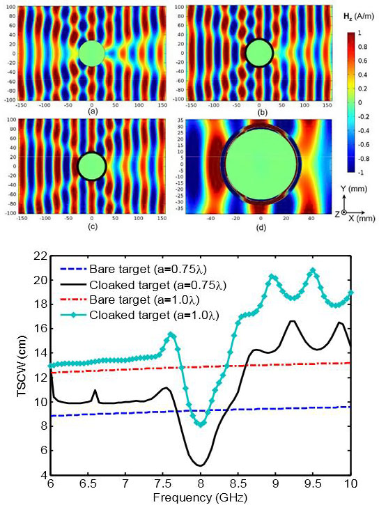
Top: Color online) Snap-shots of Hz distributions for fwork=8 GHz at TM wave incidence on: (a) bare metallic cylinder with the radius 0.75 λwork; (b) the same target cloaked by the shell with material parameters prescribed by Eq. (7); (c) the same target cloaked by the multi-layer dielectric shell; (d) zoomed-in view of (c).
Bottom: (Color online) The TSCW spectra obtained by integrating the simulated far-fields for the cloaked and bare targets of two different sizes. Dimensional parameters of the multi-layer dielectric cloaks in both cases were fixed for the wavelength corresponding to fwork=8 GHz.

Using a new kind of cloak that uses a very thin multilayer dielectric coating made of natural material, not metamaterial, researchers at Michigan Technological University demonstrated better cloaking efficiency than a similarly sized metamaterial cloak designed by using the transformation optics relations.

Michigan Technological University’s invisibility cloak researchers have done it again. They’ve moved the bar on one of the holy grails of physics: making objects invisible.

Just last month, Elena Semouchkina, an associate professor of electrical and computer engineering at Michigan Tech, and her graduate student, Xiaohui Wang, reported successful experimental demonstration of the use of non-conductive ceramic metamaterials to cloak cylindrical objects from microwave-length electromagnetic waves. Previously, Semouchkina had designed a non-conductive glass metamaterial cloak that worked with infrared frequency waves, which are shorter than microwaves.

Then, scarcely was the ink dry on their report in the IEEE Microwave and Wireless Components Letters, a journal published by the Institute of Electrical and Electronics Engineers, when they developed a different cloaking approach and published it in the American Institute of Physics journal, Applied Physics Letters.

This time, they used ordinary dielectric materials such as ceramics having differing dielectric permittivity—a measure of the response of a substance to an electrical field— instead of metamaterials, which are artificial materials with properties not found in nature. They found that they were able to cloak larger cylindrical objects and cloak them more effectively than they had using metamaterials.

This research, just published online and supported by the National Science Foundation (NSF), also won Wang an honorable mention in an IEEE student papers competition. Wang’s paper will be listed in the Technical Program Booklet at an upcoming IEEE International Symposium on Antennas and Propagation. He received $1000 IEEE grant to travel to the symposium to present this work.

According to their report in the Applied Physics Letters article, Semouchkina and Wang designed a new kind of cloak that uses a very thin multilayer dielectric coating made of natural material, not metamaterial. They compared it through mathematical analysis and computer simulations to a metamaterial cloak of similar size but based on a different principle, called transformation optics.

The new cloak demonstrated better cloaking efficiency than did a similarly sized metamaterial cloak designed by using transformation optics relations,” said Semouchkina.

The new cloak outperforms previous cloaks, which caused more reflection and more shadows, as well as distortion of the electromagnetic waves, the researchers reported. It is eight to nine times thinner than metamaterial cloaks and much simpler to make, they noted.

The multi-layer dielectric cloak could easily be scaled to work in a variety of frequency ranges,” Semouchkina said. “The design procedure developed in this work could be used to further advance the cloak parameters and for adjusting it to practical needs.

Cloaking may sound like a magic trick or something out of a movie, but it could prove useful in national security, law enforcement or other applications.

Publication: Xiaohui Wang and Elena Semouchkina, “A route for efficient non-resonance cloaking by using multilayer dielectric coating,” Appl. Phys. Lett. 102, 113506 (2013); DOI:10.1063/1.4796171


Source: Jennifer Donovan, Michigan Technological University

Image: Michigan Technological University; Xiaohui Wang and Elena Semouchkina, DOI:10.1063/1.4796171

jueves, 7 de marzo de 2013

Breakthrough could lead to drugs that better combat 'superbugs'

February 28, 2013

NDM-1, present in a number of pathogenic bacteria, including Klebsiella pneumonia and Escherichia coli, is able to defeat many of the world’s most widely used antibiotics, including penicillin derivatives, cephalosporins, monobactams and carbapenems.
LEMONT, Ill. – In the never-ending battle between antibiotic developers and the bacteria they fight, scientists at the U.S. Department of Energy’s (DOE) Argonne National Laboratory have made a key breakthrough that could allow for the development of new drugs to more effectively combat antibiotic-resistant “superbugs.”

An Argonne team led by Youngchang Kim of the Structural Biology Center, in collaboration with researchers from the Midwest Center for Structural Genomics, the University of Texas-Pan American and Texas A&M University, recently determined the structure of NDM-1, a harmful enzyme able to overcome several antibiotics. The team used a combination of X-ray crystallography at Argonne’s Advanced Photon Source (APS), biochemical assays, and computational modeling using resources at two Texas universities.

Structure of NDM-1 in complex with two cadmium ions and ampicillin. The intermediate (int-amp) is superposed with the modeled unhydrolyzed ampicillin (yellow).
NDM-1, present in a number of pathogenic bacteria, including Klebsiella pneumonia and Escherichia coli, is able to defeat many of the world’s most widely used antibiotics, including penicillin derivatives, cephalosporins, monobactams and carbapenems. The enzyme works by effectively binding to and breaking – in a process known as hydrolysis – a structure called a β-lactam ring, which is necessary for antibiotics to function.

The traditional view of enzymes, particularly those most essential for the bacteria to survive, is that they have a very specific substrate (or target – in this case the antibiotic) to bind and act on, and they interact with this target directly by forming a well-fitted lock-and-key-like complex. However NDM-1, and others like it, can act on a broad range of substrates.

These kinds of enzymes can recognize many different targets,” said Andrzej Joachimiak, head of Argonne’s Structural Biology Center and the Midwest Center for Structural Genomics.

“The appearance of NDM-1 among pathogenic bacteria represents a major concern because the enzyme can inactivate so many of the antibiotics that we use to treat infections,” said Charles Edmonds of the National Institutes of Health’s National Institute of General Medical Sciences, which partially supported the study. “This work, by providing a detailed understanding of the structure of the enzyme and its mechanism of action, brings an invaluable tool to the design of new drugs to combat this significant threat to public health.

The act of NDM-1 binding to the antibiotic, however, does not represent the entire story, because for the enzyme to effectively overcome the antibiotic, it must also cleave the β-lactam ring. The researchers found that certain metals, including zinc, manganese and cadmium, can bind to the NDM-1 active site – and to the β-lactam ring. While zinc provides the most favorable environment for cleavage to occur, cadmium tends to inhibit the enzyme’s ability to cleave the ring.

The next step in the research is to look for inhibitors that we can create that would block the functioning of the enzyme,” Joachimiak said. “If we can stop the enzyme from cutting the ring, the antibiotics stand a much better chance of staying effective.

The results of the research were recently published in The FASEB Journal in an article titled “NDM-1, the ultimate promiscuous enzyme: substrate recognition and catalytic mechanism.

The research was supported by the National Institutes of Health and the National Science Foundation. The APS and the Argonne Structural Biology Center are supported by DOE’s Office of Science, while the Argonne Midwest Center for Structural Genomics is supported by the National Institutes of Health. The researchers also used computational resources at the University of Texas at Austin and the University of Texas-Pan American.

Argonne National Laboratory seeks solutions to pressing national problems in science and technology. The nation's first national laboratory, Argonne conducts leading-edge basic and applied scientific research in virtually every scientific discipline. Argonne researchers work closely with researchers from hundreds of companies, universities, and federal, state and municipal agencies to help them solve their specific problems, advance America's scientific leadership and prepare the nation for a better future. With employees from more than 60 nations, Argonne is managed by UChicago Argonne, LLC for the U.S. Department of Energy's Office of Science.- See more at: http://www.anl.gov/articles/breakthrough-could-lead-drugs-better-combat-superbugs#sthash.exRUc0SD.dpuf