Mostrando entradas con la etiqueta Virus. Mostrar todas las entradas

martes, 5 de enero de 2016

Scientists develop hydrogen 'nano reactor' by hiding bacteria inside a virus shell

Trevor Douglas/Indiana University
This could be the key to clean and cheap biofuel.
Researchers in the US have developed a virus-like biomaterial that catalyses the formation of hydrogen inexpensively and cleanly, which could lead to new environmentally friendly ways of producing biofuel.

The biomaterial is produced by placing a modified enzyme inside the protective protein shell of a virus, called a capsid. The resulting 'nano reactor' catalyst is 150 times more efficient than the unaltered form of the enzyme.

"Essentially, we've taken a virus's ability to self-assemble myriad genetic building blocks and incorporated a very fragile and sensitive enzyme with the remarkable property of taking in protons and spitting out hydrogen gas," said Trevor Douglas, a chemist at Indiana University. "The end result is a virus-like particle that behaves the same as a highly sophisticated material that catalyses the production of hydrogen."

To create the biomaterial, known as "P22-Hyd", the team co-opted two genes from the common bacteria Escherichia coli. These genes, called hyaA and hyaB, encode key subunits of the enzyme, hydrogenase. The enzyme is then inserted into the protective capsid of a bacterial virus known as bacteriophage P22.

The resulting fusion of hydrogenase and a virus shell is not only vastly more efficient at catalysing the production of hydrogen – it's also easy to create, requiring only a simple fermentation process. And compared to other means of creating fuel cells, such as using platinum to catalyse hydrogen, it's cheap and won't take a toll on the environment.

"This material is comparable to platinum, except it's truly renewable," said Douglas. "You don't need to mine it; you can create it at room temperature on a massive scale using fermentation technology; it's biodegradable. It's a very green process to make a very high-end sustainable material."

In addition to breaking the chemical bonds of water to produce hydrogen, P22-Hyd also catalyses the reaction the other way around, recombining hydrogen and oxygen to generate power.

The form of hydrogenase the researchers are using – NiFe (nitrogen-iron) – is one of only three occurring in nature, but it's the most suited to the task, easily integrating into biomaterials and tolerating exposure to oxygen. In its altered, protected form, the enzyme is vastly superior to unaltered NiFe enzymes thanks to greater resistance to heat and chemicals in the environment.

"No one's ever had a way to create a large enough amount of this hydrogenase despite its incredible potential for biofuel production," said Douglas. "But now we've got a method to stabilise and produce high quantities of the material – and enormous increases in efficiency."

The research has been reported in Nature Chemistry.

ORIGINAL: Science Alert
PETER DOCKRILL
5 JAN 2016

jueves, 9 de enero de 2014

Viruses Build Piezoelectric Nanogenerator Through Self Assembly

Image: KAIST

Nanotechnology has opened up the possibility of building things like nature does: on the nanoscale. As such, biomimicry has been a guiding principle of nanomanufacturing.

But unlike natural processes, artificial synthesis of nanoscale structures has often required toxic and expensive conditions. Now researchers at the Korea Advanced Institute of Science and Technology (KAIST) say they've developed a synthesis process that can be done in a more natural way without the costly and extreme environments previously required.

What the researchers came up with uses a harmless, man-made virus, known as the M13 viral gene. The researchers modified it so that it acted as a template for a piezoelectric material, barium titanate (BaTiO3).

The research, which was published in the journal ACS Nano (“Virus-Directed Design of a Flexible BaTiO3 Nanogenerator”), demonstrated that they could build a high-performance, flexible nanogenerator from the piezoelectric material using the M13 viral gene as template for guiding self-assembly of the device.

"This is the first time to introduce a bio-templated inorganic piezoelectric material to a self-powered energy harvesting system, which can be realized through eco-friendly and efficient material syntheses," said Professor Keon Jae Lee from the Department of Material Science and Engineering at KAIST in a press release.

But, of course, using man-made viruses to guide the self-assembly of devices has long been the purview of Angela Belcher at MIT for over a decade. Nonetheless, we can’t quibble that this marks the first time that a virus template was used to create a nanogenerator. And it has a pretty respectable electrical output performance, claimed in the research paper to be about 300 nanoampere and 6 volts.

In fact, the real breakthrough of the research may be that the biosynthetic method that the KAIST researchers developed could open up new possibilities in bio-inspired self-assembly for applications ranging from thermoelectrics to biofuel cells.

Learn More KAISTbiomimicrynanogeneratorsnanotechnologypiezoelectricself-assemblyvirus

ORIGINAL: IEEE Spectrum
By Dexter Johnson
5 Dec 2013

jueves, 5 de diciembre de 2013

Viruses Build Piezoelectric Nanogenerator Through Self Assembly


Image: KAIST
Nanotechnology has opened up the possibility of building things like nature does: on the nanoscale. As such, biomimicry has been a guiding principle of nanomanufacturing.

But unlike natural processes, artificial synthesis of nanoscale structures has often required toxic and expensive conditions. Now researchers at the Korea Advanced Institute of Science and Technology (KAIST) say they've developed a synthesis process that can be done in a more natural way without the costly and extreme environments previously required.

What the researchers came up with uses a harmless, man-made virus, known as the M13 viral gene. The researchers modified it so that it acted as a template for a piezoelectric material, barium titanate (BaTiO3).

The research, which was published in the journal ACS Nano (“Virus-Directed Design of a Flexible BaTiO3 Nanogenerator”), demonstrated that they could build a high-performance, flexible nanogenerator from the piezoelectric material using the M13 viral gene as template for guiding self-assembly of the device.

"This is the first time to introduce a bio-templated inorganic piezoelectric material to a self-powered energy harvesting system, which can be realized through eco-friendly and efficient material syntheses," said Professor Keon Jae Lee from the Department of Material Science and Engineering at KAIST in a press release.

But, of course, using man-made viruses to guide the self-assembly of devices has long been the purview of Angela Belcher at MIT for over a decade. Nonetheless, we can’t quibble that this marks the first time that a virus template was used to create a nanogenerator. And it has a pretty respectable electrical output performance, claimed in the research paper to be about 300 nanoampere and 6 volts.

In fact, the real breakthrough of the research may be that the biosynthetic method that the KAIST researchers developed could open up new possibilities in bio-inspired self-assembly for applications ranging from thermoelectrics to biofuel cells.

ORIGINAL: IEEE Spectrum
By Dexter Johnson
Posted 5 Dec 2013

miércoles, 25 de septiembre de 2013

Google Science Fair winners: Flashlights without batteries, ‘bat signals,’ non-viral viruses

September 24, 2013

Google
Google Science Fair is clearly where minor miracles happen. Flashlights that work without batteries, traffic that moves itself out of the way of an oncoming emergency vehicle, and new flu medicines — designed on a computer — that are effective even against pandemic strains.

And all, of course, from teenagers.

Google announced the winners of its 2013 science fair today, and three teens from Australia, Canada, and the USA have emerged from the 18 finalists that Google invited to Mountain View, and won their age categories.

Bat signal for ambulances


In the 13-14 age category, Aussie teen Viney Kumar designed a system that allows ambulances and other emergency vehicles to send out a GPS-informed “bat signal” to cars within 500 meters (a third of a mile) notifying them via cellular connection that an ambulance or fire truck is coming, and requesting that they pull over.

Kumar designed the system, he said, when he saw an ambulance stuck in traffic in India, unable to move, and wondered how many people were dying for lack of prompt emergency response.

Flashlight without batteries

Canadian teen Ann Makosinski won the 15-16 year-old category with a novel flashlight idea that requires no battery. The “Hollow Flashlight” runs solely on the heat of your hand, Makosinski’s flashlight uses Peltier tiles that capture energy from the flow of heat to cooler areas, such as the surrounding air. Incredibly efficient, the light can produce up to 5.4 mW and generate a light that is five foot-candles in brightness.

Since a foot-candle is basically the brightness of a lit candle at one foot distance (shocker), her battery-less device is producing more light than five candles — impressive.

The no-viral flu virus
Tim Evanson  Model of an HIV virus


Eric Chen of the USA was both the 17-18 category winner and the grand prize winner for an extremely ambitious project with the goal of stopping the flu virus before it goes viral. Chen realized that the influenza endonuclease, or enzymes, are essential for viral growth. To stop the flu, therefore, scientists need only stop its reproduction, killing the infection by not allowing it to spread.

To find the best way of doing so, Chen set up computational models and computer-aided designs of flu viruses and the endonuclease components, and “virtually screened” potential medications that could molecularly bind to the enzymes, rendering them inert, and killing the flu dead.

It sound like science fiction, or at least something the Center for Disease Control is doing in some billion-dollar lab in Atlanta, but this teen pulled it off, finding “a number of novel, potent endonuclease inhibitors,” and laying the groundwork for further study and optimization.

This teen isn’t just smart at science — Chen has filed a patent on this discovery.

The winners all received prizes from Google and CERN, Lego, National Geographic, and Scientific American. The grand prize winner, Chen, will receive an all-expenses-paid trip to the Galapagos Islands, and $50,000 in scholarships.

domingo, 18 de agosto de 2013

Bizarre Behavior in Endangered Tigers Traced to Dog Virus

ORIGINAL: Live Science
By Tanya Lewis, Staff Writer
August 16, 2013

Marty is an endangered Amur tiger. Credit: Jan Morse | RZSS

A rare breed of tigers in Russia has been spotted behaving bizarrely for more than a decade, and now researchers say they may know why.

The majestic, endangered felines have stumbled into villages and roads, emaciated and unafraid of people. Eventually, most of the tigers died, or had to be killed.

Now a new study shows these Amur tigers (also called Siberian tigers) suffered from a fatal neurological infection called canine distemper virus.


The virus, which normally infects domestic dogs, may be responsible for the deaths of at least 1 percent of Amur tigers since 2009, the researchers said. [Gallery: Rare and Beautiful Amur Leopards]

"Losing 1 percent of an endangered population is pretty significant," study researcher Denise McAloose, a pathologist at the Wildlife Conservation Society in the Bronx, New York, said in a statement.

McAloose and her colleagues examined tissue samples from five wild Amur tigers that died or were killed as a result of a neurological disease. Canine distemper virus was responsible for the deaths of two of the big cats, and caused a severe infection in a third, the team reportedin the journal mBio.

The brains of the two tigers were full of lesions, suggesting they suffered from severe viral encephalitis, which explains the animals' strange stumbling behavior. Molecular analysis of proteins and antibodies confirmed the presence of the virus, and a gene associated with the virus was found in the third tiger.

The afflicted tigers were found across the Russian Far East, throughout the range where Amur tigers live. The virus appears to be a recent threat, because blood taken from tigers before the year 2000 tested negative for viral antibodies, McAloose said.

The researchers said they suspect the tigers might have contracted the virus from unvaccinated domestic dogs in Russia, or possibly from raccoon dogs or foxes.

To determine the source of the tiger infections, McAloose and her team are now collecting samples from dogs and small wild carnivores in the region.

McAloose called the situation for Amur tigers "quite serious."

Once found throughout the Russian Far East, northern China and the Korean peninsula, the animals were nearly driven to extinction by hunting. By the 1940s, just 40 tigers were living, according to the World Wildlife Fund. Thanks to conservation efforts, the population has increased to around 450 today.

Follow Tanya Lewis on Twitter and Google+. Follow us @livescience, Facebook & Google+. Original article on LiveScience.


Editor's Recommendations
Gallery: Tiger Species of the World
Images: One-of-a-Kind Places on Earth
Acadia National Park: Glacier-Carved Beauty
Author Bio
Add caption

Tanya Lewis Tanya joined the LiveScience staff in 2013. She received a graduate certificate in science communication from the University of California, Santa Cruz in 2012. Before that, she earned a bachelor's degree in biomedical engineering from Brown University. She has interned previously at Wired.com, Science News, Stanford Medical School, and the radio program Big Picture Science. To find out what her latest project is, you can follow Tanya on

jueves, 1 de agosto de 2013

Learning from a Virus: Keeping Genes Under Wraps


Cytomegalovirus particles as seen through an electron microscope: While some of the protein capsules are empty, others contain tightly packaged DNA, visible as dark structures. The diameter of a human hair would cover a line of about 600 virus particles. (Photo: Farah Bughio, Felicia Goodrum)

July 30, 2013 — An international collaboration of researchers including Felicia Goodrum of the University of Arizona's immunobiology department has studied how a human herpes virus carried by the majority of the population packages its genetic information during infection.

The discoveries improve the chances of developing more targeted therapies in place of existing drugs, which do not always work or come with side effects.

Experts estimate that 60 to 90 percent of the world's population carry the human cytomegalovirus, or CMV, which is one of the eight herpes viruses that infect humans.

In healthy individuals, the virus lies dormant and does not cause overt disease. However, it poses a significant risk when contracted by unborn children -- whose immune system has not matured yet -- and individuals with compromised immune function.

CMV is the leading cause of birth defects resulting from any infectious agent
. It affects one in 150 births in the US and most commonly results in hearing loss, but can also cause cognitive or physical anomalies and cerebral palsy. Once infected, the virus stays in the body for life and flares up only when the immune system is suppressed, for example in AIDS patients, transplant patients and cancer patients undergoing intensive chemotherapy.

For the study, published in the scientific journal Proceedings of the National Academy of Sciences (PNAS), Goodrum teamed up with collaborators in Germany and Israel.

The researchers investigated how a fundamental aspect of the human cell regulates the virus: the mechanism by which genetic information is packaged and stored. Understanding how the viral DNA behaves in the human host cells during dormancy and reactivation of the virus provides the basis for the development of drugs that could prevent the virus from "waking up" and causing disease.

"The human immune system is very sophisticated, and the way this virus has managed to stealthily integrate into our biology to ensure its own survival is no small feat," said Goodrum, also a member of the UA's BIO5 Institute.

"CMV is a master of human cell biology. From transcribing DNA into blueprints for proteins to the manufacturing of those proteins, from cell division to cellular metabolism, there is not a process this virus has not tweaked," Goodrum also said.

Virus factory: The top image shows uninfected, normal human cells with cell nuclei in blue and the cellular protein processing apparatus in red. In the infected cells (bottom image), the virus has produced proteins (green) that take over and reorganize the cellular processing apparatus into a virus factory. (Credit: Photo: Farah Bughio, Felicia Goodrum)
That mastery, she explained, is the reason the virus is so elusive to vaccine, and there currently is no way to eradicate it. Goodrum noted that with other herpes viruses, like Epstein-Barr or chicken pox, the infection is obvious. But that is not the case with CMV.

"From the perspective of a virus, that is the pinnacle of mastery -- to infect without ever making its presence known," Goodrum said.

"To develop more effective antiviral strategies, we must understand the biology of the virus infection and how the virus manages to persist for our lifetimes," she said. "We are trying to understand how our cellular mechanisms are being used by this virus and discover targets for drugs to control it."

Each human cell contains a thread of DNA that is about 6 feet long, stowed away in its nucleus and tightly packaged by proteins called histones. One such package of genetic material is called a chromosome.

"You can imagine histones as a spool, and the thread is DNA that wraps around the spool," Goodrum said. "This accomplishes two important objectives: first, it condenses DNA so that it can be packed into the cell nucleus and second, it provides the cell with a mechanism to regulate the activity of the genes encoded on that DNA."

The association between histones and DNA is very dynamic and acts as a key mechanism used by cells to control which genes are expressed and which are not, Goodrum explained.

For example, when DNA is wound tightly around histones, it is not accessible to enzymes specializing in making copies of genes in a process called transcription, which subsequently serve as blueprints to manufacture proteins.

"When a virus like CMV infects our cells, its DNA is packaged by histones just as if it were the cell's own DNA," Goodrum said.

"The question is, how does this happen, and does the virus have any choice in the matter? Our work maps the deposition of histones across the viral DNA chromosome and shows that the virus encodes a mechanism to reorganize these histones to favor the expression of genes from the viral chromosome."

Michael Nevels of the Institute for Medical Microbiology and Hygiene at the University of Regensburg, Germany, said: "Now that we have determined the positions of the nucleosomes, we can study how transcription is regulated, and from that others can start developing therapies."

For example, if researchers can identify molecules that play key roles in the process, they can design new drugs that target those molecules.

"Since we can't eliminate the virus, the goal is to keep it in check," said Nevels, who led the study together with Eran Segal and Einat Zalckvar of the Weizmann Institute of Science in Rehovot, Israel.

"The idea is to target the virus on the level of its DNA structure and to reduce the gene activity back to the dormancy levels," Nevels said.

One such molecular target identified in this study is a viral protein called IE1. Because it regulates the packaging and unpacking of the viral DNA, it could potentially be a target for new therapies.

Cautioning that many more steps will be necessary before this could be achieved, Nevels said that "if we could inhibit IE1, the virus genome would be packed more tightly with histones, which leaves less DNA accessible and prevents genes from becoming active."

Another strategy takes the opposite direction by deliberately awaking the virus from its dormant state so it becomes vulnerable to antiviral drugs. This could be an option for patients about to undergo an organ transplantation, which requires immune-suppressing drugs to prevent the new organ from being rejected. Suppressing the immune system allows the virus to reactivate.

"There are antivirals that target the active, replicating virus but they can't target latent virus because in that state, it's really just a piece of DNA in the cell nucleus," Goodrum said. "Before those patients enter a immunosuppressed state, you would target the viral reservoirs and force the virus out of latency."

Note: Materials may be edited for content and length. For further information, please contact the source cited above.

Journal Reference: E. Zalckvar, C. Paulus, D. Tillo, A. Asbach-Nitzsche, Y. Lubling, C. Winterling, N. Strieder, K. Mucke, F. Goodrum, E. Segal, M. Nevels. Nucleosome maps of the human cytomegalovirus genome reveal a temporal switch in chromatin organization linked to a major IE protein. Proceedings of the National Academy of Sciences, 2013; DOI: 10.1073/pnas.1305548110

lunes, 11 de marzo de 2013

Bedeviled by Dengue

ORIGINAL: The Scientist
By Beth Marie Mole
March 1, 2013

The global spread of dengue virus has immunologists and public-health experts debating the best way to curb infection.

DAVID SCHARF/GETTY IMAGES©
In 1961, during the first dengue outbreak physician Scott Halstead ever witnessed, children poured into Bangkok’s hospitals, passing and vomiting blood, faint from blisteringly high fevers. Twenty percent of the children would die within a few days as doctors scrambled to find treatments, with some in nearby Vietnam even plunging children into ice baths in an attempt to hold down their soaring temperatures.
 width="820"
The deadly illness caused by dengue virus wasn’t yet known as dengue in Thailand; doctors there referred to it as “Chinese medicine poisoning” based on a demographic quirk. Although half the city’s population was Chinese, the only time Thai doctors—who practiced Western medicine—treated Chinese children was when the children had been stricken with this mysterious, deadly illness. Thus, doctors imagined that a horrific poisoning caused by Eastern remedies was responsible for the influx of Chinese patients. Instead, Halstead explains, Chinese parents had quickly learned that the hospital, rather than traditional medicine, was the best bet—however slim the chances—for defeating dengue.

Halstead who was drafted into the US Army after World War II and originally sent to Japan in 1957 to study encephalitis, had just settled into a lab across the street from the Children’s Hospital in Bangkok. “Everything I’ve ever done,” he says in retrospect, “is related to the treatment of dengue.” In the years that followed, he and his colleagues identified dengue as the cause of the outbreak and began tracking the four different versions of the virus, each transmitted by mosquitoes. Among their seminal discoveries, the researchers learned that the hemorrhagic disease that they saw in 1961 was most common when a child is infected with a second type of dengue—a finding that would prove pivotal in the decades-long search for a vaccine that continues today.

The World Health Organization estimates that more than 2.5–3 billion people, or more than 40 percent of the world’s population, are now at risk of being infected with dengue—including some in the developed world.http://www.the-scientist.com/images/March2013/dengue_virus.jpg

But as research efforts have evolved, so has the reach of dengue infections. The disease has now become prevalent in more than 100 countries, causing as many as 100 million infections per year. And it’s still spreading. The World Health Organization (WHO) estimates that more than 2.5–3 billion people, or more than 40 percent of the world’s population, are now at risk of being infected with dengue—including some in the developed world. In the past few years, cases of dengue have popped up in Texas, Florida, France, and Croatia. Of the 500,000 cases of severe dengue requiring hospitalization and the roughly 24,000 deaths they cause each year, most are in children, according to the WHO. And control measures have been met with challenges. One high-profile vaccine trial conducted by the French vaccine company Sanofi Pasteur partially failed last year, leaving investigators scratching their heads. The defeat comes amid fears of some researchers that vaccines have the potential to exacerbate a dengue infection rather than protect against it. “We’re in a mess,” Halstead says bluntly.


Going viral
When Halstead started digging into deng width="820" width="820"ue in the 1960s, only a handful of countries were home to all four types of the virus, which represent four separate viral jumps from monkeys to humans between 100 to 800 years ago, according to the US Centers for Disease Control and Prevention (CDC). But in the decades since, the four types—simply called dengue 1, 2, 3, and 4—have independently made their way around the globe. Poor urban planning and warming climates are partly to blame, having opened new territories to the mosquitoes that carry the virus, and global travel and trade have provided the necessary transit.

Tires may be the best example of mosquito and dengue transport: shipped across the globe on barges, tires collect water rings that offer mosquitoes first-class tickets to new locations. Consequently, dengue’s vectors, Aedes aegypti (the yellow fever mosquito) and A. albopictus (the tiger mosquito), have surged in new locations in Central and South America, Australia, and even in the lower United States. The yellow fever mosquito is particularly insidious, biting during the day, and able to spawn in water-filled crannies no larger than a small cup. Its eggs can withstand drought conditions, allowing the populat width="820" width="820" width="820" width="820"ion to quickly bounce back after a dry spell. It is now a common pest of dense urban areas—bringing disease with it.

After a cluster of dengue infections struck continental Europe, the European Union (EU) provided funding in 2011 for researchers to assess where dengue would strike next, amid fear that dengue would continue to spread in the developed world. The disease has traveled so quickly that “we only have estimates of the global burden of dengue, which is astonishing,” says epidemiologist Simon Hay of Oxford University, who is part of a consortium that’s developing risk maps of future dengue spread. The group is currently working on a map of where the disease exists now; only later will it work on how warming climates and city sprawl might change the map, says Jane Messina, the head medical geographer on the project.

Concerns have flared over the possibility that dengue could easily become endemic in Europe and in the United States, which had its own cluster of infections in 2012 in Texas and Florida. Ae. aegypti is now found in 23 states and A. albopictus in 26. “Dengue can occur anywhere the mosquito vectors occur,” says Ronald Rosenberg, the associate director of the CDC’s division of vector-borne diseases and a member of the WHO’s committee on neglected tropical diseases.

Concerns have flared over the possibil width="820" width="820"ity that dengue could easily become endemic in Europe and in the United States.
If experience from abroad is any indication, the economic impact of a dengue epidemic could be huge. Though severe disease is usually rare, it creates a big burden on health-care systems, says microbiologist and dengue expert Aravinda de Silva, of the University of North Carolina at Chapel Hill, who works in Sri Lanka where dengue has long been endemic. “Hemorrhagic fever is a massive concern. Every parent is terrified of their child getting it,” he explains. Parents often bring their children into the hospital at the first sign of dengue infection, which leads to many unnecessary hospitalizations—and the costs add up. With precautionary care as well as treatments for those who do get severe disease, dengue outbreaks weigh heavily on local economies—an outbreak in Thailand in 1994, for instance, cost an estimated $51 million, not including dengue prevention programs. Such an economic strain has brought the infection to the top of priority lists in health ministries around the world.
 width="820"
The global spread of dengue over the past 3 decades, however, has driven more resources to research on how the virus spreads and how it can be defeated.

Catching a virus
Before the name “dengue” caught on in the Americas, it was often referred to as “breakbone fever,” or la quebradora in Spanish. The nickname refers to the crippling aches that come with infection. Once delivered by a mosquito, dengue virus hijacks skin and some immune cells and hitchhikes through the lymphatic system, infecting other tissues and organs and loading the bloodstream with viral replicates. The body’s immune system counters with blazing inflammation, causing sharp pains in muscles. Eighteenth-century reports of dengue infection describe the wretched gaits of patients as they staggered on swollen limbs, often in a stupor from fever.

For the lucky, those will be the worst of their symptoms. Others, however, can develop life-threatening disease. If the inflammation firestorm gets out of hand, it can damage the lining of capillaries—the smallest blood vessels—causing gaps through which blood plasma can seep. Systemic internal leaking quickly leads to a drop in blood pressure, followed by shock, organ failure, and massive bleeding.

Between 2005 and 2007, Managua, Nicaragua, saw a mysterious spike in the number of children entering hospitals with dengue hemorrhagic fever and shock syndrome. Since the 1980s, the neighborhoods around the city had been riddled with Ae. aegypti as well as the four dengue types they carry, but no one knew why so many more children were suddenly developing severe disease. Infectious disease researcher Eva Harris and her team from the University of California, Berkeley, and the Nicaraguan Ministry of Health had been working on infectious disease there for decades, and noticed severe dengue disea width="820"se occurred in children reinfected with a second type of the virus, similar to the pattern Halstead saw in the 1960s with second infections.

Simona Zompi, an immunologist working on Harris’s team, was dissecting the immune system’s response to dengue. When a person is initially infected, the dengue viral particles attack skin and immune cells by latching onto receptors on the cell surface. The cells enclose the viruses in a sac—a process called receptor-mediated endocytosis—that would normally digest the viral captives with enzymes in an acidic milieu, like a piece of food in the stomach. But dengue virus particles escape digestion by rearranging their envelope proteins to fuse with the sac’s membrane, opening a channel through which the capsid-encoated viral genome is released into the infected cell’s cytoplasm. Once free, the virus usurps the cell’s machinery to create a viral factory, triggering a full-blown dengue infection.

Zompi and the team focused on B cells, which react to the infection by creating antibodies against the infecting virus. Once the virus is defeated, some of these cells go into hibernation as memory B cells, which become quickly reactivated upon reinfection with dengue for the rest of the person’s life. Those antibodies normally foil infection by blanketing the viral particle, which prevents the virus from binding to cell receptors and entering cells. The antibody-coated virus is then taken up by monocytes or macrophages, which, after endcytosis, can digest the invader because the coated particle is unable to escape the digestive sac. (See illustration below.)

FREQUENT CHECKS: A young girl is examined by a military doctor 45 miles away from Rio de Janeiro, Brazil. The Brazilian military set up three field hospitals to help manage patients with dengue infections.© ANTONIO SCORZA/AFP/GETTY IMAGES
However, this protective immune response depends on whether antibodies can bind strongly enough to coat and disable—or neutralize—the virus, Zompi explains. Some antibodies bind poorly to the viral particle and, as a result, only a few manage to cling to the virus, which is just enough to entice macrophages and monocytes to engulf them. However, without complete antibody coverage of the viral coat, the virus can still escape from the endocytic sacs of the macrophages or monocytes and take over the very cells that have engulfed it. (See illustration below.) Now, the virus has an additional pathway to enter cells: all of the immune cells recruited and activated by the antibodies could potentially become virus-producing factories. Thus, antibodies with poor binding affinity and neutralizing potential effectively boost the infection, and the inflammatory response can crank to inferno levels, causing hemorrhagic fever and shock syndromes. Scientists refer to this explanation for severe disease as “antibody-dependent enhancement” (ADE) of the immune response.

Most antibodies generated against a specific dengue type, dengue 1 for example, will bind well and be able to thwart future infections of that type. But when a new type of dengue invades—dengue 2, for instance—the reactivated dengue 1 antibodies may only partially recognize the virus and thus lead to ADE. Indeed, Harris and Zompi found that as the predominant circulating dengue virus type switched in Managua, patients hospitalized with dengue made the strongest antibody response to a dengue type from a previous infection, not the current infecting dengue type. And most countries are infested with the four types of dengue that can fluctuate in prevalence.

Some researchers worry that vaccines designed to spur antibody production might instead trigger ADE if the B cells generate low-affinity antibodies rather than those that completely blanket the virus. If that were the case, being vaccinated could predispose children to ADE.

Vaccine development dissension
But not all dengue researchers are convinced that severe disease can be explained by the ADE response, says de Silva. There are two competing hypotheses about how severe hemorrhagic and shock diseases might unfurl: other immune cells, such as T cells, may present different fragments of the virus to the immune system, stimulating a greater inflammatory response; or, more virulent subtypes of each of the dengue types may trigger the more severe immune reaction characteristic of ADE. But the antibody hypothesis is certainly the front-runner, de Silva says—especially after his recent discovery of how dengue-neutralizing antibodies work.

During initial infection, the human immune system makes a range of antibodies with different affinities to the virus. The antibodies capable of neutralizing the virus apparently latch onto a wedge between two adjacent molecules of an envelope protein on the coat of the intact virion. The new finding is significant because it casts doubt on past immunology and vaccine-development studies that have focused on generating an antibody response against only a small fragment of the envelope protein in isolation. Antibodies that don’t neutralize the virus by binding to the wedge may be candidate ADE generators, says de Silva.

Yet many researchers in the field are reluctant to acknowledge the new complication, says Halstead. “A lot of people don’t like to have to address [the possibility of ADE] head on,” he says.

Indeed, current recommendations by the WHO on conducting clinical trials of candidate dengue vaccines don’t include specific guidelines for ADE. In fact, ADE and its potential causes are considered a hypothetical concern that should not interfere with development of a vaccine, according to the WHO.

“[ADE] may not be a concern, in my own opinion,” says infectious-disease researcher Claire Huang, of the CDC’s division of vector-borne diseases, echoing the WHO’s stance. Along with a team of researchers at the CDC, Huang has been toiling for years to develop an effective vaccine, and is working with the commercial vaccine developer Inviragen on a vaccine now in Phase 2 clinical trials. It may be more complicated, she adds, but most antibody responses are protective and robust.

The vaccine she’s been working on is tetravalent, meaning it’s designed to generate antibodies against all four types of dengue. Huang and her team borrowed the genetic backbone of an earlier successful vaccine against the type 2 virus, and dressed that genome, with its attenuating mutations, in the other three viral coats, creating four recombinant viruses. “From the outside, they all look like either dengue 1, dengue 2, dengue 3, or dengue 4, but inside they’re all the same attenuated virus,” she explains. The team hopes that the vaccine will produce a suite of neutralizing antibodies against all types of dengue.

So far, the results look promising. The vaccine sailed through Phase 1 trials, proving safe in healthy children and adults in Colombia and the U.S. who had no prior exposure to dengue. But the earlier tetravalent vaccine made by Sanofi Pasteur also had great early results. After buzzing through initial safety tests and Phase 2 trials, that vaccine moved to a Phase 2b trial, which enlisted 4,000 school-age children in Thailand’s Ratchaburi Province. The vaccine required three shots over the course of a year, and in September the company revealed that only 30 percent might be protected, although none of the results were statistically significant.

“I looked at the vaccine results and wondered if they had given people water,” Halstead says. “It was very surprising,” echoes de Silva. It’s still unclear what went wrong, he says. The vaccine showed 80 to 90 percent protection against dengue types 3 and 4, and around 60 percent protection against dengue 1. But it failed at protecting against dengue 2—the dengue that was circulating that year at high levels. The question we’re left with, de Silva says, is whether that virus was just a mismatch with the vaccine, or if the dengue 2 portion of the vaccine simply didn’t generate a good antibody response in the vaccinated children.

The only triumph of the trial was that it didn’t show an increase in severe disease. Of the 4,000 children vaccinated, only five developed severe disease. “The vaccine was still very safe,” says Dan Stinchcomb, cofounder and CEO of Inviragen. “[The Sanofi] vaccine is somewhat similar to ours, and the trial laid to rest one of the biggest concerns,” he says. The study demonstrated “that if you’re not fully protected against all four viruses, then you’re not more susceptible to severe disease,” or ADE.

But Halstead points out that the trial only followed children for a year—not long enough for them to become infected a second time. In that time frame, it’s impossible to know if the risks of ADE are actually diminished, he says.


A different angle
In the meantime, others are looking to non-vaccine strategies to contain the disease. Despite the potential for spread in the developed world, most global health experts are focused on developing countries. With advanced water-management systems, responsive public-health programs, and effective disease monitoring, dengue outbreaks in the U.S. or Europe have a good chance of being quashed quickly, says Rosenberg. “The risk pales in comparison to the daily risk of getting dengue in the tropics,” he adds.

Because of logistical barriers in developing countries, some experts doubt that vaccines are the best answer to the dengue problem. “Implementing a vaccine is difficult,” says Rosenberg. For example, 

Tags virology, vaccine design, vaccine, public health, microbiology, infectious disease, immunology, dengue virus and dengue infectionif the vaccine requires multiple boosters, most people in low-income communities will have difficulty receiving and/or affording all of the doses. “It’s unlikely that the vaccine is going to be the panacea for controlling dengue,” he says.

KIDS AT RISK: Young victims of dengue fever crowd the children’s ward of the government-run Quirino Memorial Hospital in Manila on September 11, 2010. © AFP/GETTY IMAGES
In the absence of an effective vaccine or a halt to mosquito breeding, clinicians have honed the art of treating severe disease. With no infection-specific protocol, doctors treat severe disease by compensating for lost fluids, which is akin to constantly pumping up a punctured tire. The challenge is to maintain a patient’s blood pressure at a high enough level to circulate blood without going too far and “popping” the system, thus causing life-threatening edemas that saturate the lungs or brain. This is tricky to do in adults, but even trickier in infants and children, who are more likely to develop the disease.

However, more and more doctors are beginning to learn best practices for controlling the disease. In Bangkok, where Thai clinicians are at the forefront of clinical case management, a 20 percent mortality rate of dengue-infected patients in the 1960s is now down to just 0.1 percent. Doctors and nurses convene after each death to discuss in painstaking detail what went wrong. Clinicians now have such a fine understanding of the physiology of the disease—and how to track and control it by monitoring vital signs and urine output, and administering delicate fluid therapies while monitoring plasma volume—that death is avoidable, says de Silva.

In Nicaragua, Harris and UC Berkeley-based researcher Josefina Coloma are working with an international nonprofit group to spur grass-roots community projects to educate residents about the mosquito life cycle, and to motivate them to eliminate standing water that can be a breeding ground for Ae. aegypti. Their preliminary findings showed that community efforts were able to reduce dengue infection. Simply informing people of the link between standing water and disease transmission has had an important impact, Harris says.

Indeed, the inability to keep dengue from spreading is a shame, Halstead says. Despite advanced research, he argues, dengue is a disease of medieval sanitation and water systems, irresponsible urbanization, and a lack of basic education about disease spread. “If we stop dengue by immunizing,” without mosquito controls and other prevention methods, Halstead says, “then I would say that human beings have copped out. If dengue [only] ends because of a dengue vaccine, then we’ve failed in our public-health efforts.”

viernes, 18 de enero de 2013

Descrita una nueva ruta de señalización clave en la transferencia de genes virulentos entre bacterias

ORIGINAL: DCYT

El trabajo, publicado en la revista Molecular Cell, asigna a una enzima una nueva función señalizadora
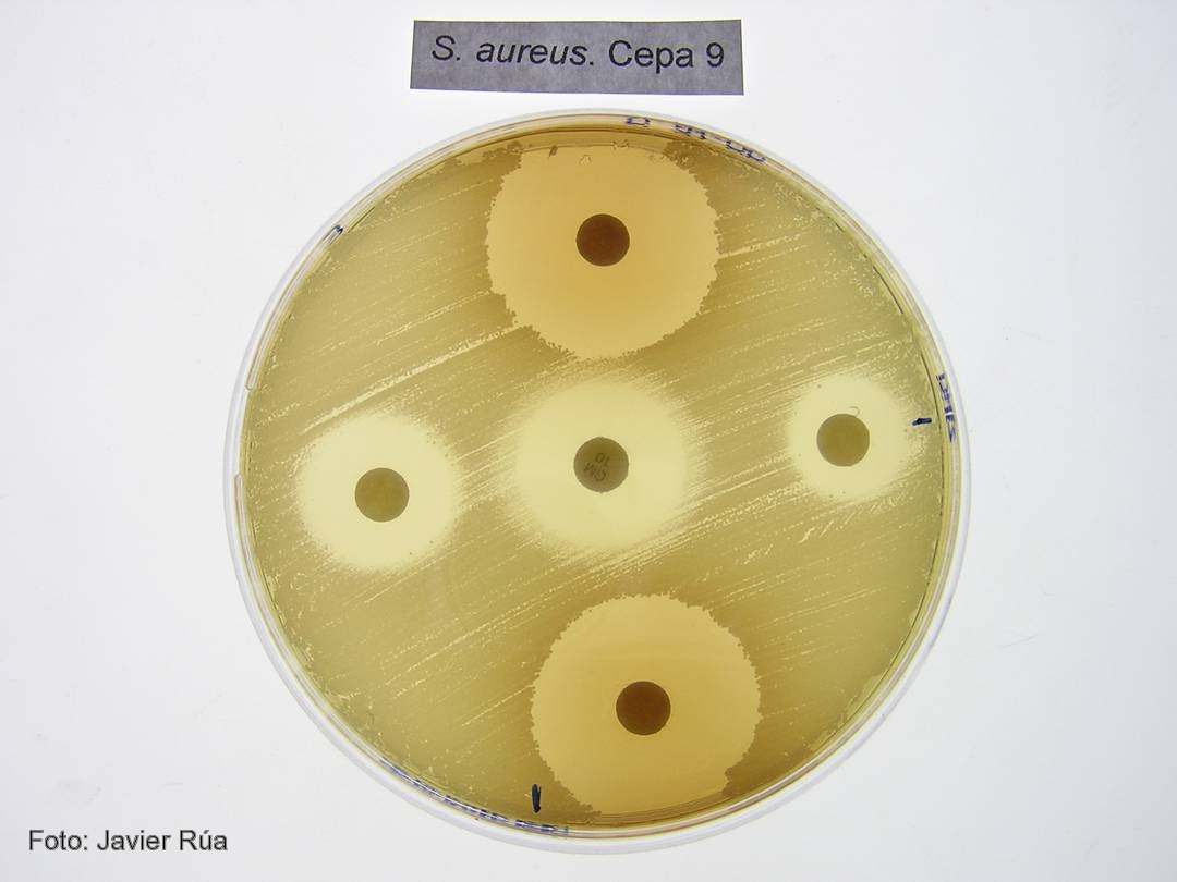
Cultivo de 'Staphylococcus aureus' tratado con los fenoles BHA (derecha) e hidroquinona (arriba).

CSIC/DICYT Un trabajo con participación del Consejo Superior de Investigaciones Científicas (CSIC) ha asignado a una enzima presente en todos los organismos vivos una nueva función en la transferencia de genes virulentos entre bacterias, un proceso que acaba provocando una infección. Los resultados, que aparecen publicados en el último número de Molecular Cell, y que han empleado como modelo la bacteria Staphylococcus aureus, la más frecuente en las infecciones adquiridas en hospitales, establecen el mecanismo de actuación de estas moléculas.

Las bacterias son capaces de transferir material genético entre sí mediante mecanismos de transferencia horizontal de genes. Cuando estos genes son virulentos, las bacterias que los reciben adquieren la capacidad de provocar enfermedades. “Algunos de los genes que codifican para toxinas y otros factores de virulencia están presentes en unas regiones denominadas islas de patogenicidad. Estas islas se transfieren de unas bacterias a otras utilizando virus que infectan bacterias, los llamados bacteriófagos”, explica el investigador del CSIC José Rafael Penadés, que trabaja en el Instituto de Biomedicina de Valencia.

El equipo de investigadores, formado también por científicos del Centro de Investigación y Tecnología Animal y la Universidad CEU Cardenal Herrera, en Valencia, han descubierto que las enzimas dUTPasas son capaces de despertar a las islas de patogenicidad para que detecten que la bacteria está siendo atacada por un virus. Antes de que la bacteria muera infectada, las islas inician su replicación y se transfieren a otras bacterias inocuas, a las que convierten en virulentas.

“El proceso evolutivo ha hecho que las islas detecten que un virus está infectando a las bacterias, lo que producirá su muerte, y utilicen la presencia del bacteriófago para activarse e iniciar su ciclo. Esto ocurre porque algunas proteínas del fago se unen a un represor que bloquea la isla”, explica el investigador del CSIC.

Proteínas G protooncogénicas
Los resultados confirman que las dUTPasas son moléculas señalizadoras que emplean un mecanismo similar al descrito para una familia de proteínas presentes en células eucariotas: las proteínas G protooncogénicas. “Las dUTPasas son activas como señalizadoras cuando se unen a un nucleótido dUTP. Es entonces cuando cambian su conformación y, una vez cumplida su función, degradan el nucleótido y pasan a estar apagadas. Este mecanismo de encendido y apagado es el mismo que el empleado por los protooncogenes”, destaca Penadés.

Según el investigador del CSIC Alberto Marina, el estudio sugiere por primera vez que las dUTPasas cumplen una función señalizadora no sólo en la mayoría de los virus, sino además en organismos vivos complejos como los eucariotas superiores. “Nuestros resultados aportan una visión completamente nueva del mecanismo de actuación de estas enzimas, que depende de una serie de características presentes en las enzimas de los bacteriófagos de Staphylococcus aureus y que están también presentes en otras muchas dUTPasas de un gran número de organismos vivos. Todo ello sugiere que el mecanismo descrito es universal”, concluye el investigador del CSIC.

Referencia bibliográfica 
María Ángeles Tormo-Más, Jorge Donderis, María García-Caballer, Aaron Alt, Ignacio Mir-Sanchís, Alberto Marina y José R. Penadés. Phage dUTPases Control Transfer of Virulence Genes by a Proto-Oncogenic G Protein-like Mechanism. Molecular Cell. DOI: 10.1016.

viernes, 11 de enero de 2013

Video: Virus caught in the act of infecting a cell

ORIGINAL: LabSpaces
Friday, January 11, 2013

Researchers found that the T7 virus has six tail fibers that are folded back against its capsid. The fibers extend as the virus locates a suitable host and as it “walks” across its host cell surface to find a site to infect. Credit: From Hu et. al. 2013. Science Express.

The top images are tomograms of the virus in action. The illustrations show T7 using its fibers to “walk” across the cell surface and infect the cell. Credit: From Hu et. al. 2013, Science Express.
The detailed changes in the structure of a virus as it infects an E. coli bacterium have been observed for the first time, report researchers from The University of Texas at Austin and The University of Texas Health Science Center at Houston (UT Health) Medical School this week in Science Express.

To infect a cell, a virus must be able to first find a suitable cell and then eject its genetic material into its host. This robot-like process has been observed in a virus called T7 and visualized by Ian Molineux, professor of biology at The University of Texas at Austin, and his colleagues.

The researchers show that when searching for its prey, the virus briefly extends — like feelers — one or two of six ultra-thin fibers it normally keeps folded at the base of its head.

Once a suitable host has been located, the virus behaves a bit like a planetary rover, extending these fibers to walk randomly across the surface of the cell and find an optimal site for infection.

At the preferred infection site, the virus goes through a major change in structure in which it ejects some of its proteins through the bacterium's cell membrane, creating a path for the virus's genetic material to enter the host.

After the viral DNA has been ejected, the protein path collapses and the infected cell membrane reseals.

"Although many of these details are specific to T7," said Molineux, "the overall process completely changes our understanding of how a virus infects a cell."

For example, the researchers now know that most of the fibers are usually bound to the virus head rather than extended, as was previously thought. That those fibers are in a dynamic equilibrium between bound and extended states is also new.

Molineux said that the idea that phages "walk" over the cell surface was previously proposed, but their paper provides the first experimental evidence that this is the case.

This is also the first time that scientists have made actual images showing how the virus's tail extends into the host — the very action that allows it to infect a cell with its DNA.

"I first hypothesized that T7 made an extended tail more than 10 years ago," said Molineux, "but this is the first irrefutable experimental evidence for the idea and provides the first images of what it looks like."

The researchers used a combination of genetics and cryo-electron tomography to image the infection process. Cryo-electron tomography is a process similar to a CT scan, but it is scaled to study objects with a diameter a thousandth the thickness of a human hair.

###

University of Texas at Austin: http://www.utexas.edu

Thanks to University of Texas at Austin for this article.

sábado, 1 de septiembre de 2012

Un grupo de científicos logra controlar los cerebros de dos monos mediante la luz

POR SUSAN YOUNG
TRADUCIDO POR LÍA MOYA (OPINNO)
LUNES, 30 DE JULIO DE 2012

El nuevo estudio, el primero que demuestra la efectividad de la optogenética sobre el comportamiento de los primates, acerca un poco más esta técnica hacia sus posibilidades terapéuticas.

Por primera vez, los científicos han sido capaces de influir en el comportamiento de un primate usando la optogenética, una técnica en la que estimulan mediante la luz neuronas modificadas genéticamente. El estudio, publicado el jueves pasado en la revista científica Current Biology, prepara el campo para usar esta potente y novedosa herramienta en el estudio de cómo el cerebro permite la cognición compleja en los primates y, más adelante, usar la técnica para tratar enfermedades.

Fuente: Kasper Bosmans
Para controlar los impulsos eléctricos de una neurona los científicos usan un virus que transporta un gen a las células del cerebro. El gen está diseñado para producir una proteína que responde a la luz. Dependiendo del tipo de proteína fotosensible que se utilice, la modificación genética activa o silencia una neurona en respuesta a un color de luz específico, que se transmite a través de fibras ópticas insertadas en el cerebro.

El campo de la optogenética, que solo cuenta con 7 años de antigüedad, ha proporcionado a los investigadores una herramienta más precisa para examinar las conexiones entre los grupos de neuronas que forman los circuitos neuronales. Estos circuitos cerebrales controlan comportamientos como el movimiento y la emoción y, cuando fallan, pueden producir enfermedades que van desde la depresión al párkinson.

Los científicos ya han usado la optogenética para controlar el comportamiento en ratones. Y aunque distintos grupos, incluyendo uno liderado por Edward Boyden, neurocientífico del Instituto Tecnológico de Massachusetts (EE.UU.) y coinventor de la optogenética, han demostrado que la técnica podría controlar la actividad neuronal en monos, ninguno había observado un efecto conductista aún.

“La gente empezaba a preguntarse si iba a ser un reto conseguir cambios en la conducta de un primate”, explica Boyden, que ha colaborado en el nuevo estudio. “Es algo muy importante tanto si lo que quieres estudiar es la cognición avanzada como si quieres pensar en potenciales usos clínicos en humanos”, afirma.

El comportamiento que se estudia en el artículo es bastante sutil: se entrenó a dos monos para que movieran los ojos expresamente hacia un objetivo en una pantalla cuando les dieran una señal. Pero al estimular las neuronas modificadas por la optogenética con la luz procedente de fibras ópticas insertadas en su cerebro, el circuito neuronal responsable se aceleraba y los monos podían completar la tarea en menos tiempo.

“Es una tarea sencilla, pero es una tarea cognitiva”, sostiene el autor sénior del estudio, Wim Vanduffle, que comparte su tiempo entre la Facultad de Medicina de Harvard (EE.UU.) y la Universidad de Leuven (Bélgica). “Es un trampolín”, afirma, uno que abre nuevas investigaciones para comprender cómo funciona el cerebro.

“En el futuro la optogenética quizá sea útil para usos terapéuticos, porque si eres capaz de activar o desactivar tipos celulares muy específicos, podrás actuar sobre circuitos que son importantes en distintas enfermedades con mucha más precisión de la que es posible ahora mismo mediante la medicación o la estimulación eléctrica”, afirma Vanduffle. “Pero aún queda un largo camino por recorrer para llegar hasta ahí”.

lunes, 28 de mayo de 2012

Powerful new approach to attack flu virus

ORIGINAL: eScienceNews

An international research team has manufactured a new protein that can combat deadly flu epidemics. MSU photographer, Kurt Stepnitz
An international research team has manufactured a new protein that can combat deadly flu epidemics. The paper, featured on the cover of the current issue of Nature Biotechnology, demonstrates ways to use manufactured genes as antivirals, which disable key functions of the flu virus, said Tim Whitehead, assistant professor of chemical engineering and materials science at Michigan State University.

"Our most potent design has proven effective on the vulnerable sites on many pandemic influenza viruses, including several H1N1 (Spanish flu, Swine flu) and H5N1 (Avian flu) subtypes," said Whitehead, the paper's co-lead author. "These new therapeutics are urgently needed, so we were especially pleased to see that it neutralizes H1N1 viruses with potency."

From its earlier research, the team used computer-aided design to engineer proteins that targeted vulnerable sites on the highly adaptable virus. From there, researchers optimized their designer proteins by comprehensively mapping the mutations that gave the proteins a strong advantage when attacking the viruses' targeted areas.

The team improved its proteins through a process called "DNA deep sequencing." This allowed Whitehead and his colleagues to simultaneously sequence millions of variants of their manufactured proteins, identify and keep the beneficial mutations and optimize the proteins' performance.

"By taking only the best mutations, we can reprogram our proteins to burrow into viruses at key locations and render them harmless," he said. "Our work demonstrates a new approach to construct therapeutic proteins, which we hope will spur development of new protein drugs by the biopharmaceutical industry."

This research also laid the groundwork for future treatments of all flu viruses as well as other diseases such as smallpox, Whitehead added.

Whitehead's co-authors included researchers from the University of Washington, the Scripps Research Institute (La Jolla, Calif.), Naval Health Research Center (San Diego) and the Weizmann Institute of Science (Rehovot, Israel).

The research was funded by Defense Research Projects Agency, the Defense Threat Reduction Agency, the National Institutes of Health, the National Institute of Allergy and Infectious Diseases and the National Institute of General Medical Sciences.

sábado, 28 de abril de 2012

Hot spring yields hybrid genome

ORIGINAL: Nature
19 Apr 2012

Image Credit: Creative Commons
In the hostile environment of a bubbling volcanic hot spring, a team of researchers at Portland State University in Oregon has discovered a new viral genome that seems to be the product of recombination between a DNA virus and an RNA virus — a natural chimaera not seen before. Their findings appeared on 19 April in the journal Biology Direct.

“It’s a mythological beast of a virus, but it actually exists,” says virologist Ken Stedman, a co-author of the study.

In the bacterial communities that populate the acidic waters of Boiling Springs Lake in northern California’s Lassen Volcanic National Park, “viruses are the only predators”, Stedman says. To get a better handle on what types of viruses are present there, he and his colleagues performed a metagenomic analysis of hundreds of thousands of viral sequences from a lake sample. The results included something unexpected: a piece of DNA coding for a protein that until now has only been seen in the capsid or ‘head’ of RNA viruses. By comparing the sequence with those of other genetic fragments, they were able to arrive at a complete viral genome. In the genome, the RNA-like sequence sat adjacent to another sequence for a replication protein that is unique to DNA viruses.

The resultant single-stranded circular genome, dubbed BSL RDHV (short for Boiling Springs Lake RNA–DNA hybrid virus), seems to be the result of a recombination event between two completely unrelated virus groups. The RNA gene did not come from a virus that could produce reverse transcriptase, the enzyme required to change RNA into DNA, so it’s unclear how the RNA gene ended up in the DNA genome. “We have no idea how it happened, but we know it happened,” says Stedman.

Stedman suggests two possible explanations. 
In one scenario, an RNA virus, a DNA virus and a retrovirus infected the same cell at the same time; the retrovirus used its reverse transcriptase enzyme to translate the RNA gene into a DNA copy, which got lumped in with the DNA virus genome. Such a scenario might also work if there was free-floating reverse transcriptase present in the environment. 
Alternatively, a special kind of viral ligase protein — which glues nucleic acids together — may have joined the dissimilar DNA and RNA strands, with the resultant hybrid code then replicated into DNA. Both scenarios are possible, if a bit far-fetched, says Stedman.

To see whether similar viruses exist in other environments, the team screened the Global Ocean Survey’s sequence database, and found three virus sequences containing the same two genes. “There were lots of cases that matched the sequence for either the DNA protein or the RNA protein, so there could actually be quite a few out there,” says Stedman. “But we can’t link them together, so we can’t say for sure.”

Metagenomic analyses can give researchers a picture only of the virus’s genome, not the virus itself or its host. “The downside of the approach is that little can be inferred about the biology of viruses discovered in environmental samples,” says Cédric Feschotte, an evolutionary geneticist at the University of Texas at Arlington. “It is unclear whether these viruses are good representatives even of their own group of viruses.” Stedman acknowledges these limitations, and now plans to investigate these questions with further sampling.

But, the fact that the unique virus exists suggests that recombination can happen between lineages that diverged billions of years ago. “It has been hypothesized that a key aspect of the earliest stages in the evolution of genetic systems involved rampant recombination and fusion between small, diverse — to the point of having different strategies of replication and expression — virus-like genetic elements,” says Eugene Koonin, an evolutionary geneticist at the National Institutes of Health in Bethesda, Maryland. “Stedman’s findings show that such recombination between diverse viruses indeed takes place and could be an important route of virus evolution.”

“The discovery of an apparent hybrid between DNA and RNA viruses blurs the traditional boundaries between two major viral groups, further highlighting the plasticity of the viral world and the seemingly unlimited possibilities of chimaerism among viral entities,” says Feschotte. If so, there could be more chimeras out there, just waiting for virologists to find them.

Correction: An earlier version of this blog post incorrectly stated that Ken Stedman presented the finding at NASA’s Astrobiology Conference this week. It was Stedman’s co-author, Geoffrey Deimer, a doctoral candidate at Portland State University, who presented the finding.