Mostrando entradas con la etiqueta Genes. Mostrar todas las entradas

lunes, 14 de septiembre de 2015

Designer molecule shines a spotlight on mysterious four-stranded DNA

Confocal microscopy image of the new molecule inside human bone cancer cells

A small fluorescent molecule has shed new light on knots of DNA thought to play a role in regulating how genes are switched on and off.

DNA is typically arranged in a double helix, where two strands are intertwined like a coiled ladder, but previous research has shown the existence of unusual DNA structures called quadruplexes, where four strands are arranged in the form of little knots.

Now researchers at Imperial College London led by Dr Marina Kuimova and Professor Ramon Vilar are unravelling the mysteries of these four-stranded DNA structures. They have created a fluorescent molecule that can reveal the presence of these structures in living cells.

This could be a game changer to accelerate research into these DNA structures.
– Professor Ramon Vilar

The team used the glowing molecule to target quadruplex DNA inside human bone cancer cells grown in the laboratory. Together with colleagues from Kings College London, they studied the interactions between the two in real time, using powerful microscopes. 

Quadruplexes can form when a strand of DNA rich in guanines – one of the four building blocks in DNA - folds over onto itself. Several distinct quadruplex structures have been found in the human genome but their exact role remains unclear. Recent studies have shown they are particularly prevalent in regions nearby oncogenes – genes that have the potential to cause cancer.
Structure of a G-quadruplex DNA
highlighting one of the guanine
tetrads
There is mounting evidence that quadruplexes are involved in switching genes on and off because of where they are usually positioned within the genome, says Professor Vilar, from Imperial's Department of Chemistry.

If this can be proved, it would make quadruplexes an extremely important target for treating diseases such as cancer. But to understand what role they play, we need to be able to study them in living cells. Our new fluorescent molecule allows us to do this by directly monitoring the behaviour of quadruplexes inside living cells in real time.

The team designed the fluorescent molecule to glow more intensely when attached to DNA. Using powerful microscopes they discovered that they could distinguish between the molecules binding to the more common double helical DNA and quadruplex DNA because it glowed for much longer when bound to quadruplexes.

HUNT FOR NEW COMPOUNDS
The researchers were also able to visualise the fluorescent molecule being displaced from quadruplex DNA by another molecule known to be a very good quadruplex binder. This suggests that the Imperial molecule could be used to hunt for new compounds that can bind to quadruplexes.

Co-author Arun Shivalingam, who worked on the study during his PhD at Imperial, says: “Until now, to image quadruplexes in cells researchers have had to hold the cells in place using chemical fixation. However, this kills them and brings into question whether the molecule really interacts with quadruplexes in a dynamic environment.

Professor Vilar adds: “We’ve shown that our molecule could be potentially used to verify in live cells and in real time whether potential quadruplex DNA binders are hitting their target. This could be a game changer to accelerate research into these DNA structures.

-
'The interactions between a small molecule and G-quadruplexes are visualized by fluorescence lifetime imaging microscopy' (DOI: 10.1038/ncomms9178) is published in Nature Communications on 09 September 2015.

09 September 2015

sábado, 12 de julio de 2014

The Man Who Rewrote the Tree of Life

Carl Woese may be the greatest scientist you’ve never heard of. “Woese is to biology what Einstein is to physics,” says Norman Pace, a microbiologist at the University of Colorado, Boulder. A physicist-turned-microbiologist, Woese specialized in the fundamental molecules of life—nucleic acids—but his ambitions were hardly microscopic. He wanted to create a family tree of all life on Earth.

Woese certainly wasn’t the first person with this ambition. The desire to classify every living thing is ageless. The Ancient Greeks and Romans worked to develop a system of classifying life. The Jewish people, in writing the Book of Genesis, set Adam to the task of naming all the animals in the Garden of Eden. And in the mid-1700s, Swedish botanist Carl von Linné published Systema Naturae, introducing the world to a system of Latin binomials—Genus speciesthat scientists use to this day.
Carl Woese in his later years

What Woese was proposing wasn’t to replace Linnaean classification, but to refine it. During the late 1960s, when Woese first started thinking about this problem as a young professor at the University of Illinois, biologists were relying a lot on guesswork to determine how organisms were related to each other, especially microbes. At the time, researchers used the shapes of microbes—their morphologies—and how they turned food into energy—their metabolisms—to sort them into bins. Woese was underwhelmed. To him, the morphology-metabolism approach was like trying to create a genealogical history using only photographs and drawings. Are people with dimples on their right cheeks and long ring fingers all members of the same family? Maybe, but probably not.

If you wanted to build a tree of life prior to what Woese did, there was no way to put something together that was based upon actual data,” says Jonathan Eisen, an evolutionary microbiologist at the University of California Davis.

Just as outward appearances aren’t the best way to determine family relations, Woese believed that morphology and metabolism were inadequate classifiers for life on Earth. Instead, he figured that DNA could sketch a much more accurate picture. Today, that approach may seem like common sense. But in the late 60s and early 70s, this was no easy task. Gene sequencing was a time-consuming, tedious task. Entire PhDs were granted for sequencing just one gene. To create his tree of life, Woese would need to sequence the same gene in hundreds, if not thousands, of different species. “When Woese first announced his results, I thought he was exaggerating at first.

So Woese toiled in his lab, sometimes with his postdoc George Fox but often alone, hunched over a light box with a magnifying glass, sequencing genes nucleotide by nucleotide. It took more than a decade. “When Woese first announced his results, I thought he was exaggerating at first,” Fox recalls. “Carl liked to think big, and I thought this was just another of his crazy ideas. But then I looked at the data and the enormity of what we had discovered hit me.

Woese and Fox published their results in 1977 in a well-respected journal, the Proceedings of the National Academy of Science. They had essentially rewritten the tree of life. But Woese still had a problem: few scientists believed him. He would spend the rest of his life working to convince the biological community that his work was correct.

Animal, Vegetable, Mineral

Following the publication of Linnaeus’s treatise in the 18th century, taxonomy progressed incrementally. The Swedish botanist had originally sorted things into three “kingdoms” of the natural world: animal, vegetable, and mineral. He placed organisms in their appropriate cubbyholes by looking at similarities in appearance. Plants with the same number of pollen-producing stamens were all lumped together, animals with the same number of teeth per jaw were grouped, and so on. With no knowledge of evolution and natural selection, he didn’t have a better way to comprehend the genealogy of life on Earth. Woese believed that DNA could unlock the hidden relationships between different organisms.

The publication of Darwin’s On the Origin of Species in 1859, combined with advances in microscopy, forced scientists to revise Linnaeus’s original three kingdoms to include the tiniest critters, including newly visible ones like amoebae and E. coli. Scientists wrestled with how to integrate microbial wildlife into the tree of life for the next 100 years. By the mid-20th century, however, biologists and taxonomists had mostly settled on a tree with five major branches: protists, fungi, plants, animals, and bacteria. It’s the classification system that many people learned in high school biology class.

Woese and other biologists weren’t convinced, though. Originally a physics major at Amherst College in Massachusetts and having received a PhD in biophysics from Yale in 1953, Woese believed that there had to be a more objective, data-driven way to classify life. Woese was particularly interested in how microbes fit into the classification of life, which had escaped a rigorous genealogy up until that point.

He arrived at the University of Illinois Urbana-Champaign as a microbiologist in the mid-1960s, shortly after James Watson and Francis Crick won the Nobel prize for their characterization of DNA’s double-helix form. It was the heyday of DNA. Woese was enthralled. He believed that DNA could unlock the hidden relationships between different organisms. In 1969, Woese wrote a letter to Crick, stating that:

…this can be done by using the cell’s ‘internal fossil record’—i.e., the primary structures of various genes. Therefore, what I want to do is to determine primary structures for a number of genes in a very diverse group of organisms, on the hope that by deducing rather ancient ancestor sequences for these genes, one will eventually be in the position of being able to see features of the cell’s evolution.

This type of thinking was “radically new,” says Norman Pace, a microbiologist at the University of Colorado, Boulder. “No one else was thinking in this direction at the time, to look for sequence-based evidence of life’s diversity.”
 
Evolution’s Timekeeper

Although the field of genetics was still quite young, biologists had already figured out some of the basics of how evolution worked at the molecular level. When a cell copies its DNA before dividing in two, the copies aren’t perfectly identical. Mistakes inevitably creep in. Over time, this can lead to significant changes in the sequence of nucleotides and the proteins they code for. By finding genes with sites that mutate at a known rate—say 4 mutations per site per million years—scientists could use them as an evolutionary clock that would give biologists an idea of how much time had passed since two species last shared a common ancestor.

To create his evolutionary tree of life, then, Woese would need to choose a gene that was present in every known organism, one that was copied from generation to generation with a high degree of precision and mutated very slowly, so he would be able to track it over billions of years of evolution.

This would let him make a direct measure of evolutionary history,” Pace says. “By tracking these gene sequences over time, he could calculate the evolutionary distance between two organisms and make a map of how life on Earth may have evolved.” The choice was especially fortuitous.

Some of the most ancient genes are those coding for molecules known as ribosomal RNAs. In ribosomes, parts of the cell that float around the soupy cytoplasm, proteins and ribosomal RNA, or rRNA, work together to crank out proteins. Each ribosome is composed of large and small subunits, which are similar in both simple, single-celled prokaryotes and more complex eukaryotes. Woese had several different rRNA molecules to choose from in the various subunits, which are classified based on their length. At around 120 nucleotides long, 5S rRNA wasn’t big enough to use to compare lots of different organisms. On the other end of the spectrum, 23S rRNA was more than 2300 nucleotides long, making it far too difficult for Woese to sequence using the technologies of the time. The Goldilocks molecule—long enough to allow for meaningful comparisons but not too long and difficult to sequence—was 16S rRNA in prokaryotes and its slightly longer eukaryotic equivalent, 18S rRNA. Woese decided to use these to create his quantitative tree of life.

His choice was especially fortuitous, Eisen says, because of several factors inherent in 16S rRNA that Woese couldn’t have been aware of at the time, including its ability to measure evolutionary time on several different time scales. Certain parts of the 16S rRNA molecule mutate at different speeds. Changes to 16S rRNA are, on the whole, still extremely slow (humans share about 50% of their 16S rRNA sequence with the bacterium E. coli), but one portion mutates much more slowly than the other. It’s as if the 16S rRNA clock has both an hour hand and a minute hand. The very slowly evolving “hour hand” lets biologists study the long-term changes to the molecule, whereas the more quickly evolving “minute hand” provides a more recent history. “This gives this gene an advantage because it lets use ask questions about deep evolutionary history and more recent history at the same time,” Eisen says.

Letter by letter

Selecting the gene was just Woese’s first challenge. Now he had to sequence it in a variety of different organisms. In the late 60s and early 70s, when Woese began his work, DNA sequencing was far from automated. Everything, down to the last nucleotide, had to be done by hand. Woese used a method to catalog short pieces of RNA developed in 1965 by British scientist Frederick Sanger, which used enzymes to chop RNA into small pieces. These small pieces were sequenced, and then scientists had to reassemble the overlapping pieces to determine the overall sequence of the entire molecule—a process that was tedious, expensive, and time-consuming, but that was seen as a minor annoyance to a workhorse like Woese, Fox says. “All he cared about was getting the answer.

Woese started with prokaryotes, the single-celled organisms that were his primary area of interest. He and his lab started by growing bacteria in a solution of radioactive phosphate, which the cells incorporated into backbones of their RNA molecules. This made the 16S rRNA radioactive. Then, Woese and Fox extracted the RNA from the cells and chopped it into smaller pieces using enzymes that acted like scissors. The enzymatic scissors would only cut at certain sequences. If a sequence was present in one organism but missing in a second, the scissors would pass over the second one’s sequence. Its fragment would be longer. “To Carl, each spot was a puzzle that he would solve.”

Since RNA’s sugar-phosphate backbone is negatively charged, the researchers could use a process known as electrophoresis to separate the different length pieces. As electricity coursed through gels containing samples, it pulled the smaller, lighter bits farther through the gels than the longer, heavier chunks. The result was distinct bands of different lengths of RNA. Woese and Fox then exposed each gel to photographic paper over several days. The radioactive bands in the gel transferred marks to the paper. This created a Piet Mondrian-esque masterpiece of black bands on a white background. Each different organism left its own mark. “To Carl, each spot was a puzzle that he would solve,” Fox says.

After developing each image, Woese and Fox returned to the gel and neatly cut out each individual blotch that contained fragments of a certain length. They then chopped up these fragments with another set of enzymes until they were about five to 15 nucleotides long, a length that made sequencing easier. For some of the longer fragments, it took several iterations of the process before they were successfully sequenced. The sequences were then recorded on a set of 80-column IBM punch cards. The cards were then run through a large computer to compare band patterns and RNA sequences among different organisms to determine evolutionary relationships. At the beginning, it took Woese and Fox months to obtain a single 16S rRNA fingerprint.

This process was a huge breakthrough,” says Peter Moore, an RNA chemist at Yale University who worked with Woese on other research relating to RNA’s structure. “It gave biologists a tool for sorting through microorganisms and giving them a conceptual way to understand the relationship between them. At the time, the field was just a total disaster area. Nobody knew what the hell was going on.
RNA is so fundamental to life that some scientists think it's the spark that started it all. To learn more about RNA, visit NOVA’s RNA Lab.


By the spring of 1976, Woese and Fox had created fingerprints of a variety of bacterial species when they turned to an oddball group of prokaryotes: methanogens. These microbes produce methane when they break down food for energy. Because even tiny amounts of oxygen are toxic to these prokaryotes, Woese and Fox had to grow them under special conditions.

After months of trial and error, the two scientists were finally able to obtain an RNA fingerprint of one type of methanogen. When they finally analyzed its fingerprint, however, it looked nothing like any of the other bacteria Woese and Fox had previously analyzed. All of the previous bacterial gels contained two large splotches at the bottom. They were entirely absent from these new gels. Woese knew instantly what this meant.

To fellow microbiologist Ralph Wolfe, who worked in the lab next door, Woese announced, “I don’t even think these are bacteria, Wolfe.

He dropped the full bombshell on Fox. “The methanogens didn’t have any of the spots he was expecting to see. When he realized this wasn’t a mistake, he just went nuts. He ran into my lab and told me we had discovered a new form of life,” Fox recalls.

The New Kingdom

The methanogens Woese and Fox had analyzed looked superficially like other bacteria, yet their RNA told a different story, sharing more in common with nucleus-containing eukaryotes than with other bacteria. After more analysis of his RNA data, Woese concluded that what he was tentatively calling Archaea (from Latin, meaning primitive) wasn’t a minor twig on the tree of life, but a new main branch. It wasn’t just Bacteria and Eukarya any more .

To prove to their critics that these prokaryotes really were a separate domain on the tree of life, Woese and Fox knew the branch needed more than just methanogens. Fox knew enough about methanogen biology to know that their unique RNA fingerprint wasn’t the only thing that made them strange. For one thing, their cell walls lacked a mesh-like outer layer made of peptidoglycan. Nearly every other bacterium Fox could think of contained peptidoglycan in its cell wall—until he recalled a strange fact he had learned as a graduate student—another group of prokaryotes, the salt-loving halophiles, also lacked peptidoglycan. 

Grand Prismatic Spring in Yellowstone National Park is home to many species of thermophilic archaea.

Fox turned to the research literature to search for other references to prokaryotes that lack peptidoglycan. He found two additional examples: Thermoplasma and Sulfolobus. Other than the missing peptidoglycan, these organisms and the methanogens seemed nothing alike. Methanogens were found everywhere from wetlands to the digestive tracts, halophiles flourished in salt, Thermoplasma liked things really hot, and Sulfolobus are often found in volcanoes and hot, acidic springs.

Despite their apparent differences, they all metabolized food in the same, unusual way—unlike anything seen in other bacteria—and the fats in the cell membrane were alike, too. When Woese and Fox sequenced the 16S rRNA of these organisms, they found that these prokaryotes were most similar to the methanogens.

Once we had the fingerprints, it all fell together,” Fox says.

Woese believed his findings were going to revolutionize biology, so he organized a press conference when the paper was published in PNAS in 1977. It landed Woese on the front page of the New York Times, and created animosity among many biologists. “The write-ups were ludicrous and the reporters got it all wrong,” Wolfe says. “No biologists wanted anything to do with him.

It wasn’t just distaste for what looked like a publicity stunt that was working against Woese. He had spent most of the last decade holed up in his third floor lab, poring over RNA fingerprints. His reclusive nature had given him the reputation of a crank. It also didn’t help that he had single-handedly demoted many biologists’ favorite species. Thanks to Woese, Wolfe says, “Microbes occupy nearly all of the tree. Then you have one branch at the very end where all the animals and plants were. And the biologists just couldn’t believe that all the plants and all the animals were really just one tiny twig on one branch.” “He was a brash, iconoclastic outsider, and his message did not go down well.

Although some specialists were quick to adopt Woese’s new scheme, the rest of biology remained openly hostile to the idea. It wasn’t until the mid-1980s that other microbiologists began to warm to the idea, and it took well over another decade for other areas of biology to follow suit. Woese had grown increasingly bitter that so many other scientists were so quick to reject his claims. He knew his research and ideas were solid. But he was left to respond to what seemed like an endless stream of criticism. Shying from these attacks, Woese retreated to his office for the next two decades.

He was a brash, iconoclastic outsider, and his message did not go down well,” says Moore, the Yale RNA chemist.

Woese’s cause wasn’t helped by his inability to engage critics in dialogue and discussion. Both reticent and abrupt, he preferred his lab over conferences and presentations. In place of public appearances to address his detractors, he sent salvos of op-eds and letters to the editor. Still, nothing seemed to help. The task of publicly supporting this new tree of life fell to Woese’s close colleagues, especially Norman Pace.

But as technology improved, scientists began to obtain the sequences of an increasing number of 16S rRNAs from different organisms. More and more of their analyses supported Woese’s hypothesis. As sequencing data poured in from around the world, it became clear to nearly everyone in biology that Woese’s initial tree was, in fact, been correct.

Now, when scientists try to discover unknown microbial species, the first gene they sequence is 16S rRNA. “It’s become one of the fundamentals of biology,” Wolfe says. “After more than 20 years, Woese was finally vindicated.”

Woese died on December 30, 2012, at the age of 84 of complications from pancreatic cancer. At the time of his death, he had won some of biology’s most prestigious awards and had become one of the field’s most respected scientists. Thanks to Woese’s legacy, we now know that most of the world’s biodiversity is hidden from view, among the tiny microbes that live unseen in and around us, and in them, the story of how life first evolved on this planet.

Tell us what you think on Twitter #novanext, Facebook, or email.

Photo credits: Jason Lindsey/University of Illinois, Tim Bocek/Flickr (CC BY-NC-SA)
Carrie Arnold
Twitter

Carrie Arnold is a freelance science writer living in Virginia. She has written about many aspects of the living world for publications including Scientific American, Discover, New Scientist, Science News, and more.

ORIGINAL: PBS
30 Apr 2014

viernes, 31 de enero de 2014

The genetic contribution Neanderthal man made to modern humanity is clearer

Kissing cousins

HOW Neanderthal are you? That question sounds vaguely insulting. But unless you are African, or of recent African ancestry, the answer is likely to be 1-3%.

Though Homo sapiens is the only type of human around at the moment, that was not true until recently. Sixty thousand years ago, when modern humans first left Africa, they encountered other species of humanity, such as Neanderthals (imagined above, in an artist’s interpretation), in Europe and Asia. In some cases, they interbred with them. The genetic traces of those encounters remain in modern human genomes. And two studies, one just published in Nature, and one in Science, have now looked in detail at this miscegenation, and tried to understand its consequences.


The Nature study, conducted by Sriram Sankararaman of Harvard Medical School and his colleagues, looked at the genomes of 1,004 living people of European and Asian descent and compared them with Neanderthal DNA from a 50,000-year-old toe bone found in a Siberian cave, and also with the genomes of 176 west Africans. This latter group, Dr Sankararaman assumed, could have little Neanderthal DNA in them because Neanderthals, as far as can be determined from the fossil record, lived only in Europe and western Asia.

Dr Sankararaman and his colleagues certainly did find plenty of DNA which seems to have come from Neanderthals in their Eurasians. Tellingly, it was not sprinkled evenly throughout the modern human genome. That let them make educated guesses about the effects it is having on those who carry it. For instance, genes affecting the production of keratin—an important component of hair and skin—showed more Neanderthal influence than most. Neanderthals, whose homeland was much colder then than it is now because of the ice age, were hairier (and thus better insulated) than Homo sapiens. Retaining Neanderthal traits of this sort, in an African species that was trying to make good in sub-Arctic conditions, would thus be encouraged by natural selection.

More surprisingly, Dr Sankararaman also found Neanderthal DNA in genes associated with diabetes, Crohn’s disease, lupus and even the propensity to smoke. This does not necessarily mean such DNA was bad for those who inherited it. A gene which increases the risk of diabetes in modern circumstances of abundant food might, for example, have had benefits in a more austere environment.

Indeed, truly deleterious DNA would be expected to be noticeable by its absence, because natural selection would have worked to eliminate it in the 30,000 years since Neanderthals died out. And Dr Sankararaman found evidence for exactly that, as well.

There is, for example, little Neanderthal DNA on the X chromosome (which, along with the Y chromosome, determines an individual’s sex). Nor is there much in genes that are expressed in the testicles. Studies from other hybrid animals, which are frequently sterile, suggest genes which reduce male fertility are often found on the X chromosome. Since few things are a bigger evolutionary no-no than being unable to produce children, tremendous selective pressure would have existed to remove the offending DNA from the hybrid descendants of Neanderthals and Homo sapiens.

The study published in Science, by Benjamin Vernot and Joshua Akey of the University of Washington, in Seattle, reaches similar conclusions to Dr Sankararaman’s. Dr Vernot and Dr Akey hunted down Neanderthal DNA in the genomes of 665 Europeans and East Asians. They, too, found evidence of its having inserted itself into genes associated with the skin, and that not all of the newly arrived genetic material is helpful to its current bearers.

They made other discoveries, too. With the help of computer models, they concluded that there were probably several pulses of interbreeding over the millennia, rather than a steady stream of it. Both they and Dr Sankararaman also found that, on average, East Asians have more Neanderthal DNA than Europeans do—which is odd, because Neanderthals are not known to have lived in East Asia.

The ghost in the machine
Dr Vernot and Dr Akey also used their data to try to improve understanding of the Neanderthal genome itself, by combining the bits and bobs scattered among modern humans. Though both their study and Dr Sankararaman’s depended on being able to identify what was Neanderthal by comparing modern human genomes with fossil DNA, the fossil material available is imperfect. Looking at the exact sequence of DNA “letters” (the chemical bases which carry the genetic message) in areas identified as Neanderthal in modern genomes can therefore improve understanding of the Neanderthal original.

Crucially, though the amount of Neanderthal DNA in any individual is small, the exact bits vary a lot from person to person. Look at enough people, then, and it becomes possible to rebuild quite large swathes of the Neanderthal genome. Dr Vernot and Dr Akey reckon that from their sample of 665 they have recovered around 20% of it.

This is an impressive figure for an extinct species. It shows just how much the concept of a “species” is a construct of human thinking rather than a truly natural category. Technically, Neanderthals may be gone. But their DNA ghosts linger on.

From the print edition: Science and technology


ORIGINAL: The Economist
Feb 1st 2014

jueves, 12 de diciembre de 2013

Scientists discover double meaning in genetic code

Scientists have discovered a second code hiding within DNA. This second code contains information that changes how scientists read the instructions contained in DNA and interpret mutations to make sense of health and disease.

Genome scientist Dr. John Stamatoyannopolous led
a team that discovered a second code hidden in DNA
A research team led by Dr. John Stamatoyannopoulos, University of Washington associate professor of genome sciences and of medicine, made the discovery. The findings are reported in the Dec. 13 issue of Science. The work is part of the Encyclopedia of DNA Elements Project, also known as ENCODE. The National Human Genome Research Institute funded the multi-year, international effort. ENCODE aims to discover where and how the directions for biological functions are stored in the human genome.

Since the genetic code was deciphered in the 1960s, scientists have assumed that it was used exclusively to write information about proteins. UW scientists were stunned to discover that genomes use the genetic code to write two separate languages.
  • One describes how proteins are made, and the 
  • other instructs the cell on how genes are controlled
One language is written on top of the other, which is why the second language remained hidden for so long.

For over 40 years we have assumed that DNA changes affecting the genetic code solely impact how proteins are made,” said Stamatoyannopoulos. “Now we know that this basic assumption about reading the human genome missed half of the picture. These new findings highlight that DNA is an incredibly powerful information storage device, which nature has fully exploited in unexpected ways.”

The genetic code uses a 64-letter alphabet called codons. The UW team discovered that some codons, which they called duons, can have two meanings, 
  • one related to protein sequence, and 
  • one related to gene control
These two meanings seem to have evolved in concert with each other. The gene control instructions appear to help stabilize certain beneficial features of proteins and how they are made.
The discovery of duons has major implications for how scientists and physicians interpret a patient’s genome and will open new doors to the diagnosis and treatment of disease.

The fact that the genetic code can simultaneously write two kinds of information means that many DNA changes that appear to alter protein sequences may actually cause disease by disrupting gene control programs or even both mechanisms simultaneously,” said Stamatoyannopoulos.

Grants from the National Institutes of Health U54HG004592, U54HG007010, and UO1E51156 and National Institute of Diabetes and Digestive and Kidney Diseases FDK095678A funded the research.

In addition to Stamatoyannopoulos, the research team included Andrew B. Stergachis, Eric Haugen, Anthony Shafer, Wenqing Fu, Benjamin Vernot, Alex Reynolds, and Joshua M. Akey, all from the UW Department of Genome Sciences, Anthony Raubitschek of the UW Department of Immunology and Benaroya Research Institute, Steven Ziegler of Benaroya Research Institute, and Emily M. LeProust, formerly of Agilent Technologists and now with Twist Bioscience.

ORIGINAL: U of Washington
December 12, 2013

lunes, 25 de febrero de 2013

piRNAs Key Role in Coordinating Biological Activity

ORIGINAL: SciTechDaily
by Staff
February 25, 2013 

Image: DNA from Shutterstock

A team of Yale researchers discovered that specialized RNAs called piRNAs guide epigenetic factors to numerous sites throughout the genome of the fruit fly Drosophila, where these switches work to turn genes on or off.

If a genome is the blueprint for life, then the chief architects are tiny slices of genetic material that orchestrate how we are assembled and function, Yale School of Medicine researchers report February 21 in the journal Developmental Cell.

The study pinpoints the molecular regulators of epigenetics — the process by which unchanging genes along our DNA are switched on and off at precisely right time and place.

“Our genome is like a landscape with lakes, mountains, and rivers, but it is not yet a community or a city full of buildings,” said Haifan Lin, director of the Yale Stem Cell Center and senior author of the study. “What this system does is decide where and when to send out the masons, carpenters, and electricians to build a city or a community.

In the past 20 years, scientists have discovered that some proteins, called epigenetic factors, traverse the static genome and turn the genes on or off. The staggering number of potential combinations of active and inactive genes explains why a relatively small number of genes can carry out such a wide range of functions. But what guides these epigenetic factors to their target? The answer, the Yale team has found, is specialized RNAs called piRNAs.

In the latest study, the Yale team discovered that piRNAs guide epigenetic factors to numerous sites throughout the genome of the fruit fly Drosophila, where these switches work to turn genes on or off. The dramatic change in gene expression patterns found illustrated piRNAs key role in coordinating biological activity.

This is the first major mechanism discovered that controls where epigenetic factors —the gene switches — are to be placed in the genome,” Lin said.

Several types of cancers appeared to be triggered when the wrong kinds of piRNAs guide epigenetic factors to activate the wrong genes. Blocking the action of these piRNAs should become a new opportunity to treat cancers, Lin said.

Xiao A. Huang and Hang Yin of Yale are co-lead authors of the paper.

The research was funded by a National Institutes of Health Pioneer Award to Haifan Lin and a grant from Connecticut Stem Cell Research Fund to Lin and former Yale professor and co-author Michael Snyder, now of Stanford University.

Publication: Haifan Lin, et al., “A Major Epigenetic Programming Mechanism Guided by piRNAs,” Developmental Cell, 21 February 2013; DOI: 10.1016/j.devcel.2013.01.023

martes, 22 de enero de 2013

10 Incredible Ways Genes Control Our Lives


Feeling tipsy? Carrying a few extra pounds? Maybe it’s in your genes. Presenting 10 amazing ways our genes can change our lives.

viernes, 18 de enero de 2013

Descrita una nueva ruta de señalización clave en la transferencia de genes virulentos entre bacterias

ORIGINAL: DCYT

El trabajo, publicado en la revista Molecular Cell, asigna a una enzima una nueva función señalizadora
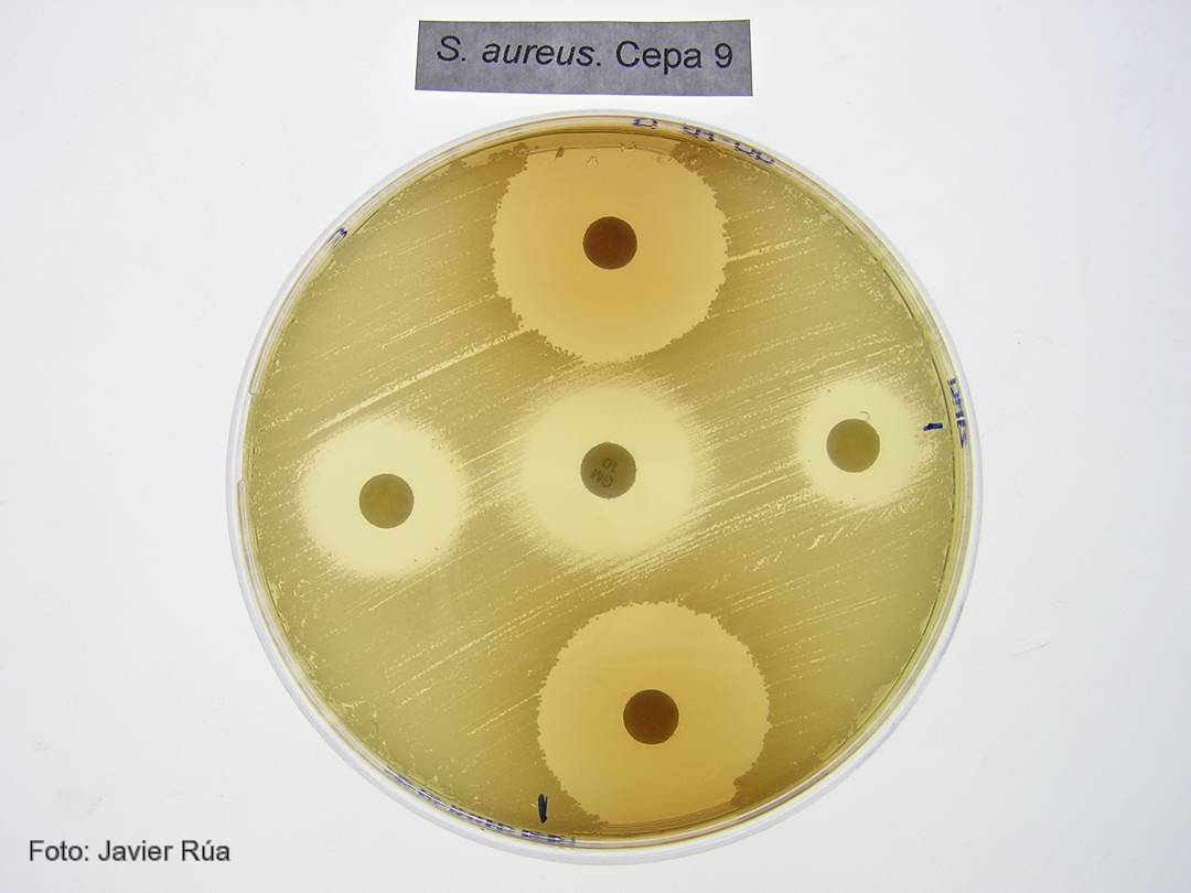
Cultivo de 'Staphylococcus aureus' tratado con los fenoles BHA (derecha) e hidroquinona (arriba).

CSIC/DICYT Un trabajo con participación del Consejo Superior de Investigaciones Científicas (CSIC) ha asignado a una enzima presente en todos los organismos vivos una nueva función en la transferencia de genes virulentos entre bacterias, un proceso que acaba provocando una infección. Los resultados, que aparecen publicados en el último número de Molecular Cell, y que han empleado como modelo la bacteria Staphylococcus aureus, la más frecuente en las infecciones adquiridas en hospitales, establecen el mecanismo de actuación de estas moléculas.

Las bacterias son capaces de transferir material genético entre sí mediante mecanismos de transferencia horizontal de genes. Cuando estos genes son virulentos, las bacterias que los reciben adquieren la capacidad de provocar enfermedades. “Algunos de los genes que codifican para toxinas y otros factores de virulencia están presentes en unas regiones denominadas islas de patogenicidad. Estas islas se transfieren de unas bacterias a otras utilizando virus que infectan bacterias, los llamados bacteriófagos”, explica el investigador del CSIC José Rafael Penadés, que trabaja en el Instituto de Biomedicina de Valencia.

El equipo de investigadores, formado también por científicos del Centro de Investigación y Tecnología Animal y la Universidad CEU Cardenal Herrera, en Valencia, han descubierto que las enzimas dUTPasas son capaces de despertar a las islas de patogenicidad para que detecten que la bacteria está siendo atacada por un virus. Antes de que la bacteria muera infectada, las islas inician su replicación y se transfieren a otras bacterias inocuas, a las que convierten en virulentas.

El proceso evolutivo ha hecho que las islas detecten que un virus está infectando a las bacterias, lo que producirá su muerte, y utilicen la presencia del bacteriófago para activarse e iniciar su ciclo. Esto ocurre porque algunas proteínas del fago se unen a un represor que bloquea la isla”, explica el investigador del CSIC.

Proteínas G protooncogénicas
Los resultados confirman que las dUTPasas son moléculas señalizadoras que emplean un mecanismo similar al descrito para una familia de proteínas presentes en células eucariotas: las proteínas G protooncogénicas. “Las dUTPasas son activas como señalizadoras cuando se unen a un nucleótido dUTP. Es entonces cuando cambian su conformación y, una vez cumplida su función, degradan el nucleótido y pasan a estar apagadas. Este mecanismo de encendido y apagado es el mismo que el empleado por los protooncogenes”, destaca Penadés.

Según el investigador del CSIC Alberto Marina, el estudio sugiere por primera vez que las dUTPasas cumplen una función señalizadora no sólo en la mayoría de los virus, sino además en organismos vivos complejos como los eucariotas superiores. “Nuestros resultados aportan una visión completamente nueva del mecanismo de actuación de estas enzimas, que depende de una serie de características presentes en las enzimas de los bacteriófagos de Staphylococcus aureus y que están también presentes en otras muchas dUTPasas de un gran número de organismos vivos. Todo ello sugiere que el mecanismo descrito es universal”, concluye el investigador del CSIC.

Referencia bibliográfica 
María Ángeles Tormo-Más, Jorge Donderis, María García-Caballer, Aaron Alt, Ignacio Mir-Sanchís, Alberto Marina y José R. Penadés. Phage dUTPases Control Transfer of Virulence Genes by a Proto-Oncogenic G Protein-like Mechanism. Molecular Cell. DOI: 10.1016.

domingo, 10 de junio de 2012

Birth Control Pills Affect Women's Taste in Men

How synthetic hormones change desire in women--and their choice in a mate


Image: © iStockPhoto / Ceneri
This year 2.25 million Americans will get married—and a million will get divorced. Could birth control be to blame for some of these breakups? Recent research suggests that the contraceptive pill—which prevents women from ovulating by fooling their body into believing it is pregnant—could affect which types of men women desire. Going on or off the pill during a relationship, therefore, may tempt a woman away from her man.

It’s all about scent. Hidden in a man’s smell are clues about his major histocompatibility complex (MHC) genes, which play an important role in immune system surveillance. Studies suggest that females prefer the scent of males whose MHC genes differ from their own, a preference that has probably evolved because it helps offspring survive: couples with different MHC genes are less likely to be related to each other than couples with similar genes are, and their children are born with more varied MHC profiles and thus more robust immune systems.

A study published in August in the Proceedings of the Royal Society B, however, suggests that women on the pill undergo a shift in preference toward men who share similar MHC genes. The female subjects were more likely to rate these genetically similar men’s scents (via a T-shirt the men had worn for two nights) as pleasant and desirable after they went on the pill as compared with before. Although no one knows why the pill affects attraction, some scientists believe that pregnancy—or in this case, the hormonal changes that mimic pregnancy—draws women toward nurturing relatives.

Women who start or stop taking the pill, then, may be in for some relationship problems. A study published last year in Psychological Science found that women paired with MHC-similar men are less sexually satisfied and more likely to cheat on their partners than women paired with MHC-dissimilar men. So a woman on the pill, for example, might be more likely to start dating a MHC-similar man, but he could ultimately leave her less sexually satisfied. Then if she goes off the pill during the relationship, the accompanying hormonal changes will draw her even more strongly toward more MHC-dissimilar men. These immune genes may have a “powerful effect in terms of how well relationships are cemented,” says University of Liverpool psychologist Craig Roberts, co-author of the August paper.