Mostrando entradas con la etiqueta Scripps Research. Mostrar todas las entradas

martes, 24 de enero de 2017

TSRI Scientists Create First Stable Semisynthetic Organism


LA JOLLA, CA – January 23, 2017 – Life’s genetic code has only ever contained four natural bases. These bases pair up to form two “base pairs”—the rungs of the DNA ladder—and they have simply been rearranged to create bacteria and butterflies, penguins and people. Four bases make up all life as we know it.

Until now. Scientists at The Scripps Research Institute (TSRI) have announced the development of the first stable semisynthetic organism. Building on their 2014 study in which they synthesized a DNA base pair, the researchers created a new bacterium that uses the four natural bases (called A, T, C and G), which every living organism possesses, but that also holds as a pair two synthetic bases called X and Y in its genetic code.

TSRI Professor Floyd Romesberg and his colleagues have now shown that their single-celled organism can hold on indefinitely to the synthetic base pair as it divides. Their research was published January 23, 2017, online ahead of print in the journal Proceedings of the National Academy of Sciences.

We’ve made this semisynthetic organism more life-like,” said Romesberg, senior author of the new study.

While applications for this kind of organism are still far in the future, the researchers say the work could be used to create new functions for single-celled organisms that play important roles in drug discovery and much more.

Building a Unique Organism
When Romesberg and his colleagues announced the development of X and Y in 2014, they also showed that modified E. coli bacteria could hold this synthetic base pair in their genetic code. What these E. coli couldn’t do, however, was keep the base pair in their code indefinitely as they divided. The X and Y base pair was dropped over time, limiting the ways the organism could use the additional information possessed in their DNA.

Your genome isn’t just stable for a day,” said Romesberg. “Your genome has to be stable for the scale of your lifetime. If the semisynthetic organism is going to really be an organism, it has to be able to stably maintain that information.

Romesberg compared this flawed organism to an infant. It had some learning to do before it was ready for real life.

In stepped TSRI Graduate Student Yorke Zhang and Brian Lamb, an American Cancer Society postdoctoral fellow in the Romesberg lab at the time of the study. Together, they helped develop the means for the single-celled organism to retain the artificial base pair.

First, Zhang and Lamb, co-first authors of the study, optimized a tool called a nucleotide transporter, which brings the materials necessary for the unnatural base pair to be copied across the cell membrane. The transporter was used in the 2014 study, but it made the semisynthetic organism very sick,” Zhang explained. The researchers discovered a modification to the transporter that alleviated this problem, making it much easier for the organism to grow and divide while holding on to X and Y.

Next, the researchers optimized their previous version of Y. The new Y was a chemically different molecule that could be better recognized by the enzymes that synthesize DNA molecules during DNA replication. This made it easier for cells to copy the synthetic base pair.

A New Use for CRISPR-Cas9
Finally, the researchers set up a “spell check” system for the organism using CRISPR-Cas9, an increasingly popular tool in human genome editing experiments. But instead of editing a genome, the researchers took advantage of CRISPR-Cas9’s original role in bacteria.

The genetic tools in CRISPR-Cas9 (a DNA segment and an enzyme) originated in bacteria as a kind of immune response. When a bacterium encounters a threat, like a virus, it takes fragments of the invader genome and pastes them into its own genome—a bit like posting a “wanted” poster on the off chance it sees the invader again. Later, it can use those pasted genes to direct an enzyme to attack if the invader returns.

Knowing this, the researchers designed their organism to see a genetic sequence without X and Y as a foreign invader. A cell that dropped X and Y would be marked for destruction, leaving the scientists with an organism that could hold on to the new bases. It was like the organism was immune to unnatural base pair loss.

We were able to address the problem at a fundamental level,” said Lamb, who now serves as a research scientist at Vertex Pharmaceuticals.

Their semisynthetic organism was thus able to keep X and Y in its genome after dividing 60 times, leading the researchers to believe it can hold on to the base pair indefinitely.

We can now get the light of life to stay on,” said Romesberg. “That suggests that all of life’s processes can be subject to manipulation.

A Foundation for Future Research
Romesberg emphasized that this work is only in single cells and is not meant to be used in more complex organisms. He added that the actual applications for this semisynthetic organism are “zero” at this point. So far, scientists can only get the organism to store genetic information.

Next, the researchers plan to study how their new genetic code can be transcribed into RNA, the molecule in cells needed to translate DNA into proteins. “This study lays the foundation for what we want to do going forward,” said Zhang.
Professor Floyd Romesberg (right) and Graduate Student Yorke Zhang led the new study at The Scripps Research Institute, along with Brian Lamb (not pictured).(Photo by Madeline McCurry-Schmidt.)

Additional authors of the study, “A semisynthetic organism engineered for the stable expansion of the genetic alphabet,” were Aaron W. Feldman and Anne Xiaozhou Zhou of TSRI; Thomas Lavergne of the University of Grenoble; and Lingjun Li of Henan Normal University.

The study was supported by the National Institutes of Health (grant GM060005), a National Science Foundation Graduate Research Fellowship (grant DGE-1346837), the National Natural Science Foundation of China (grant 21472036), a Labex ARCANE grant (ANR-11-LABX-0003-01), NanoBio-ICMG platforms (FR 2607) and a postdoctoral fellowship from the American Cancer Society, Illinois Division.

About The Scripps Research Institute
The Scripps Research Institute (TSRI) is one of the world's largest independent, not-for-profit organizations focusing on research in the biomedical sciences. TSRI is internationally recognized for its contributions to science and health, including its role in laying the foundation for new treatments for cancer, rheumatoid arthritis, hemophilia, and other diseases. An institution that evolved from the Scripps Metabolic Clinic founded by philanthropist Ellen Browning Scripps in 1924, the institute now employs more than 2,500 people on its campuses in La Jolla, CA, and Jupiter, FL, where its renowned scientists—including two Nobel laureates and 20 members of the National Academies of Science, Engineering or Medicine—work toward their next discoveries. The institute's graduate program, which awards PhD degrees in biology and chemistry, ranks among the top ten of its kind in the nation. In October 2016, TSRI announced a strategic affiliation with the California Institute for Biomedical Research (Calibr), representing a renewed commitment to the discovery and development of new medicines to address unmet medical needs. For more information, see www.scripps.edu.

# # #

For information:
Office of Communications 
Tel: 858-784-2666 
Fax: 858-784-8118 

jueves, 19 de febrero de 2015

Scripps Florida Scientists Announce Anti-HIV Agent So Powerful It Can Work in a Vaccine

JUPITER, FL – February 18, 2015 – In a remarkable new advance against the virus that causes AIDS, scientists from The Scripps Research Institute (TSRI) have announced the creation of a novel drug candidate that is so potent and universally effective, it might work as part of an unconventional vaccine.

The research, which involved scientists from more than a dozen research institutions, was published February 18 online ahead of print by the prestigious journal Nature.

Michael Farzan. Michael Farzan Biosketch
The Scripps Research Institute (TSRI)
The study shows that the new drug candidate blocks every strain of HIV-1, HIV-2 and SIV (simian immunodeficiency virus) that has been isolated from humans or rhesus macaques, including the hardest-to-stop variants. It also protects against much-higher doses of virus than occur in most human transmission and does so for at least eight months after injection.

Our compound is the broadest and most potent entry inhibitor described so far,” said Michael Farzan, a professor on TSRI's Florida campus who led the effort. “Unlike antibodies, which fail to neutralize a large fraction of HIV-1 strains, our protein has been effective against all strains tested, raising the possibility it could offer an effective HIV vaccine alternative.

Blocking a Second Site
When HIV infects a cell, it targets the CD4 lymphocyte, an integral part of the body’s immune system. HIV fuses with the cell and inserts its own genetic material—in this case, single-stranded RNA—and transforms the host cell into a HIV manufacturing site.

The new study builds on previous discoveries by the Farzan laboratory, which show that a co-receptor called CCR5 contains unusual modifications in its critical HIV-binding region, and that proteins based on this region can be used to prevent infection.

With this knowledge, Farzan and his team developed the new drug candidate so that it binds to two sites on the surface of the virus simultaneously, preventing entry of HIV into the host cell. “When antibodies try to mimic the receptor, they touch a lot of other parts of the viral envelope that HIV can change with ease,” said TSRI Research Associate Matthew Gardner, the first author of the study with Lisa M. Kattenhorn of Harvard Medical School. “We’ve developed a direct mimic of the receptors without providing many avenues that the virus can use to escape, so we catch every virus thus far.

The team also leveraged preexisting technology in designing a delivery vehicle—an engineered adeno-associated virus, a small, relatively innocuous virus that causes no disease. Once injected into muscle tissue, like HIV itself, the vehicle turns those cells into “factories” that could produce enough of the new protective protein to last for years, perhaps decades, Farzan said.

Data from the new study showed the drug candidate binds to the envelope of HIV-1 more potently than the best broadly neutralizing antibodies against the virus. Also, when macaque models were inoculated with the drug candidate, they were protected from multiple challenges by SIV.

This is the culmination of more than a decade’s worth of work on the biochemistry of how HIV enters cells,” Farzan said. “When we did our original work on CCR5, people thought it was interesting, but no one saw the therapeutic potential. That potential is starting to be realized.

In addition to Farzan, Gardner and Kattenhorn, authors of the study, “AAV-expressed eCD4-Ig provides durable protection from multiple SHIV challenges,” include Hema R. Kondur, Tatyana Dorfman, Charles C. Bailey, Christoph H. Fellinger, Vinita R. Josh and Brian D. Quinlanand of TSRI; Dennis R. Burton of TSRI, the International AIDS Vaccine Initiative (IAVI) and Ragon Institute; Pascal Poignard of IAVI’s Neutralizing Antibody Center at TSRI; Jessica J. Chiang, Michael D. Alpert, Annie Y. Yao and Ronald C. Desrosiers of Harvard Medical School; Kevin G. Haworth and Paula M. Cannon of the University of Southern California; Julie M. Decker and Beatrice H. Hahn of the University of Pennsylvania; Sebastian P. Fuchs and Jose M. Martinez-Navio of the University of Miami Miller School of Medicine; Hugo Mouquet and Michel C. Nussenzweig of The Rockefeller University; Jason Gorman, Baoshan Zhang and Peter D. Kwong of the National Institutes of Health; Michael Piatak Jr. and Jeffrey D. Lifson of the Frederick National Laboratory for Cancer Research; Guangping Gao of the University of Massachusetts Medical School; David T. Evans of the University of Wisconsin; and Michael S. Seaman of Beth Israel Deaconess Medical Center.

The work was supported by the National Institutes of Health (grants R01 AI091476, R01 AI080324, P01 AI100263, RR000168 and R01AI058715).

About The Scripps Research Institute The Scripps Research Institute (TSRI) is one of the world's largest independent, not-for-profit organizations focusing on research in the biomedical sciences. TSRI is internationally recognized for its contributions to science and health, including its role in laying the foundation for new treatments for cancer, rheumatoid arthritis, hemophilia, and other diseases. An institution that evolved from the Scripps Metabolic Clinic founded by philanthropist Ellen Browning Scripps in 1924, the institute now employs about 3,000 people on its campuses in La Jolla, CA, and Jupiter, FL, where its renowned scientists—including two Nobel laureates—work toward their next discoveries. The institute's graduate program, which awards PhD degrees in biology and chemistry, ranks among the top ten of its kind in the nation. For more information, see www.scripps.edu.

# # #

For information:
Office of Communications
Tel: 858-784-2666
Fax: 858-784-8136
press@scripps.edu

ORIGNAL: Scripps

viernes, 7 de marzo de 2014

The dawning of the age of genomic medicine, finally

Craig Venter (R) speaks with Eric Topol, Scripps Health chief academic officer and director of the Scripps Translational Science Institute, during a symposium on ''The Future of Genomic Medicine'' at Scripps Seaside Forum in La Jolla, California March 6, 2014. Credit: Reuters/Sam Hodgson


Director of the Cardiovascular Research Institute Dr. Elizabeth McNally (L) looks on as Megan Puckelwartz prepares DNA from human patients at the University of Chicago in Chicago, March 4, 2014. Picture taken March 4, 2014.


(Reuters) - When President Bill Clinton announced in 2000 that Craig Venter and Dr. Francis Collins of the National Human Genome Research Institute had succeeded in mapping the human genome, he solemnly declared that the discovery would "revolutionize" the treatment of virtually all human disease.

The expectation was that this single reference map of the 3 billion base pairs of DNA -- the human genetic code -- would quickly unlock the secrets of Alzheimer's, diabetes, cancer and other scourges of human health.

As it turns out, Clinton's forecast was not unlike President George Bush's "mission accomplished" speech in the early days of the Iraq war, said Dr. Eric Topol of Scripps Translational Science Institute, which is running a meeting On the Future of Genomic Medicine here March 6-7.

Thirteen years after Clinton's forecast, even Venter acknowledges that mapping the human genome has had little clinical impact. "Yes, there's been progress, but we all would have hoped it would have been more rapid," he said in an interview in his offices this week.

But that is finally changing.

"We are at an inflection point," said Collins, who now directs the National Institutes of Health. In a telephone interview, he said he never expected an "overnight, dramatic impact" from sequencing the human genome, in part because of cost.

Recently, a combination of lower-cost sequencing technology and a growing list of wins in narrow corners of medicine are starting to show that genomic medicine is on the verge of delivering on at least some of those early claims.

Recent advances in sequencing have been "pretty stunning" and genomics is "just on the threshold" of delivering results, Venter told Reuters.

Although much is left to be learned about the genome, scientists believe knowing a person's genetic code will lead to highly personalized treatments for cancer, better predictions for diseases in babies and help unlock the puzzle of mysterious genetic diseases that currently go undiagnosed and untreated.

Venter is staking his latest entrepreneurial venture on that expectation. Earlier this week, he announced formation of a new company, Human Longevity Inc., to undertake a massive project: sequencing 40,000 human genomes a year in a search for new therapies to preserve health and fight off diseases, including cancer, heart disease and Alzheimer's.

To do that, Human Longevity will use two HiSeq X Ten machines and has an option to buy three more. The sequencers, made by Illumina Inc., can map a single genome for as little as $1,000.

Collins' government-funded Human Genome Project spent $3 billion and took 13 years to sequence the human genome.

Breaching the $1,000 genome could prove to be a watershed. At that cost, said Illumina Chief Executive Jay Flatley, ambitious projects like Venter's are economically feasible and clinical results more achievable.

"We've still only scratched the surface of what the genome holds," he said. "What we need to do now is get hundreds of thousands to millions of genomes in databases with clinical information," he added.

MAKING A DIFFERENCE
Advances in sequencing equipment and the advent of next-generation sequencing has transformed the work Dr. Elizabeth McNally does as director of the Cardiovascular Genetics Clinic at the University of Chicago.

In seven short years, she said, her group has gone from testing just one gene at a time to testing 60 to 70 genes and she is moving quickly into whole genome sequencing.

McNally points to the case of Jeanne Sambrookes - a patient who is alive today because of these advances.

As a child, Sambrookes often noticed the distinct, hunched posture of her mother, her aunt and her grandmother as they struggled to climb a flight of stairs.

Sambrookes had been very athletic as a young teen, but as she matured, she noticed a heaviness in her legs. By age 20, running left her tired. At 40, she needed a pacemaker, just like her mother did at that age.

"I started thinking there is something to this," said Sambrookes, now 56, who lives in Michigan City, Indiana.

After some dead ends, she found McNally, who cast a wide net, testing for more than two dozen genes that could account for Sambrookes' heart and muscle problems.

The culprit turned out to be a mutation in a gene called Lamin that causes Limb-girdle muscular dystrophy. The disease can cause weakness and wasting of the muscles between the shoulders and knees. The mutation can also cause electrical disturbances of the heart.

McNally recommended Sambrookes replace her pacemaker with an implantable cardiac defibrillator that could protect against sudden cardiac death.

That proved to be the right call. Last August, Sambrookes' heart stopped three times. Each time, the defibrillator shocked her back to life.

"She literally tried to die three times," McNally recalls of her patient. "It still takes my breath away."

Although McNally uses panels of 70 to 80 genes in her clinic, she has started experimenting with whole genomes. With the reduced cost of gene mapping, whole gene sequencing is a potentially cheaper, more powerful tool.

The reduced cost of mapping is cutting the cost of research, too -- another factor that could speed clinical outcomes. McNally's team recently published a paper in the journal Bioinformatics in which she used Beagle, a supercomputer housed at Argonne National Laboratory, to analyze 240 full genomes in about two days. Such an endeavor normally takes months.

"That dramatically decreases the cost associated with analysis because we sped up the time," said McNally.

CORNERS OF MEDICINE
Dr. Jay Shendure, associate professor of Genome Sciences at the University of Washington in Seattle, said the impact of gene sequencing is beginning to emerge in specific areas -- after a startup period that was longer and narrower than expected.

"I do think there are these corners of medicine, which are important ones, that may happen relatively quickly," he said.

A key example is the use of a pregnant woman's blood to see if her fetus may have trisomies -- chromosomal abnormalities associated with Down syndrome and other disorders.

"Almost overnight, sequencing is in the process of taking over as the primary means of screening for trisomies in at-risk populations, and maybe eventually to everyone," Shendure said.

The clinical results are promising. A trial of Illumina's test published last week in the New England Journal of Medicine found about 3.6 percent of standard tests for trisomies had false positive results, compared with 0.3 percent with Illumina's Verify test.

That means fewer women would need to go through invasive follow-up diagnostic tests using amniocentesis or chorionic villus sampling, both of which can cause miscarriages.

If the tests become routine practice, Goldman Sachs analyst Issac Ro estimates the market could reach $6 billion a year.

Venter's new company, Human Longevity, has picked cancer as its first sequencing target. Working with the University of -California, San Diego, the company plans to sequence the genomes, as well as the tumors, of every cancer patient treated at UCSD's Moores Cancer Center.

Collins calls cancer a "disease of the genome" and notes that genomics has revealed cancer to be a collection of different mutations, all of which contribute to its growth.

Drug companies have responded with treatments that block aberrant pathways, an approach called precision medicine.

"That's happened pretty quickly because of this window that DNA sequencing has provided," said Collins.

(Reporting by Julie Steenhuysen; Editing by David Greising and Dan Grebler)

ORIGINAL:
Reuters
Mar 6, 201

martes, 20 de agosto de 2013

Being Printed, Living Tissue

ORIGINAL: NYTimes
By HENRY FOUNTAIN
August 18, 2013

Being Printed, Living Tissue: At labs around the world, researchers have been experimenting with bioprinting, but there are many formidable obstacles to overcome.


SAN DIEGO — Someday, perhaps, printers will revolutionize the world of medicine, churning out hearts, livers and other organs to ease transplantation shortages. For now, though, Darryl D’Lima would settle for a little bit of knee cartilage.

Printing Out a Biological Machine (August 20, 2013)


Darryl D'Lima, an orthopedic specialist, worked with a bioprinter in his research on cartilage at Scripps Clinic in San Diego. . Sandy Huffaker for The New York Times

Dr. D’Lima, who heads an orthopedic research lab at the Scripps Clinic here, has already made bioartificial cartilage in cow tissue, modifying an old inkjet printer to put down layer after layer of a gel containing living cells. He has also printed cartilage in tissue removed from patients who have undergone knee replacement surgery.

There is much work to do to perfect the process, get regulatory approvals and conduct clinical trials, but his eventual goal sounds like something from science fiction: to have a printer in the operating room that could custom-print new cartilage directly in the body to repair or replace tissue that is missing because of injury or arthritis.

Just as 3-D printers have gained in popularity among hobbyists and companies who use them to create everyday objects, prototypes and spare parts (and even a crude gun), there has been a rise in interest in using similar technology in medicine. Instead of the plastics or powders used in conventional 3-D printers to build an object layer by layer, so-called bioprinters print cells, usually in a liquid or gel. The goal isn’t to create a widget or a toy, but to assemble living tissue.

At labs around the world, researchers have been experimenting with bioprinting, first just to see whether it was possible to push cells through a printhead without killing them (in most cases it is), and then trying to make cartilage, bone, skin, blood vessels, small bits of liver and other tissues. There are other ways to try to “engineer” tissue — one involves creating a scaffold out of plastics or other materials and adding cells to it. In theory, at least, a bioprinter has advantages in that it can control the placement of cells and other components to mimic natural structures.

But just as the claims made for 3-D printing technology sometimes exceed the reality, the field of bioprinting has seen its share of hype. News releases, TED talks and news reports often imply that the age of print-on-demand organs is just around the corner. (Accompanying illustrations can be fanciful as well — one shows a complete heart, seemingly filled with blood, as the end product in a printer).

The reality is that, although bioprinting researchers have made great strides, there are many formidable obstacles to overcome.

“Nobody who has any credibility claims they can print organs, or believes in their heart of hearts that that will happen in the next 20 years,” said Brian Derby, a researcher at the University of Manchester in Britain who reviewed the field last year in an article in the journal Science.

For now, researchers have set their sights lower. Organovo, for instance, a San Diego company that has developed a bioprinter, is making strips of liver tissue, about 20 cells thick, that it says could be used to test drugs under development.

A lab at the Hannover Medical School in Germany is one of several experimenting with 3-D printing of skin cells; another German lab has printed sheets of heart cells that might some day be used as patches to help repair damage from heart attacks. A researcher at the University of Texas at El Paso, Thomas Boland, has developed a method to print fat tissue that may someday be used to create small implants for women who have had breast lumpectomies. Dr. Boland has also done much of the basic research on bioprinting technologies. “I think it is the future for regenerative medicine,” he said.

Dr. D’Lima acknowledges that his dream of a cartilage printer — perhaps a printhead attached to a robotic arm for precise positioning — is years away. But he thinks the project has more chance of becoming reality than some others.

Printing a whole heart or a whole bladder is glamorous and exciting,” he said. “But cartilage might be the low-hanging fruit to get 3-D printing into the clinic.

One reason, he said, is that cartilage is in some ways simpler than other tissues. Cells called chondrocytes sit in a matrix of fibrous collagens and other compounds secreted by the cells. As cells go, chondrocytes are relatively low maintenance — they do not need much nourishment, which simplifies the printing process.

Keeping printed tissue nourished, and thus alive, is one of the most difficult challenges facing researchers. Most cells need to be within a short distance — usually a couple of cell widths — of a source of nutrients. Nature accomplishes this through a network of microscopic blood vessels, or capillaries.

But trying to emulate capillaries in bioprinted tissue is difficult. With his fat tissue, Dr. Boland’s approach is to build channels into the degradable gel containing the fat cells, and line the channels with the kind of cells found in blood vessels. When the printed fat is implanted, the tubes “start to behave as micro blood vessels,” he said.

The body naturally produces chemical signals that would cause it to start growing small blood vessels into the implant, Dr. Boland said, but the process is slow. With his approach, he said, “we expect this will be sped up, and hopefully keep the cells alive.”

With cartilage, Dr. D’Lima does not need to worry about blood vessels — the chondrocytes get the little nourishment they need through diffusion of nutrients from the joint lining and bone, which is aided by compression of the cartilage as the joints move. Nor does he need to be concerned with nerves, as cartilage lacks them.

But there is still plenty to worry about. Although it is less than a quarter of an inch thick, cartilage of the type found in the knee or hip has a complex structure, with several layers in which collagen and other fibrous materials are oriented differently.

The printing demands change with every layer,” Dr. D’Lima said. “Most 3-D printers just change the shape. We are changing the shape, the composition, the type of cells, even the orientation of the cells.

Dr. D’Lima has been involved in orthopedic research for years; one of his earlier projects, a sensor-laden knee-replacement prosthesis called the electronic knee, has provided invaluable data about the forces that act on the joint. So he was aware of other efforts to make and repair cartilage. “But we didn’t want to grow tissue in the lab and then figure how to transplant it into the body,” he said. “We wanted to print it directly in the body itself.

He and his colleagues began thinking about using a thermal inkjet printer, in which tiny channels containing the ink are heated, producing a vapor bubble that forces out a drop. The technology is very reliable and is used in most consumer printers, but the researchers were wary because of the heat produced. “We thought it would kill the cells,” Dr. D’Lima said.

But Dr. Boland, then at Clemson University, and others had already done the basic research that showed that the heat pulse was so rapid that most cells survived the process.

Dr. D’Lima’s group soon discovered another problem: the newest thermal inkjets were too sophisticated for their work. “They print at such high resolution that the print nozzles are too fine for cells to squeeze through,” he said.

They found a 1990s-era Hewlett-Packard printer, a Deskjet 500, with bigger nozzles. But that printer was so old that it was difficult finding ink cartridges; the researchers finally located a supplier in China who had some.

Their idea was to replace the ink in the cartridges with their cartilage-making mixture, which consisted of a liquid called PEG-DMA and the chondrocytes. But even that created a problem — the cells would settle out of the liquid and clog the printhead. So the researchers had to devise a way to keep the mixture stirred up.

The mixture also has to be liquid to be printed, but once printed it must become a gel — otherwise the end product would just be a watery mess. PEG-DMA becomes a gel under ultraviolet light, so the solution was to keep the print area constantly exposed to UV light to harden each drop as it was printed. “So now you’re printing tissue,” Dr. D’Lima said.

But Dr. D’Lima and his group are investigating other materials for their gel. While PEG-DMA is biocompatible (and approved for use by the Food and Drug Administration), it would remain in the body and might eventually cause inflammation. So they are looking for substances that could degrade over time, to be replaced by the matrix produced by the chondrocytes. The printed material could be formulated to degrade at the same rate as the natural matrix is produced.

There are plenty of other challenges as well, Dr. D’Lima said, including a basic one — how to get the right kinds of cells, and enough of them, for the printer. It would not make much sense to use a patient’s own limited number of cartilage cells from elsewhere in the body. So his lab is investigating the use of stem cells, precursor cells that can become chondrocytes. “The advantage of stem cells is that it would mean a virtually unlimited supply,” Dr. D’Lima said.

Dr. D’Lima’s team is investigating other technologies that might be used in combination with bioprinting, including electrospinning, a method of creating the fibers in the matrix, and nanomagnetism, a way to orient the fibers. His lab takes a multidisciplinary approach — he even attends Siggraph, the large annual computer graphics convention, to get ideas. “They’re like 10 years ahead of medical technology,” he said.

Meanwhile, the lab has upgraded its printing technology. The Deskjet is still around, but it has not been used in more than a year. It has been supplanted by a much more sophisticated device from Hewlett-Packard — essentially a programmable printhead that allows the researchers to adjust drop size and other characteristics to optimize the printing process.

Dr. D’Lima said the biggest remaining hurdles were probably regulatory ones — including proving to the F.D.A. that printed cartilage can be safe — and that most of the scientific challenges had been met. “I think in terms of getting it to work, we are cautiously optimistic,” he said.

lunes, 24 de junio de 2013

Two Mutations Found to Have Triggered Evolutionary Leap 500 Million Years Ago In Humans

ORIGINAL: 33rdSquare
June 24, 2013

Add caption
A research team led by a University of Chicago scientist has discovered two key mutations that sparked a hormonal revolution 500 million years ago.

Evolution, it seems, sometimes jumps instead of crawls.

A research team led by a University of Chicago scientist has discovered two key mutations that sparked a hormonal revolution 500 million years ago.

In a feat of "molecular time travel," the researchers resurrected and analyzed the functions of the ancestors of genes that play key roles in modern human reproduction, development, immunity and cancer. By re-creating the same DNA changes that occurred during those genes' ancient history, the team showed that two mutations set the stage for hormones like estrogen, testosterone and cortisol to take on their crucial present-day roles.

"Changes in just two letters of the genetic code in our deep evolutionary past caused a massive shift in the function of one protein and set in motion the evolution of our present-day hormonal and reproductive systems," said Joe Thornton, PhD, professor of human genetics and ecology & evolution at the University of Chicago, who led the study.

"If those two mutations had not happened, our bodies today would have to use different mechanisms to regulate pregnancy, libido, the response to stress, kidney function, inflammation, and the development of male and female characteristics at puberty," Thornton said.

The findings were published online in the Proceedings of the National Academy of Sciences.

lunes, 29 de abril de 2013

Scripps Research Institute Scientists Discover How a Protein Finds Its Way


Katrin Karbstein.
Photo: TSRI
JUPITER, FL, April 29, 2013 – Proteins, the workhorses of the body, can have more than one function, but they often need to be very specific in their action or they create cellular havoc, possibly leading to disease.

Scientists from the Florida campus of The Scripps Research Institute (TSRI) have uncovered how an enzyme co-factor can bestow specificity on a class of proteins with otherwise nonspecific biochemical activity.

The protein in question helps in the assembly of ribosomes, large macromolecular machines that are critical to protein production and cell growth. This new discovery expands scientists’ view of the role of co-factors and suggests such co-factors could be used to modify the activity of related proteins and their role in disease.

In ribosome production, you need to do things very specifically,” said TSRI Associate Professor Katrin Karbstein, who led the study.Adding a co-factor like Rrp5 forces these enzymes to be specific in their actions. The obvious possibility is that if you could manipulate the co-factor, you could alter protein activity, which could prove to be tremendously important.

The new study, which is being published the week of April 29, 2013, in the online Early Edition of the Proceedings of the National Academy of Science, sheds light on proteins called DEAD-box proteins, a provocative title actually derived from their amino acid sequence. These proteins regulate all aspects of gene expression and RNA metabolism, particularly in the production of ribosomes, and are involved in cell metabolism. The link between defects in ribosome assembly and cancer and between DEAD-box proteins and cancer is well documented.

The findings show that the DEAD-box protein Rok1, needed in the production of a small ribosomal subunit, recognizes the RNA backbone, the basic structural framework of nucleic acids. The co-factor Rrp5 then gives Rok1 the ability to target a specific RNA sequence by modulating the structure of Rok1.

Despite extensive efforts, the roles of these DEAD-box proteins in the assembly of the two ribosomal subunits remain largely unknown,” Karbstein said. “Our study suggests that the solution may be to identify their cofactors first.

The first author of the study, “Cofactor-Dependent Specificity of a DEAD-box Protein,” is Crystal L. Young. Also a co-author of the paper is Sohail Khoshnevis.

The study was supported by National Institutes of Health Grant R01-GM086451 and the American Heart Association.

About The Scripps Research Institute
The Scripps Research Institute (TSRI) is one of the world's largest independent, not-for-profit organizations focusing on research in the biomedical sciences. TSRI is internationally recognized for its contributions to science and health, including its role in laying the foundation for new treatments for cancer, rheumatoid arthritis, hemophilia, and other diseases. An institution that evolved from the Scripps Metabolic Clinic founded by philanthropist Ellen Browning Scripps in 1924, the institute now employs about 3,000 people on its campuses in La Jolla, CA, and Jupiter, FL, where its renowned scientists—including three Nobel laureates—work toward their next discoveries. The institute's graduate program, which awards PhD degrees in biology and chemistry, ranks among the top ten of its kind in the nation. For more information, see www.scripps.edu.

# # #

For information:
Office of Communications 
Tel: 858-784-2666
Fax: 858-784-8136

viernes, 28 de diciembre de 2012

SMART’ Teens Pair Up with TSRI Mentors

By Cindy Brauer

Staff Scientist Marc-Andre Elsliger shows robotics equipment to students from Murietta Mesa and Frances Parker high schools. (Photo by Jennifer Vela.)
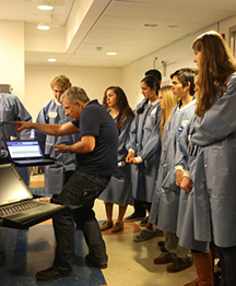
Dressed in blue lab coats and safety glasses, 40 enthusiastic San Diego high school students toured laboratories, examined high-tech x-ray and electron microscopy equipment, and fired volleys of questions at researchers. These teenagers were participants in the recent Students Modeling A Research Topic (SMART) Team‘s Mentor Match Event hosted by The Scripps Research Institute (TSRI) California campus.

Now in its fifth year at TSRI, the community outreach initiative, which has been run jointly with the University of California, San Diego (UCSD) Super Computer Center, brings the reality of science directly to the students, according to Stephen Connelly, a staff scientist in the Wilson/Kelly labs. Connelly and Ange Mason of the Super Computer Center are coordinating this year’s San Diego SMART Team program.

Over the course of the academic year, SMART Teams work to complete a number of academic tasks in their groups before working with volunteer mentor scientists from TSRI on research projects that focus on understanding the structure-function relationship of a protein studied within the mentor’s lab. Trained in computer visualization software, the teams design and build 3D models of their proteins to help tell their specific research story. Finally, they create oral presentations explaining their work to a lay audience and a poster to present to a scientific audience.

With additional support from their high school teachers, who themselves received special training, and SMART program coordinators, students explore the molecular world and experience science as a process and not just a collection of facts, said Connelly.

The day-long Mentor Match Event, pairing teams with their research mentors, was organized as a “mini-science conference,” said Connelly, “with students squarely at the center of all the activity.” The student teams represented Francis Parker, El Capitan, Canyon Crest Academy and Murietta Mesa high schools.

TSRI Professor Ian Wilson, Hansen Professor of Structural Biology and long-time SMART Team supporter, provided one of the morning’s highlights as he shared thoughts on his life’s work in scientific research and recent developments in high-throughput structural biology.

The teens also got close-up views of the advanced crystallization robotics suite and the electro-microscopy suite during tours of the Joint Center for Structural Genomics and the Center for Integrative Molecular Biosciences at TSRI.

Following presentations on their proposed scientific projects, the SMART teams were “matched’ with TSRI mentor scientists. This year’s match-ups are:

  • Ryan Hoffman, research associate in the Ward lab, with El Capitan students, working on troponin, a protein that converts calcium fluctuation’s in the body into movement
  • Jintang Du, research fellow in the Gottesfeld lab, with the Canyon Crest Academy team, researching sirtuins, proteins that help regulate biologic pathways in bacteria.
  • Peter Lee, graduate student in the Wilson lab, with Francis Parker students, studying hemagglutinin, a protein found on the surface of influenza viruses
  • Daniel Murin, graduate student in the Sapphire/Ward labs, with Murietta Mesa students, working on a glycoprotein involved with the Ebola virus
  • Jessica Bruhn-Johannsen, graduate student in the Sapphire lab, with Murietta Mesa team members, studying the V protein, important in the body’s immune system response to viruses
Ronnie Fournier, an El Capitan ninth-grader summed up students’ response to the day’s activities. "It was the best science experience I could imagine having,” he said. “The scientists, labs tours and meeting everyone. No, really… it was amazing!"

UCSD, which is currently applying for funding for the program, will assume leadership of the San Diego SMART Team program next year. However, said Connelly, TSRI researchers will be able to continue serving as mentors.

The SMART Teams program has its roots in a 2008 partnership between the Milwaukee School of Engineering (MSOE) Center for BioMolecular Modeling (CBM) and the University of Wisconsin School of Medicine and Public Health, which then disseminated the SMART Team program to eight top research institutions across the United States. The San Diego program has received funding by the Howard Hughes Medical Institute, Biogen Idec, the National Institutes of Health and the National Science Foundation. Connelly also acknowledged Marisela Chevez, who helped establish and build the San Diego SMART Teams program; Professor Ian Wilson and Associate Professor David Goodsell for their continued support and assistance; and Dawn Eastmond, Nancy De Monte and Jennifer Vela who were especially helpful with the Mentor Match Event.


domingo, 3 de junio de 2012

La basura en los océanos

2012-05-29

Algunas criaturas prosperan con la presencia de fragmentos microscópicos de plástico 
¿Alguna vez has oído hablar de la Gran Placa de Basura del Pacífico (PBP)? 

Se trata de una región en el norte del océano Pacífico donde las corrientes marinas del norte y las corrientes de aire del sur chocan al fluctuar en dirección contraria, creando una región circular y tranquila llamada Giro del Pacífico Norte.

Es en el centro de esta región donde yacen toneladas de basura.

Muchas imágenes se han distribuido donde aparece una enorme placa sólida de basura, principalmente plástico PET, donde un hombre trata de remar en su canoa. Una imagen de este lugar, que se le adjudica a la PBP, está disponible abajo.

¿Alarmante? No te preocupes, esta es una de las tantas interpretaciones erróneas que el público tiene sobre lo que pasa con el plástico en los océanos (la imagen es de un depósito en Manila), debido a la desinformación que se ha hecho en pro de una consciencia ambiental.

"¡Esa foto del hombre en la canoa me ha estado siguiendo toda mi carrera!" dijo Miriam Goldstein, bióloga marina del Instituto Scripps, y quien ha completado un estudio del cómo el plástico está cambiando el ecosistema en el Giro del Pacífico Norte.

Goldstein habló con la publicación online io9.com sobre los mitos y verdades sobre la PBP.

"Creo que es un ejemplo de la multimedia telefónica, donde alguien quiere una imagen dramática para ilustrar su historia, y con la magia de Internet, la imagen es malinterpretada" dijo Goldstein.

La especialista en biología marina ha realizado múltiples trabajos de campo en la PBP, a mil 600 kilómetros de la costa californiana, nadando incluso en la placa. Según Goldstein, nunca había visto nada parecido con lo de la fotografía.

A continuación, se desmienten los siguientes mitos:

Mito: Existe una isla gigante de basura sólida flotando en el Pacífico.

Hecho: hay millones de microscópicas piezas de plástico, cerca de 0.4 piezas por cada metro cúbico, flotando sobre apenas 2 mil 736 kilómetros cuadrados de superficie en el océano Pacífico

Esta cantidad ha aumentado significativamente los últimos 40 años. Y aunque el tamaño de estos fragmentos es el de una falange, la cantidad es lo amenazante 

Mito: Todo este plástico está matando a los animales.

Hecho: algunos animales están siendo heridos, pero otros están prosperando. Éste vendría siendo el problema.

Nadie argüiría que un ecosistema marino no se vería afectado por el plástico, pero es difícil determinar si las aves y peces que se comen el material mueren por esta causa.

Además, existen animales como insectos marinos, pequeños cangrejos, percebes y algunos invertebrados que están emergiendo debido a la influencia del plástico contaminante.

Los percebes e invertebrados llegan a causar daños a ecosistemas que invaden, además de afectar los cascos de barcos. Como estas criaturas viven en el fondo marino, no se ven afectadas por el plástico fragmentado. 

Mito: La capa de plástico está matando al océano.

Hecho: La placa de plástico es una ecosistema fuera de balance.

La "plasticosfera" es un término acuñado por el biólogo marino Eric Zettler para describir a la criaturas que viven y prosperan en un ambiente de superficies duras en el agua. Son similares a los animales que usan superficies como los cascos de barcos para hacerlos su nicho marino.

El problema con la plasticosfera es que está cambiando radicalmente el balance del ecosistema de mares profundos, antes precedido principalmente por peces.

Entre más plástico y basura se distribuya por estas partes oceánicas, más invadirán los ecosistemas los animales de la plasticosfera, desequilibrándolos con efectos irreversibles.

viernes, 16 de marzo de 2012

Escultura interactiva, puente entre Arte y Ciencia

ORIGINAL: Scripps
Auto-Line Assembly
La ciencia se une al arte en una nueva escultura interactiva, Línea de Auto-Ensamblaje: Del Caos al Orden, recientemente instalado en el campus de California del Instituto de Investigación Scripps, en las afueras de la entrada principal Edificio del Instituto Skaggs / Biología Molecular. Es tanto una obra de arte y una demostración científica, la escultura es una colaboración entre el Profesor de Investigación Scripps Art Olson y Tibbitts Skylar, un diseñador y arquitecto del Laboratorio de Medios del  Instituto de Tecnología de Massachusetts.

La escultura muestra las interacciones al azar en el proceso de auto-ensamblaje, dijo Olson. La estructura es una gran jaula blanca  que rota, dónde caen piezas más pequeñas de color rojo en el interior de forma aleatoria. Las piezas interiores se basan en la estructura de las unidades de montaje de los poliovirus, cuya la estructura atómica se determinó por Scripps Research en la década de 1980.


Luego de que TIbbits viera uno de los videos de Olson en YouTube (http://www.youtube.com/watch?feature=player_embedded&v=X-8MP7g8XOE), los dos colaboraron para construir una versión a gran escala del científico "del virus de auto-ensamblaje en una botella" para la presentación potencial en los lugares de la ciencia y museos. La escultura está fabricada en plástico de polietileno, espuma de poliuretano e imanes.

Originalmente encargado para exhibirse en la Conferencia TED 2012 celebrada 27 febrero-2 marzo en Long Beach, California, la obra de arte, que no pudo ser desmontada, fue trasladado a su actual ubicación en el campus después de la conferencia. "Dado que la pieza está concebida para ser un prototipo para instalarse, tanto en  lugares de arte y museos de ciencias, hemos querido tenerla aquí en Scripps para su análisis posterior, fortalecimiento, y mejora", dijo Olson.

Auto-Line Assembly ha sido recibida con curiosidad y entusiasmo en el campus. "Una vez que la gente se da cuenta de que está destinado a ser rotado, y que las piezas individuales en el interior, cuando caen, se ensamblan para formar una esfera completa, están encantados con él", dijo Olson. Cuando se ve las piezas interiores de la instalación montadas, se introduce la mano en la escultura para separarlas. "La próxima vez que pasan, las piezas se vuelven a montar, así que obviamente las personas están participando muy activamente con ella",

Los fondos adicionales para la obra de arte fue proporcionado por la semilla Media Group, cuyo brazo de investigación, Phyllotax Lab, creó una página web para describir la obra en detalle.