

|
| What lies within the H atom? |
Information flow
The wave function is a central tenet of quantum theory – put simply, it contains the maximum knowledge that is available about the state of a quantum system. More specifically, the wavefunction is the solution to the Schrödinger equation. The square of the wavefunction describes the probability of where exactly a particle might be located at a given time. Although it features prominently in quantum theory, directly measuring or observing the wavefunction is no easy task, as any direct observation destroys the wavefunction before it can be fully observed.
In the past, "Rydberg wavepacket" experiments have tried to observe the wavefunction using ultrafast laser pulses. In these experiments, the atoms are in a superposition of their highly excited "Rydberg states". These experiments show that the periodic electron orbitals around nuclei are described by coherent superpositions of quantum-mechanical stationary states. The wavefunction of each of these states is a standing wave with a nodal pattern (a "node" is where there is zero probability of finding an electron) that reflects the quantum numbers of the state. While previous experiments have attempted to capture the elusive wavefunction or the nodal patterns, the methods used were not successful. Direct observation of the nodal structure of a single atom being most difficult to achieve.
Plotting waves
In the new work, Aneta Stodolna, of the FOM Institute for Atomic and Molecular Physics in the Netherlands, along with Marc Vrakking at the Max-Born-Institute in Berlin, Germany, and other colleagues in Europe and the US have shown that photoionization microscopy can directly obtain the nodal structure of the electronic orbital of a hydrogen atom placed in a static electric field. In the experiment, the hydrogen atom is placed in the electric field E and is excited by laser pulses. The ionized electron escapes from the atom and follows a particular trajectory to the detector – a dual microchannel plate (MCP) detector – that is perpendicular to the field itself. Given that there are many such trajectories that reach the same point on the detector, interference patterns can be observed, which the team magnify by a factor of more than 20,000 using an electrostatic zoom lens. The interference pattern directly reflects the nodal structure of the wavefunction. The experiments were carried out with both resonant ionization involving a Rydberg state and non-resonant ionization.
The team chose the hydrogen atom thanks to its unique properties. "These [hydrogen atoms] are very peculiar...as hydrogen has only one electron, which interacts with the nucleus via a purely Coulombic interaction, it has a particular structure when we place it in a DC electric field," says Vrakking. He goes on to explain that thanks to its single-electron status, hydrogen's wavefunction can be written as the product of two wavefunctions, which describe how it changes as a function of two coordinates – the so-called parabolic coordinates. That is, the Hamiltonian of the hydrogen atom (in an external electric field) describes a splitting of its energy levels, which is known as the "Stark effect". More importantly, though, this "Stark Hamiltonian" is exactly separable in terms of the two parabolic coordinates, which are linear combinations of the distance of the electron from the hydrogen nucleus r and the displacement of the electron along the electric-field axis z.
Vrakking told physicsworld.com that the shape of the two parabolic wavefunctions is therefore "completely independent of the strength of the field, and so it is invariable – it stays the same as the electron travels for more than half a metre in the experiment – all the way from where the ionization occurs up to the 2D detector". This, he explains, is crucial to scaling up the spatial distribution to magnify the nodal patterns to millimetre-scale dimensions, where they can be observed with the naked eye on the 2D detector and recorded with a camera system. "What you see on the detector is what exists in the atom," he says. The group observed several hundreds of thousands of ionization events to obtain the results, with the same preparation of the wavefunction for each.
What lies within
The figure at the top of this article shows the team's main result – the raw camera data for four measurements, where the hydrogen atoms were excited to states with zero, one, two and three nodes in the wavefunction for one of the parabolic coordinates. "If you look at the measured projections on the detector, you can easily recognize the nodes, and see their radial, ring-like structure," says Vrakking.
 |
| Eye of the atom |
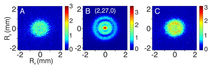
He also points out the "striking difference" between images recorded following resonant excitation and images recorded following non-resonant excitation – this is seen in the image to the right, where a comparison is given between a measurement taken for one resonant and two non-resonant nodes. Images (A) and (C) were taken after non-resonant ionization, while for the central image, (B), the laser was tuned to a resonance with two nodes in the wavefunction. For the resonant ionization, the outermost ring extends significantly further radially, compared with the other two images – something that could be explained by a special kind of tunnelling effect taking place.
Vrakking says that the ultimate goal of the research was to study and visualize the hydrogen atom. Future experiments may look at how the atom would react within a magnetic field, study time-resolved electron dynamics, investigate holographic interference microscopy and perhaps even observe molecules using photoionization microscopy.
Helium under the microscope
Currently, however, the researchers are studying and analysing a helium atom using photoionization microscopy, and a paper on this will be published in the coming months. "As there are two electrons in a helium atom, we are getting some very interesting information," says Vrakking. He says that while in some aspects the responses of the helium atom are very similar to that of hydrogen, there are also some major differences. "Although one of the helium electrons is very tightly bound to the nucleus, and the other one is very highly excited, we can see that the electrons know of each other's existence and that they 'talk to each other'," says Vrakking, explaining that this could allow the team to "see" entanglement of the electrons.
The research is published in Physical Review Letters.
ORIGINAL: Physics World
May 23, 2013
No hay comentarios:
Publicar un comentario
Nota: solo los miembros de este blog pueden publicar comentarios.