ORIGINAL:
The Guardian
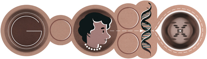 |
| 25 July 2013 Google Doodle: Rosalind Franklin (25 July 1920 - 16 April 1958). Today's Google Doodle honours pioneering British biophysicist and x-ray crystallographer, Rosalind Franklin |
 |
| Rosalind Franklin. Wikipedia |
Today is the 93rd anniversary of the birth of British biophysicist Rosalind Franklin, who was a pioneering x-ray crystallographer who is being honoured today with a special
Google Doodle.
Nearly everyone who has heard about Dr Franklin knows that her work was critical to elucidating the structure and function of DNA -- a discovery that was recognised by the 1962 Nobel Prize in Physiology or Medicine four years after her tragic death from ovarian cancer. (Dr Franklin was not nominated for this award; Nobel Prizes are not awarded posthumously.)
Dr Franklin was an
x-ray crystallographer.
To do her work, she would purify a molecule, such as DNA, grow it into crystals and bombard the crystal with x-rays. After photographing the x-ray diffraction pattern created by collisions with atoms within the crystal (see right), Dr Franklin then could directly determine the structure of the molecule.
 |
| Photo 51 |
Dr Franklin's work, particularly "
Photo 51" (pictured, right; by Raymond Gosling), was critical for correctly determining the structure and function of DNA. According to my sources,
Maurice Wilkins, who was Dr Franklin's supervisor at King's College London, shared several of her images, including "Photo 51", with James Watson without her knowledge or consent. Up until the time when they saw this particular image,
Watson and his colleague, Francis Crick, had erroneously postulated that the DNA "backbone" was on the inside of the molecule and the nucleic acid "bases" pointed outwards, like the teeth of a comb. This image pointed out their error in logic so they quickly revised their hypothesis and published it, which then led to them being awarded the Nobel Prize a few years later.
But this is where most people's knowledge of Dr Franklin's life ends:
what did she do after her work with DNA crystals had concluded?
In fact,
after finishing her work in Wilkin's lab in 1953, Dr Franklin left King's College to study the tobacco mosaic virus (TMV) at Birkbeck College in London. This is where she spent the last five years of her life, conducting pioneering work into the structure of
viruses.
Discovered to be a virus in 1930,
TMV is both stable and structurally simple, and thus, it easily forms crystals, making it an excellent candidate for x-ray crystallography studies. TMV also is easy to work with in the lab, being a highly infectious agricultural pest that attacks a wide variety of plant species. In susceptible plants, it causes obvious symptoms but doesn't kill its host plants, which also made it attractive for scientific study.
Finding Birkbeck College to be a much more collegial environment than King's College, Dr Franklin assembled a team of talented scientists whose researches complemented her own. Based on her team's findings,
Dr Franklin hypothesized that TMV was a hollow tube made of proteins that contained a single strand of RNA that spiraled inside the length of the tube like a thread spiraling inside a donut hole -- both hypotheses were found to be correct after her death.
Solving the structure of a plant viruses meant that it would also be possible to solve the structure of other viruses -- animal viruses.
Polio viruses were the obvious choice since they had been crystallized in 1955 (doi:
10.1073/pnas.41.12.1020). For that reason,
Dr Franklin sought funding necessary to solve the structure of the polio virus.
During a visit to the United States in 1956, Dr Franklin began suffering health problems that were soon diagnosed as ovarian cancer. After two abdominal surgeries in one month, she returned to the lab and redoubled her efforts. Although she felt fine for awhile, the cancer soon returned.
Despite her illness and impending death, Dr Franklin still managed to obtain funding to keep her team going for another three years, researching the polio virus.
Shortly after her untimely death on 16 April 1958 at the age of 37 years old, two members of her team,
John Finch and
Aaron Klug, dedicated the resulting paper to her memory (doi:
10.1038/1831709a0).
Throughout her professional life,
Dr Franklin -- an unmarried female Jewish scientist -- was faced with tremendous challenges from most of her (white male) colleagues. But thanks to supportive environment at Birkbeck College, she was able to pursue her work in relative peace during her final years.
After her death,
John Bernal who was chair of the physics department at Birkbeck College, wrote obituaries published in the
New York Times and in
Nature (doi:
10.1038/182154a0) that celebrated her "
beautifully executed researches, carried out with apparently effortless skill, and her gift for organizing research projects".
"
As a scientist Miss Franklin was distinguished by extreme clarity and perfection in everything she undertook", Professor Bernal wrote.
"
Her photographs are among the most beautiful x-ray photographs of any substance ever taken."
Dr Franklin's life, Professor Bernal concluded, was a perfect example of single-minded devotion to research.
Sources:
Rosalind Franklin: My Favourite Scientist
Biology%2CChemistry%2CHealth%2CMedicine">Schaffer F.L. (1955). Crystallization of Purified MEF-1 Poliomyelitis Virus Particles, Proceedings of the National Academy of Sciences, 41 (12) 1020-1023. doi:
10.1073/pnas.41.12.1020
Bernal J.D. (1958). Dr. Rosalind E. Franklin, Nature, 182 (4629) 154-154. doi:
10.1038/182154a0
Glynn J. (2008). Rosalind Franklin: 50 years on, Notes and Records of the Royal Society, 62 (2) 253-255. doi:
10.1098/rsnr.2007.0052
Finch J.T. & Klug A. (1959). Structure of Poliomyelitis Virus, Nature, 183 (4677) 1709-1714. doi:
10.1038/1831709a0
Creager, Angela N. H. & Morgan, Gregory J. (2008). After the Double Helix: Rosalind Franklin's Research on Tobacco
mosaic virus, Isis, 99 (2) 239-272. doi:
10.1086/588626
.. .. .. .. .. .. .. .. .. .. ..
Grrlscientist can also be found here:
Maniraptora, and she lurks on social media:
facebook,
Google +,
LinkedIn,
Pinterest and of course, she's quite active on twitter: @
GrrlScientist


