Mostrando entradas con la etiqueta Biomaterial. Mostrar todas las entradas

miércoles, 23 de marzo de 2016

Creating 3-D tissue and its potential for regeneration

Bioprinting technique may provide potential for tissue repair and regenerative medicine


Researchers are one step closer to embedding vascular networks into thick human tissues, which could result in tissue repair and regeneration — and ultimately even replacement of whole organs.

A team at the Wyss Institute for Biologically Inspired Engineering at Harvard University and the Harvard John A. Paulson School for Engineering and Applied Sciences (SEAS) has invented a method for 3-D bioprinting thick vascularized tissue constructs. The vasculature network enables fluids, nutrients, and cell growth factors to be perfused uniformly throughout the tissue.

The advance was reported Monday in the journal Proceedings of the National Academy of Sciences.

This latest work extends the capabilities of our multi-material bioprinting platform to thick human tissues, bringing us one step closer to creating architectures for tissue repair and regeneration,” says the study’s senior author, Jennifer A. Lewis, who is a Wyss core faculty member and the Hansjörg Wyss Professor of Biologically Inspired Engineering at SEAS.

Printing Vascular Tissue


Printing vessel vasculature is essential for sustaining functional living tissues. Until now, bioengineers have had difficulty building thick tissues, lacking a method to embed vascular networks. Credit: Lewis Lab/ Wyss Institute at Harvard University

In the study, Lewis and her team showed that their 3-D printed, vascularized tissues could thrive and function as living tissue architectures for upwards of six weeks.

To date, scaling up human tissues built of a variety of cell types has been limited by an inability to embed life-sustaining vascular networks. Building on their earlier work, Lewis and her team have now increased the tissue thickness threshold nearly tenfold, setting the stage for future advances in tissue engineering and repair. The method combines vascular plumbing with living cells and an extracellular matrix, enabling the structures to function as living tissues.

As an example of what can be done with the technology, Lewis’ team printed 1-centimeter-thick tissue containing human bone marrow stem cells surrounded by connective tissue. By pumping bone growth factors through supporting vasculature lined with the same endothelial cells found in human blood vessels, the scientists induced the cells to develop into bone cells over the course of one month, according to the study.

This research will help to establish the fundamental scientific understanding required for bioprinting of vascularized living tissues,” said Zhijian Pei, National Science Foundation program director for the Directorate for Engineering Division of Civil, Mechanical, and Manufacturing Innovation, which funded the project. “Research such as this enables broader use of 3-D human tissues for drug safety and toxicity screening and, ultimately, for tissue repair and regeneration.

Lewis’ novel 3-D bioprinting method uses a customizable, printed silicone mold to house the printed tissue structure. Inside this mold, layers of vascular channels made of pluronic (a material that liquefies at refrigerator temperature) and living stem cells are interdigitated like locking fingers. A cellular matrix is poured around this structure, and solidifies. The entire device is then refrigerated until the pluronic turns to liquid and is sucked out by a vacuum. This creates channels through which liquid containing endothelial cells, oxygen, nutrients, and growth factors — basically, simulated blood — can flow.

The bioprinted material can be used to create living tissue cultures as well as to drive directed tissue growth such as differentiating stem cells. To achieve a variety of tissue shapes, thicknesses, and composition, the shape of the printed silicone chip can be customized and the printable cellular material can be tuned to include a wide variety of cell types. In other words, this new method creates a fully controllable, living 3-D tissue environment, researchers say.

Having the vasculature prefabricated within the tissue allows enhanced cell functionality at the deep core of the tissue, and gives us the ability to modulate those cell functions through the use of perfusable substances such as growth factors,” said David Kolesky, a graduate researcher at the Wyss Institute and SEAS and one of the study’s first authors.

Jennifer and her team are shifting the paradigm in the field of tissue engineering based on their unique bioprinting approach,” said Wyss Institute Director Donald Ingber. “Their ability to build living 3-D vascularized tissues from the bottom up provides a potential way to form macroscale functional tissue replacements that can be surgically connected to the body’s own blood vessels to provide immediate perfusion of these artificial tissues, and thus, greatly increase their likelihood of survival. This would overcome many of the problems that held back tissue engineering from clinical success in the past.

Ingber is also the Judah Folkman Professor of Vascular Biology at Harvard Medical School and the vascular biology program at Boston Children’s Hospital, and professor of bioengineering at SEAS. In addition to Lewis and Kolesky, other team members on the new study include co-first authors Kimberly Homan, research associate at the Wyss Institute, and Mark Skylar-Scott, postdoctoral fellow at the Wyss Institute.

The work was supported by the National Science Foundation and the Wyss Institute for Biologically Inspired Engineering at Harvard University.

Adapted from a Wyss Institute press release written by Kat J. McAlpine, Wyss Institute Communications.

ORIGINAL: Harvard Gazzette
March 8, 2016

miércoles, 20 de enero de 2016

IU scientists create 'nano-reactor' for the production of hydrogen biofuel

Combining bacterial genes and virus shell creates a highly efficient, renewable material used in generating power from water

BLOOMINGTON, Ind. -- Scientists at Indiana University have created a highly efficient biomaterial that catalyzes the formation of hydrogen -- one half of the "holy grail" of splitting H2O to make hydrogen and oxygen for fueling cheap and efficient cars that run on water.

A modified enzyme that gains strength from being protected within the protein shell -- or "capsid" -- of a bacterial virus, this new material is 150 times more efficient than the unaltered form of the enzyme.
An artist's rendering of P22-Hyd, a new biomaterial created by encapsulating a hydrogen-producing enzyme within a virus shell.
Photo by Trevor Douglas
 The process of creating the material was recently reported in "Self-assembling biomolecular catalysts for hydrogen production" in the journal Nature Chemistry.

"Essentially, we've taken a virus's ability to self-assemble myriad genetic building blocks and incorporated a very fragile and sensitive enzyme with the remarkable property of taking in protons and spitting out hydrogen gas," said Trevor Douglas, the Earl Blough Professor of Chemistry in the IU Bloomington College of Arts and Sciences' Department of Chemistry, who led the study. "The end result is a virus-like particle that behaves the same as a highly sophisticated material that catalyzes the production of hydrogen."
Trevor Douglas | Photo by Montana State University
Other IU scientists who contributed to the research were Megan C. Thielges, an assistant professor of chemistry; Ethan J. Edwards, a Ph.D. student; and Paul C. Jordan, a postdoctoral researcher at Alios BioPharma, who was an IU Ph.D. student at the time of the study.

The genetic material used to create the enzyme, hydrogenase, is produced by two genes from the common bacteria Escherichia coli, inserted inside the protective capsid using methods previously developed by these IU scientists. The genes, hyaA and hyaB, are two genes in E. coli that encode key subunits of the hydrogenase enzyme. The capsid comes from the bacterial virus known as bacteriophage P22.
Illustration showing the release of NiFe-hydrogenase from inside the virus shell, or "capsid," of bacteriophage P22.
Photo by Trevor Douglas
The resulting biomaterial, called "P22-Hyd," is not only more efficient than the unaltered enzyme but also is produced through a simple fermentation process at room temperature.

The material is potentially far less expensive and more environmentally friendly to produce than other materials currently used to create fuel cells. The costly and rare metal platinum, for example, is commonly used to catalyze hydrogen as fuel in products such as high-end concept cars.

"This material is comparable to platinum, except it's truly renewable," Douglas said. "You don't need to mine it; you can create it at room temperature on a massive scale using fermentation technology; it's biodegradable. It's a very green process to make a very high-end sustainable material."

In addition, P22-Hyd both breaks the chemical bonds of water to create hydrogen and also works in reverse to recombine hydrogen and oxygen to generate power. "The reaction runs both ways -- it can be used either as a hydrogen production catalyst or as a fuel cell catalyst," Douglas said.

The form of hydrogenase is one of three occurring in nature: di-iron (FeFe)-, iron-only (Fe-only)- and nickel-iron (NiFe)-hydrogenase. The third form was selected for the new material due to
  •  its ability to easily integrate into biomaterials and 
  • tolerate exposure to oxygen.
NiFe-hydrogenase also gains significantly greater resistance upon encapsulation to breakdown from chemicals in the environment, and it retains the ability to catalyze at room temperature. Unaltered NiFe-hydrogenase, by contrast, is highly susceptible to destruction from chemicals in the environment and breaks down at temperatures above room temperature -- both of which make the unprotected enzyme a poor choice for use in manufacturing and commercial products such as cars.

These sensitivities are "some of the key reasons enzymes haven't previously lived up to their promise in technology," Douglas said. Another is their difficulty to produce.

"No one's ever had a way to create a large enough amount of this hydrogenase despite its incredible potential for biofuel production. But now we've got a method to stabilize and produce high quantities of the material -- and enormous increases in efficiency," he said.

The development is highly significant according to Seung-Wuk Lee, professor of bioengineering at the University of California-Berkeley, who was not a part of the study.

"Douglas' group has been leading protein- or virus-based nanomaterial development for the last two decades. This is a new pioneering work to produce green and clean fuels to tackle the real-world energy problem that we face today and make an immediate impact in our life in the near future,” said Lee, whose work has been cited in a U.S. Congressional report on the use of viruses in manufacturing.

Beyond the new study, Douglas and his colleagues continue to craft P22-Hyd into an ideal ingredient for hydrogen power by investigating ways to activate a catalytic reaction with sunlight, as opposed to introducing elections using laboratory methods.

"Incorporating this material into a solar-powered system is the next step," Douglas said.

This research was supported by the U.S. Department of Energy.

Jan. 4, 2016

martes, 18 de noviembre de 2014

What is the 3D Print Canal House?


The 3D Print Canal House is an exhibition, research- and building site for 3D Printing Architecture. A unique project where an international team of partners collaborates in ‘research & doing’ linking science, design, construction and community, by 3D printing a canal house at an expo-site in the very heart of Amsterdam.


FREQUENTLY ASKED QUESTIONS

1. Why 3D Printing?
3D printing is a fascinating new production technique. It allows you to directly translate a digital file into a physical product. 3D printing can have huge implications for the way we fabricate things - for example the elimination of waste, transport costs and standardisation of elements - DUS architects is investigating what the implications of 3D printing are for the building industry. What better way to do this than by 3D printing an entire house?


2. Why a canal house?
The canal house is a symbol of Amsterdam. When the canal belt was built 400 years ago, Amsterdam was a prime example of innovation. Each canal house can house several functions, such as trade, storage, living, craft, and each canal house is richly ornamented and unique. A canal house is recognizable and attractive. It is interesting to investigate what this traditional architype can be in a 21st century context. 3D printing a canal house shows the world how to combine traditional local values with new innovative ideas.

3. What are some of the advantages and disadvantages of 3D printing a building?
One great advantage of 3D printing over traditional buidling techniques (such as prefabricated concrete) is the possibilities of using a high level of detail and ornament and variation. Rather than using standardized elements, 3D printed designs can each be modified and customized to fit the user's needs and taste. It will no longer be more expensive or more labour intensive to add details to for example your façade and it is easy to create unique objects.

3D printing is an additive manufacturing technique. That means the process goes straight from the raw material to the final product, thus eliminating waste. There are no transport costs, as designs can simply be transferred digitally and printed locally. This also implies that when 3D printing is used widely in each part of the world, it will no longer be cheaper to have things produced in countries like China or Bangladesh as opposed to the Netherlands. Everyone can just produce everything in their own local context.

In terms of disadvantages, it is obviously a huge challenge to create a building that complies with all the current building regulations. There is the question of insulation, fireproofing, wind loads, foundations...these, as well as the possible materials to print with (using this printer) are all things that are being researched and investigated.

4. How does the Kamermaker work?
The Kamermaker works in exactly the same way as the Ultimaker, the small desktop 3D printer, as it is simply an upscaled version. A digital design is placed in the 'brain' of the printer, a very simple computer, where it is translated into a G-code. A G-code is a file that slices a 3D model into layers. This file programs the printer to move along a path that is optimal for that design, layer by layer. 

In the 'control room' of the printer is also the material supply. We print with plastic in the form of granulate which enters an extruder via a funnel. In the extruder the granulate is heated (the material melts at 170 degrees Celsius) and pressed together to a homogeneous liquid. This is brought to the printer head by a heated tube. The printer head extrudes the melted material along the programmed path on the X and Y axes and when finished moves up one step along the Z axis. This is fairly similar to a normal printer, only with one more direction, which allows objects to be printed layer by layer.

5. What materials does the Kamermaker print with?
We are currently printing with bioplastics. The granulate that goes into the Kamermaker is called Macromelt, a type of industrial glue (Hotmelt) developed by Henkel. It is made of 80% of vegetable oil. It melts at 170 degrees Celsius. We aim to print with a material that is sustainable, of biological origin, melts at a relatively low temperature, and of course is sturdy and stable. We are also researching the possibilities of printing with recycled materials: Plastics of course, but we’re also looking into using wood pallets and natural stone waste.

Technically, the Kamermaker can print with any material that melts (at a temperature that isn't too high) and then hardens again.

7. Who are the initiators and partners? 
DUS architects is the initiator of both de KamerMaker and the 3D Print Canal House. DUS architects  is an Amsterdam based architecture office founded in 2004 by Hans Vermeulen, Hedwig Heinsman and Martine de Wit. DUS architcts builds ‘public architecture’: Architecture that influences the public domain using scale 1:1 models, urban process- and strategy design, and that ranges from temporary interiors to long-term urban transformation trajectories.  www.dusarchitects.com

DUS architects is collaborating with lots of important partners who invest in the project with knowledge and means. For example:

  • Henkel is developing a new sustainable 3D print material for the building industries. 
  • Heijmans is researching what new construction techniques are needed for 3D printing buildings. 
  • The Municipality of Amsterdam investigates the effect of the digital maker-industries on regulations and opportunities for employment.
Check the 'partners' section at this website for an actual overview of all our courageous partners!


8. How is the house financed?
The project is partially funded by the contributions of our partners and partially funded by the municipality of Amsterdam, Amsterdam Fund for the Arts and the DOEN Foundation. A lot of the sponsorship the 3D Print Canal House gets is in natura, through contributing knowledge or materials. In fact, the 3D Print Canal House is one big collaboration project, in which everybody shares and gets a share.

And of course our visitors help finance the house by paying an entrance fee!

9. How much does the house cost?
That is impossible to say since all of the materials we use have never been on the market for this purpose. The 3D Print Canal House is a research project partially funded and partially created by DUS architects and its partners. At the end of the research trajectory, we hope to be able to give an accurate estimate of what it takes to 3D print a house. The goal is to create a cost-effective building technique for building sustainable and comfortable houses.

10. What is there to do at the 3D print canal house?
The construction site of the 3D print canal house not just a building site, it is an open workplace where an international team of partners collaborates in 'research and doing (R&DO), as well as an open source expo. You can visit it like a regular museum to learn more about the techniques being researched and used, and you can watch the team at work! We have an audiotour to explain you everything you see.

The building is constantly under construction: in three years time more and more rooms will be printed and assembled on site as the design and printing techniques progresses. So evry week this museum changes!

Also there are regular workshops and events held at the expo (watch our calendar). If you wish to book a personalised tour, attend a workshop or lecture, or rent out the expo for a special event, please contact info@3dprintcanalhouse.com

11. Why do I need to pay an entrance fee?
When you visit the 3d Print Canal House, you are not just visiting a building site, you are visiting an open research workplace and exhibition. By paying €2,50 entrance fee, we can create an informative and secure tour for visitors without being of to much disturbance for the research and work progress. With the fee you pay, you are directly funding the development of the project. Don't worry, you'll be getting value for your money, such as a free audiotour.

12. How can I contribute?
The 3D Print Canal House is an open project. That means that we are learning from our audience during the project. We are always happy to hear about your ideas.

You can contribute by sharing your knowledge or simply by visiting and paying entrance fee or booking a group tour.

If you wish to make further contributions, find out how by clicking on 'Become a Friend!'

13. When will the house be finished?
The 3D Print Canal House is a 3 year research and development project. This does not mean that the expected time it will take to 3D print a building in the future will be 3 years. On the contrary: The aim for the use of 3D printing in architecture is to build faster compared to traditional building techniques. Within the 3 years research project of the 3D print Canal House, DUS architects is building the Canal House and by doing so building up new knowledge for this purpose and sharing this with the community through the website and expo center.

14. What will happen to the building after it's finished?
Most likely it will be a public building. We hope that the 3D Print Canal House will become a hub for innovation and new production techniques and materials for the building industry. And of course that many more 3D printed buildings will pop up around the globe!

15. What sets this print project apart from other large scale 3D print initiatives?
Currently, 3D printing is on the rise, and there are many other initiatives, both on a large and small scale (also see 'other 3D printing initiatives' for a bit more in depth overview) going on that we can hardly keep track.

What makes the 3D Print Canal House special is that it is a project which is 'open' in every way: The initiators, designers and builders (DUS architects) are the client: the focus is on research, experimentation and development, instead of finishing a house. The project involves many different industries, disciplines and parties tied together by a common goal. More importantly, the process is being shown to the public (not only the successes but also the possible failures it will encounter!) in order to learn and develop.

16. What are some other interesting 3D printing initiatives?
3D printing is already widely used in many industries, particularly when on a small scale a high level of unique detail is required (big chances are that if you need a crown or hearing aid, it will be 3D printed). It is a production technique that is becoming more and more widely available. The great thing is that 3D printing can easily create objects or parts of objects that are custom fit and have a high level of detail and variety.

The 3D Print Canal House is a pioneering example and the first house that is being printed on the spot with the largest portable 3D printer. However, there are quite a few other initiatives on a similar large scale around right now, and chances are there will be more and more over the next months and years.

Some interesting examples to look at are:

Examples of using ‘our’ FDM printing technique combined with a concrete like material are
and 
  • Shanghai WinSun Decoration Design Engineering Co. 
http://www.3ders.org/articles/20140401-10-completely-3d-printed-houses-appears-in-shanghai-built-in-a-day.html
  • A different technique is developed by Enrico Dini in Italy using first a layer of sand and immediately after a layer of binder (glue) - turning the sand into a solid concrete like object. This printer is also used for Universe Architecture's Landscape House.
  • Dirk van der Kooij in Zaandam produces interior objects like chairs and vases with a FDM like printing technique using recycled plastics. www.dirkvanderkooij.nl

On a smaller scale 3D printing is used in the

  • medical industry, 
  • machinery, 
  • prototyping, 
  • jewerly, 
  • art, 
  • furniture, and 
  • many more. 
Would you like to start 3D printing yourself? Visit open fablabs like Protospace or i-Fabrica (in the Netherlands). If you would like to have your digital model 3D printed by a thirt party upload your model on one of these websites:

Your 3D printed object will be shipped right to your home!There is plenty of information on 3D printing to find on the internet. An example of an easy to read overview on 3D printing can be found here. But the best way to learn more is of course to visit the expo centre!


jueves, 27 de marzo de 2014

2014 Koch Institute Image Award Winners

Last fall, we featured The Koch Institute Image Award galleries in several Cell Picture Shows. This Show furthers the collaboration, as we showcase this year’s winning submissions. Both the Koch Institute Public Galleries and the Cell Picture Show share a similar ethos: recognition and dissemination of the extraordinary imagery produced through life science research. On March 4, 2014, these winning images were unveiled at MIT’s Koch Institute for Integrative Cancer Research in Cambridge, MA. 

We congratulate the 2014 Image Award Winners and are excited to continue to the collaboration between MIT and Cell Press. This collection of stunning images offers a window into the fascinating worlds opened to us by microscopy and other biomedical imaging techniques.

Biopolymer in Bloom
Julio D’Arcy, Erik Dreaden, and Paula Hammond
Hammond Laboratory
MIT Koch Institute
A New Environment for Studying Cell Growth. Measuring cancer cells’ real-time response to external influences can be challenging. Here, engineers have created biocompatible plastic structures onto which cells can adhere and develop as they would inside the body. The electrically conductive nature of the scaffolds allows researchers to measure the properties of the growing cells. By changing the environment or introducing new substances into the system, researchers can figure out which factors promote or discourage cell growth.
Image: This image, taken with a scanning electron microscope, shows the micro- and nano-scale structures of this device

The More the Messier
Kristin Knouse
Amon Laboratory
MIT Koch Institute

Understanding Complicated Cell Division. The mitotic spindle is an array of tracks that partitions chromosomes during cell division. Most normal cells form bipolar spindles, which segregate chromosomes equally into two daughter cells. However, many cancer cells form multipolar spindles, which cause chromosome mis-segregation and genomic instability.
Image: Like many cancer cells, liver cells also form multipolar spindles during cell division. Shown here is a liver cell with a multipolar spindle (green) pulling the chromosomes (blue) in many directions. Further research into cell division in the liver could indicate how this process is exploited or disrupted in cancer, revealing novel avenues for cancer therapy.

Target Practice
Omar F. Khan and Edmond W. Zaia
Langer and Anderson Laboratories
MIT Koch Institute

Improving Gene Therapy with Nanotechnology. How can we turn off the genes that promote the development of cancer? Using specially designed nanoparticles as genetic patches, engineers can deliver customized payloads to a cell’s gel-like cytoplasm, where most cellular activity occurs, and mitigate the effects of cancer-causing genes in the cell’s nucleus.
Image: This image shows nanoparticles (red) in the cytoplasm of cervical tumor cells (green). As researchers learn more about how cells respond to these therapies, they will continue to tweak the patches to determine the appropriate distribution of synthetic and genetic material to best target different types of cancer.

Blood, Heat, and Tumors
Alex Bagley, Jeff Wyckoff, and Sangeeta Bhatia
Bhatia Laboratory
MIT Koch Institute

Improving Drug Delivery with Gold Nanorods. Blood vessels are highways through the body. They can transport drugs to cancer cells, but finding the appropriate ramp to exit the vessel can be tricky.
Image: This image shows a network of blood vessels (green) and collagen (purple) infused with gold nanorods (yellow) inside of a living tumor. When researchers heat the particles with near-infrared light, the blood vessels become leaky, making it easier to deliver a therapeutic cargo to its final destination. Because blood vessels provide a universal transport system, such combination therapy has widespread implications for treatment, regardless of cancer type or specific drug needed

The Bad Seed
Mandar Deepak Muzumdar 
Jacks Laboratory
MIT Koch Institute

Modeling the Growth of a Tumor. Small changes have big effects. Although scientists know that certain gene mutations trigger tumor formation, the subsequent cellular events that drive cancer progression are not well understood. Cell-specific fluorescent marking allows researchers to track mutated cells over the entire course of cancer development.
Image: This image shows mutated (green) and nonmutated (red and yellow) cells in a pancreas. Over time, the green cells will multiply dramatically and form a solid tumor, while the others will not. Comparing properties and behaviors of the different cell types will set the stage for earlier diagnosis, better treatment, and even chemoprevention of deadly cancers.

Rainbow Connections
Zeynep Saygin
Kanwisher Laboratory
MIT Department of Brain & Cognitive Sciences

Mapping Neural Pathways in the Brain. The human brain is massively complex. Neuroimaging techniques such as MRI provide a noninvasive tool for studying its inner workings.
Image: This image shows pathways of nerve fibers through the brain in three dimensions: up/down (blue), front/back (green), and left/right (red). By comparing these maps of connectivity with maps of neural function, researchers can begin to predict how individual brains will respond to different stimuli. That will eventually help them to understand healthy brain development and will enable earlier diagnosis and interventions for conditions such as autism and dyslexia.

Silencing Echoes
Soheil Feizi, Steven Lee (Artist), Daniel Marbach, Muriel Medard, and Manolis Kellis Computational Biology Group
MIT Computer Science and Artificial Intelligence Laboratory

Cleaning Up Networks. Are all connections meaningful? This image visualizes a new algorithm (known as "network deconvolution") for determining important relationships in complex networks. Like a filter on a camera lens, it reveals which links (lines) between interconnected elements (points) are most essential. As the lens passes over each network area, indirect links disappear and direct links become visible. Already tested on large networks mapping gene regulation, protein folding, and academic co-authorship, network deconvolution can be used to identify key drivers of biological, social, and technological systems.

Ganglion Style
Alex Norton for EyeWire
Seung Laboratory
MIT Department of Brain and Cognitive Sciences and MIT Media Lab

Crowdsourcing Science through Online Games. It's all fun and games until somebody maps a neuron! Then it’s time to move on to the next one. The online game EyeWire challenges players, most of whom have no background in neuroscience, to create virtual 3D models of actual neurons using real laboratory data.
Image: The reconstruction seen here shows ganglion cells in the retina. By comparing this gamer-generated map to previously collected data about the neurons’ firing activity, neuroscientists can create a functional model of how vision works. With more than 100,000 players, EyeWire has already helped researchers to uncover how the eye helps us perceive moving stimuli.

Something Fishy
Annie Cavanagh and David McCarthy
School of Pharmacy
University College London

The Secret Lives of Zebrafish. Humans and fish have more in common than you might expect. Since the 1970s, a tropical freshwater minnow known as the zebrafish has been used to study the genetic and physiological development of living organisms. By mapping the zebrafish genome and studying irregularities in their development, researchers have been able to create robust models of how vertebrates develop and identify genetic conditions that lead to diseases such as cancer.
Image: This image shows a false-color scanning electron micrograph of a zebrafish embryo. It appears in the Koch Institute Public Galleries as part of a partnership between the Koch Institute and Wellcome Images.

Collateral Damage
Aprotim Mazumder, Jennifer A. Calvo, and Leona D. Samson
Samson Laboratory
MIT Koch Institute, Department of Biological Engineering, Department of Biology, and Center for Environmental Health Sciences

Investigating the Side Effects of Chemotherapeutics. How much is too much? When treating cancer, it is important to balance a drug’s effectiveness at killing tumor cells with its toxicity to healthy cells elsewhere in the body.
Image: This image of brain tissue shows cerebellar granule neurons (pink), which sustain significant damage when exposed to certain DNA-damaging therapeutics, and surrounding Purkinjee cells (orange), which do not. Researchers are studying these responses to determine the cell properties and repair mechanisms that make different cell types more or less vulnerable to chemotherapy.

ORIGINAL: Cell

lunes, 13 de enero de 2014

KI Introduces the World's First Carbon-Negative Chair Made with AIrCarbon

Two childhood friends spent a decade, beginning in college, figuring out how to cheaply make plastic from carbon that's been captured from the atmosphere.

(Photo: Dan MacMedan, USA TODAY)
Story Highlights
While in college, two friends set out to turn air pollution into plastic
Their decade-long journey has led to an award-winning carbon-based product
They aim to reduce global warming by capturing heat-trapping carbon emissions

A decade ago in his Princeton dorm room, Mark Herrema had an aha moment. He read a newspaper story about the rise in heat-trapping methane emissions from dairy farms and decided to do something about it.

He thought — why not pull the carbon from the air and use it to make stuff? A politics major who also studied chemistry, he teamed up with childhood friend Kenton Kimmel, a biomedical engineering student at Northwestern University. They took odd jobs after graduation to fund their research.

"I was a bellhop and Kenton was a valet," says Herrema, recalling how they worked 14 to 16 hours every day — even holidays — for years to pay their bills and test their ideas in rented lab space.

Industry experts told them it was a fool's errand. For good reason. Scientists had spent decades trying to capture carbon and use it to make plastic but couldn't do it cheaply enough. The two friends cracked the code by developing a ten-times more efficient bio-catalyst, which strips the carbon from a liquefied gas and rearranges it into a long chain plastic molecule.

Mark Herrema shows a container of the plastic pellets that his California-based company, Newlight Technologies, makes from recycled methane gas.(Photo: Dan MacMedan, USA TODAY)

The result? Today, the 31-year-old co-founders of California-based Newlight Technologies have two factories that take methane captured from dairy farms and use it to make AirCarbon — plastic that will soon appear in the form of chairs, food containers and automotive parts. Coming next year: cellphone cases for Virgin Mobile.

"You'll be able to hold carbon in your hand," Herrema says of the products, which an independent lab says remove more carbon from the atmosphere than their manufacturing emits. By replacing oil-based plastics, he says he wants to help reduce global warming: "We actually want to change the world."

"This will be a paradigm shift in our industry," says Dick Resch, CEO of furniture maker KI, saying AirCarbon will produce the first carbon-negative furniture. KI, which has backed Newlight for eight years and holds exclusive industry rights to its product, plans next year to sell AirCarbon chairs and eventually other products.

"I wish I had been smart enough to figure this out," says William Dowd, former global director of industrial biotech research and development at Dow Chemical. He says venture capitalists asked him to look at Newlight's work, but he initially demurred, doubting it would break ground. "I was astounded by what they were able to do."

STORY: How technology can halt climate change


Dowd, who is not a Newlight investor, says AirCarbon closely resembles polypropylene and could be a cheaper alternative. He doubts it will do much to reduce global warming, citing the enormity of greenhouse gas emissions from power plants alone.

"It can't be a significant contributor to solving the (climate) problem," agrees Harvard physicist David Keith, adding the supply chain isn't big enough to absorb the 15-plus tons of carbon dioxide emitted per capita each year in the United States. Keith started Calgary-based Carbon Engineering, co-funded by Bill Gates, to capture carbon at industrial scale and use it to make low-carbon fuel.

Still, "it's a step in the right direction," says Brent Ehrlich, products editor of BuildingGreen, a company that studies the construction industry. Ehrlich says AirCarbon could replace a lot of oil-based plastic, adding: "It could potentially add up."

Herrema says his creation is much more than "a drop in the bucket" and is just starting to take off. AirCarbon was chosen as "bio-material of the year" by the 2013 International Conference on Bio-based Plastics and Composites.

The winners of the innovation award have been elected by the participants of the conference.

Though their journey had many "tough" months, Herrema says he and Kimmel had enough naivete to believe they'd succeed. "We always felt," he says, "that a breakthrough was just around the corner." 
KI introduces the world's first carbon-negative chair made with AirCarbon™—a revolutionary, paradigm-shifting thermoplastic developed by California-based Newlight Technologies. Newlight's patented manufacturing technology captures carbon that would otherwise be in the air and converts it into AirCarbon. KI will be the exclusive provider of AirCarbon in the contract furniture industry. "By using carbon that would otherwise be in the air we are breathing right now, AirCarbon turns everyday goods into products that actually improve the environment," said Mark Herrema, CEO, Newlight. "Combined with a cost profile that is more favorable than oil-based plastics, AirCarbon has the potential to change the world." Upon completion of lifecycle analysis and environmental testing in early 2014, KI will begin introducing AirCarbon into some of its most successful product lines, including the Strive and Grazie seating collections.


ORIGINAL: USA Today

jueves, 9 de enero de 2014

Viruses Build Piezoelectric Nanogenerator Through Self Assembly

Image: KAIST

Nanotechnology has opened up the possibility of building things like nature does: on the nanoscale. As such, biomimicry has been a guiding principle of nanomanufacturing.

But unlike natural processes, artificial synthesis of nanoscale structures has often required toxic and expensive conditions. Now researchers at the Korea Advanced Institute of Science and Technology (KAIST) say they've developed a synthesis process that can be done in a more natural way without the costly and extreme environments previously required.

What the researchers came up with uses a harmless, man-made virus, known as the M13 viral gene. The researchers modified it so that it acted as a template for a piezoelectric material, barium titanate (BaTiO3).

The research, which was published in the journal ACS Nano (“Virus-Directed Design of a Flexible BaTiO3 Nanogenerator”), demonstrated that they could build a high-performance, flexible nanogenerator from the piezoelectric material using the M13 viral gene as template for guiding self-assembly of the device.

"This is the first time to introduce a bio-templated inorganic piezoelectric material to a self-powered energy harvesting system, which can be realized through eco-friendly and efficient material syntheses," said Professor Keon Jae Lee from the Department of Material Science and Engineering at KAIST in a press release.

But, of course, using man-made viruses to guide the self-assembly of devices has long been the purview of Angela Belcher at MIT for over a decade. Nonetheless, we can’t quibble that this marks the first time that a virus template was used to create a nanogenerator. And it has a pretty respectable electrical output performance, claimed in the research paper to be about 300 nanoampere and 6 volts.

In fact, the real breakthrough of the research may be that the biosynthetic method that the KAIST researchers developed could open up new possibilities in bio-inspired self-assembly for applications ranging from thermoelectrics to biofuel cells.

Learn More KAISTbiomimicrynanogeneratorsnanotechnologypiezoelectricself-assemblyvirus

ORIGINAL: IEEE Spectrum
By Dexter Johnson
5 Dec 2013

viernes, 28 de junio de 2013

16-year-old student in Turkey turns bananas into plastic

ORIGINAL: Raw Story
By Agence France-Presse
June 27, 2013

There’s nothing slippery about Elif Bilgin’s idea of using banana peels as a substitute for old-school petroleum-based plastics.

The 16-year-old student from Istanbul spent two years perfecting a way to make a bioplastic out of discarded banana peels that could, in turn, be used for the electrical insulation of cables.

On Thursday, her efforts paid off when Scientific American named her the winner of its $50,000 Science in Action prize, a stepping stone to the Google Science Fair for young inventors in California this September.

In her research, Bilgin — who says “science is my calling” — determined that if starch and cellulose from such food waste as mango skins can be used to make bioplastics, then banana peels ought to do the trick, too.

For me, this means that my project actually has a potential to be a solution to the increasing pollution problem caused by petroleum-based plastic,” said Bilgin, who counts Nobel laureate Marie Curie among her heroes.

It also means that I have started the process of changing the world, which makes me feel like a winner already,” she added in an interview on the Scientific American’s website, scientificamerican.com.

jueves, 27 de junio de 2013

Forcing Patterns Of Organic Molecules On Graphene Surfaces

ORIGINAL: ACS


Materials: Polymer stamps functionalize graphene surfaces by accelerating Diels-Alder reactions between inks and the material


Forced Patterns. A stamp coated in a cyclopentadiene ink and attached to an atomic force microscope created this repeating pattern of two-by-three dots of dye on a graphene surface. The inset image shows a magnification of one of these patterns.  Credit: J. Am. Chem. Soc.
To fabricate electronic devices that take advantage of graphene’s great conductivity and strength, materials scientists first need to chemically modify the material to tweak its other properties. Attaching biomolecules, for instance, could enable new types of sensors, and patterning graphene with impurities would make the material behave like a semiconductor, needed for many electronic devices.

Unfortunately, graphene often resists such functionalization. Now chemists report that a little force does the trick (J. Am. Chem. Soc. 2013, DOI: 10.1021/ja4042077).

A team led by Adam B. Braunschweig at the University of Miami and Kendall N. Houk at the University of California, Los Angeles, used pressure to accelerate Diels-Alder reactions on a graphene surface to produce patterns of covalent modifications. Other researchers had shown that graphene can participate in Diels-Alder reactions either as the diene or the dienophile, the two classes of reactants in the ring-forming reaction (J. Am. Chem. Soc. 2011, DOI:10.1021/ja200118b). It’s also well-known that high pressure accelerates the reaction.

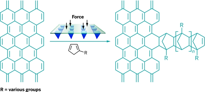
FUNCTIONAL DRAWING. Cyclopentadiene ink pushed into a graphene surface accelerates a Diels-Alder reaction.
The chemists coated a two-by-three array of 80-nm-wide polymer tips with inks containing various molecules linked to cyclopentadiene, a classic Diels-Alder reactant. For one of the inks, they attached the dye cyanine 3 to cyclopentadiene. The researchers mounted the tip array onto an atomic force microscope and gently pushed the tips into the surface of graphene. The cyclopentadienes reacted with the graphene, resulting in 20-µm × 40-µm patches of graphene decorated with a two-by-three pattern of dye dots.

The technique works at ambient temperatures and pressures, the chemists note. They are now trying to use the method to functionalize graphene with carbohydrates to make biological sensors.

Chemical & Engineering NewsISSN 0009-2347Copyright © 2013 American Chemical Society