Mostrando entradas con la etiqueta Synthetic Biology. Mostrar todas las entradas

domingo, 26 de agosto de 2018

Test Tube Artificial Neural Network Recognizes "Molecular Handwriting"

Conceptual illustration of a droplet containing an artificial neural network made of DNA that has been designed to recognize complex and noisy molecular information, represented as 'molecular handwriting.' Credit: Olivier Wyart


Test tube chemistry using synthetic DNA molecules can be utilized in complex computing tasks to exhibit artificial intelligence

Researchers at Caltech have developed an artificial neural network made out of DNA that can solve a classic machine learning problem: correctly identifying handwritten numbers. The work is a significant step in demonstrating the capacity to program artificial intelligence into synthetic biomolecular circuits.

The work was done in the laboratory of Lulu Qian, assistant professor of bioengineering. A paper describing the research (paywall) appears online on July 4 and in the July 19 print issue of the journal Nature.

"Though scientists have only just begun to explore creating artificial intelligence in molecular machines, its potential is already undeniable," says Qian. "Similar to how electronic computers and smart phones have made humans more capable than a hundred years ago, artificial molecular machines could make all things made of molecules, perhaps including even paint and bandages, more capable and more responsive to the environment in the hundred years to come."

Artificial neural networks are mathematical models inspired by the human brain. Despite being much simplified compared to their biological counterparts, artificial neural networks function like networks of neurons and are capable of processing complex information. The Qian laboratory's ultimate goal for this work is to program intelligent behaviors (the ability to compute, make choices, and more) with artificial neural networks made out of DNA.

"Humans each have over 80 billion neurons in the brain, with which they make highly sophisticated decisions. Smaller animals such as roundworms can make simpler decisions using just a few hundred neurons. In this work, we have designed and created biochemical circuits that function like a small network of neurons to classify molecular information substantially more complex than previously possible," says Qian.

To illustrate the capability of DNA-based neural networks, Qian laboratory graduate student Kevin Cherry chose a task that is a classic challenge for electronic artificial neural networks: recognizing handwriting.

Human handwriting can vary widely, and so when a person scrutinizes a scribbled sequence of numbers, the brain performs complex computational tasks in order to identify them. Because it can be difficult even for humans to recognize others' sloppy handwriting, identifying handwritten numbers is a common test for programming intelligence into artificial neural networks. These networks must be "taught" how to recognize numbers, account for variations in handwriting, then compare an unknown number to their so-called memories and decide the number's identity.

WHY DNA?
Key to creating biomolecular circuits out of DNA are the strict binding rules between molecules of DNA. A single-stranded DNA molecule is composed of smaller molecules called nucleotides—abbreviated A, T, C, and G—arranged in a string, or sequence. The nucleotides in a single-stranded DNA molecule can bond with those of another single strand to form double-stranded DNA, but the nucleotides bind only in very specific ways: An A nucleotide with a T or a C nucleotide with a G.

Taking advantage of these predictable binding rules, Qian and her colleagues can design short strands of DNA to undergo predictable chemical reactions in a test tube and thereby compute tasks, such as molecular pattern recognition. In 2011, Qian and her colleagues created the first artificial neural network made of DNA molecules that could recognize four simple patterns.

In the work described in the Nature paper, Cherry, who is the first author on the paper, demonstrated that a neural network made out of carefully designed DNA sequences could carry out prescribed chemical reactions to accurately identify "molecular handwriting." Unlike visual handwriting that varies in geometrical shape, each example of molecular handwriting does not actually take the shape of a number. Instead, each molecular number is made up of 20 unique DNA strands chosen from 100 molecules, each assigned to represent an individual pixel in any 10 by 10 pattern. These DNA strands are mixed together in a test tube.

"The lack of geometry is not uncommon in natural molecular signatures yet still requires sophisticated biological neural networks to identify them: for example, a mixture of unique odor molecules comprises a smell," says Qian.

Given a particular example of molecular handwriting, the DNA neural network can classify it into up to nine categories, each representing one of the nine possible handwritten digits from 1 to 9.

First, Cherry built a DNA neural network to distinguish between handwritten 6s and 7s. He tested 36 handwritten numbers and the test tube neural network correctly identified all of them. His system theoretically has the capability of classifying over 12,000 handwritten 6s and 7s—90 percent of those numbers taken from a database of handwritten numbers used widely for machine learning—into the two possibilities.

Crucial to this process was encoding a "winner take all" competitive strategy using DNA molecules, developed by Qian and Cherry. In this strategy, a particular type of DNA molecule dubbed the annihilator was used to select a winner when determining the identity of an unknown number.

"The annihilator forms a complex with one molecule from one competitor and one molecule from a different competitor and reacts to form inert, unreactive species," says Cherry. "The annihilator quickly eats up all of the competitor molecules until only a single competitor species remains. The winning competitor is then restored to a high concentration and produces a fluorescent signal indicating the networks' decision." 

Next, Cherry built upon the principles of his first DNA neural network to develop one even more complex, one that could classify single digit numbers 1 through 9. When given an unknown number, this "smart soup" would undergo a series of reactions and output two fluorescent signals, for example, green and yellow to represent a 5, or green and red to represent a 9.

Qian and Cherry plan to develop artificial neural networks that can learn, forming "memories" from examples added to the test tube. This way, Qian says, the same smart soup can be trained to perform different tasks.

"Common medical diagnostics detect the presence of a few biomolecules, for example cholesterol or blood glucose." says Cherry. "Using more sophisticated biomolecular circuits like ours, diagnostic testing could one day include hundreds of biomolecules, with the analysis and response conducted directly in the molecular environment."

The paper is titled "Scaling up molecular pattern recognition with DNA-based winner-take-all neural networks." Funding was provided by the National Science Foundation, the Burroughs Wellcome Fund, and the Shurl and Kay Curci Foundation.

Related:

ORIGINAL: Caltech
by Lori Dajose
07/05/2018

With Synthetic Biology Software, Geneticists Design Living Organisms From Scratch

Image: Chris Bickel
The first time geneticist Jef Boeke designed a synthetic chromosome, he sometimes wrote and edited its DNA sequence in a Microsoft Word document.

His goal was to create a slightly altered version of yeast chromosome 9, the shortest of the 16 chromosomes that make up the organism’s genome and contain all the operating instructions for life. He started with the short chromosome’s right arm, but even this task was daunting. Its DNA code consisted of 90,000 “letters,” the molecules referred to as A, C, G, and T that are arranged in particular sequence to encode biological function.

Painstakingly, Boeke went through the code, making changes that he thought would be scientifically interesting or that would make the chromosome more stable. This misery drove him to seek help from student Sarah Richardson in his neighbor Joel Bader’s lab, who wrote scripts to automate some of the most tedious steps. This was the embryonic beginning of what was to become the genome design software called BioStudio.


Once Boeke finished his design, the synthetic chromosome was constructed by taking short snippets of manufactured DNA and stringing them together. Then Boeke’s team checked the design by taking a normal yeast cell, swapping out its natural chromosome 9, and looking to see if it would keep functioning with a manmade chromosome inside. Nobody knew if it would work.

It did. The results were published in Nature in 2011, and the quest to build synthetic critters from scratch took a big step forward. Boeke’s team prepared to design the other 15 chromosomes to make a completely synthetic yeast—and the world’s first completely synthetic complex organism.

But the manual approach wasn’t scalable. The chromosome 9 project had involved 90,000 letters, a length denoted as 90 kb. The overall yeast genome was 12 million letters long, or 12Mb. “It was obvious right away that we needed something much more heavyweight,” Boeke says.

The results of their solution are now on display in the journal Science, which yesterday published seven papers from the synthetic Yeast 2.0 project. One of those papers describes their breakthrough enabling technology, the custom-built software program BioStudio.

Boeke, who leads the yeast project and serves as director of NYU’s Institute for Systems Genetics, oversaw the genome design. The papers published today describe that design process using BioStudio and also report on the completion of five new chromosomes by collaborators from around the world.


BioStudio allowed Boeke’s team to take the normal yeast genome and make the deletions, insertions, and changes they wanted, making genetic tinkering as easy as cut and paste. The program also includes a version control feature akin to Word’s track changes, recording each edit of the genome so it can easily be reversed if it’s later found to be detrimental to the yeast’s survival.

Nothing like BioStudio existed when Boeke asked Richardson to help him out. Then a PhD candidate at Johns Hopkins University (and now chief scientific officer at the synthetic biology startup MicroByre), Richardson says existing software focused on displaying long genome sequences and allowing researchers to annotate them as they laboriously figured out the purpose of various strings of DNA. When she asked around about adding an editing function to let researchers change those intricate sequences, she got shocked responses. “You would have thought I’d suggested abandoning a toddler at the mall,” she says.

Richardson worked with Boeke to create a genome editing software that was wrapped in a user-friendly web interface called Gbrowse. For a while, Boeke was the software’s only user, and he provided Richardson with plenty of frank feedback. “I’d say, it’s way too slow, it’s killing me!” he remembers. They achieved one big speed-up when they realized that every edit—even the insertion of just a few letters—was causing a cascade of updates throughout the entire genome. By localizing the update, Boeke says, the editing process got about 15 times faster. The BioDesign software makes genetic tinkering as easy as cut and paste.

Once BioStudio was fully up and running, Boeke’s team designed the full genome of what they call Sc2.0, referencing the scientific name for brewer’s yeast, Saccharomyces cerevisiae. Overall, their Sc2.0 genome design is 8 percent shorter than the original yeast genome, and it includes 1.1 Mb (or roughly a million) changes.

After finalizing this initial design, they asked collaborators around the world to take on the project of building specific chromosomes. They knew the design would continue to morph, as some of their initial changes would prove infeasible. But they also knew that all edits made by their collaborators would be captured in track changes.

The original edits came from a long list, Boeke says. “We spent something like eight months debating what changes to put on the list,” he says. “It’s fundamentally an arbitrary list of genetic changes we thought would be interesting.” But the team had to be careful not to push it too far: “We knew that with every change we made, we’d increase the risk that we’d kill the yeast,” he says.

BioStudio enabled the designers to make some major edits easily, explains Leslie Mitchell, a postdoc researcher in Boeke’s lab who took the lead on much of the genome design. With single keystrokes, she could make changes that would affect all the DNA in a chromosome. Some of these system-wide changes removed repetitive segments of DNA or took out pieces called transposons that make genomes more prone to mutation. Another added “watermarks” that would show up when the synthetic DNA was added to a normal yeast cell, making it obvious which parts of the cell were human-made.

After such broad-scale edits were done, Mitchell says, the designers could go in and look at each chromosome’s sequence in detail, making expert decisions about where they wanted to make further changes. Overall, she estimates, it took about an hour to edit 100 kb of DNA, so the 500-kb chromosome 5 took about 5 hours to design.

Richardson, the coder, remembers that the researchers had one more big ask for BioStudio, which had to do with DNA assembly. While synthetic biology companies now make it easy to order custom strings of manufactured DNA, those strings are typically fairly short. For the synthetic yeast project, the researchers would order strings of DNA that were only about 70 letters, or base-pairs, long. When those strings arrived in the lab, the researchers first assembled them into “building blocks” of about 750 bp, then put those building blocks together into into 2-4 kb “minichunks,” then constructed 10 kb “chunks,” and finally built 30-60 kb “megachunks.” Synthetic biology lends itself to engineering’s classic “design-build-test” cycle.

But there are genetic constraints on how strings of DNA can be assembled. The researchers wanted BioStudio to take any long DNA sequence and make it “modular,” chopping it up into pieces that could be ordered from the DNA-makers and then patched together in that series of assembly steps. “They wanted to be able to push a button when they were done with their edits, and have the genome slot itself into an assembly pattern,” Richardson remembers. “That was the craziest thing they asked for.”

Synthetic biology lends itself to engineering’s classic “design-build-test” cycle. For Sc2.0, megachunks of the designer chromosomes were built and inserted into normal yeast cells to test whether they interfered with its life functions. If the yeast cell died or displayed abnormal behavior, the researchers embarked on a debugging process.

In one type of debugging, they would make many yeast colonies with many different combinations of synthetic megachunks and watch to see which colonies failed, then look for the common denominator in those failures. Mitchell, who led the work on designing and debugging chromosome 6, explains that there were different sorts of bugs. The most interesting were those that arose from genome changes they’d made that they expected to be harmless—because those bugs taught the researchers something about yeast biology.

Boeke says that so far, the team has found a bug in their genome design roughly every 300 kb. “But there may be more, we may not have found them all yet!” he says. With most of the synthetic yeast chromsomes still under construction, he’s still expecting surprises. “It’s like when you release code and wait for the user feedback,” he says. Geneticists need software to help them explore this new design frontier: the design of life itself.

The synthetic yeast project is on track to complete all 16 chromosomes by the end of 2017. Then the team will turn to the task of putting all the chromosomes into a single cell, and seeing if it still functions as a yeast cell should. That process may yield still more bugs, Mitchell says. “It might be that individual changes on two chromosomes are well tolerated, but they don’t work when you put them together,” she says. “We may potentially have to track bugs across chromosomes.”

While BioStudio has been invaluable for the synthetic yeast project, the researchers aren’t sure whether it will be useful for other synthetic biology projects. “If you want to make the kinds of changes we made for yeast, it’s very straightforward,” says Mitchell, “but for other types of changes you’d have to write the code.” The software is open source, she notes, so interested parties could build on it.

Whether it’s BioStudio or another program, the fast-growing field of synthetic biology will need software to help geneticists explore this new design frontier: the design of life itself.

Some synthetic biology startups are trying to adapt simple organisms like yeast to make them produce useful products, such as biofuels, vaccines, or even perfume. Other researchers are more interested in constructing whole critters from scratch, in hopes of gaining new insights into the mechanics of life in the process.

The first completely synthetic genome was bacterial, constructed at the J. Craig Venter Institute in 2010; its single-chromosome measured 1 Mb in length. From that start, the 12-Mb yeast genome marks a big step up. And Boeke is part of a group that has proposed to scale up considerably from the single-celled yeast. Last June they called for the creation of a synthetic human genome as part of a massive project to develop DNA assembly technology; they published an article in the journal Science that suggested a $100 million investment to get the project off the ground.

The human genome clocks in at 3 billion letters, or 3 Gb. To tackle that project, genome designers and coders may have to get together for a Synthetic Bio Hackathon.
Learn More BioDesigngenomesoftwaresynthetic biologyyeast

ORIGINAL: IEEE Spectrum
By Eliza Strickland
Posted 10 Mar 2017 | 20:30 GMT

miércoles, 29 de noviembre de 2017

Semi-Synthetic Life Form Now Fully Armed and Operational

WILLIAM B. KIOSSES, PHD, THE SCRIPPS RESEARCH INSTITUTE

Could life have evolved differently? A germ with “unnatural” DNA letters suggests the answer is yes.

E. coli bacteria with an expanded genetic code could help manufacture new drugs.

Every living thing on Earth stores the instructions for life as DNA, using the four genetic bases A, G, C, and T.

All except one, that is.

In the San Diego laboratory of Floyd Romesberg—and at a startup he founded—grow bacteria with an expanded genetic code. They have two more letters, an “unnatural” pair he calls X and Y.

Romesberg, head of a laboratory at the Scripps Research Institute, first amended the genes of the bacterium E. Coli to harbor the new DNA components in 2014. Now, for the first time, the germs are using their expanded code to manufacture proteins with equally unusual components.

“We wanted to prove the concept that every step of information storage and retrieval could be mediated by an unnatural base pair,” he says. “It’s not a curiosity anymore.”

The bacterium is termed a “semi-synthetic” organism, since while it harbors an expanded alphabet, the rest of the cell hasn’t been changed. Even so, Peter Carr, a biological engineer at MIT’s Lincoln Laboratory, says it suggests that scientists are only beginning to learn how far life can be redesigned, a concept known as synthetic biology.

“We don’t know what the ultimate limits are on our ability to engineer living systems, and this paper helps show we’re not limited to four bases,” he says. “I think it’s pretty impressive.”

Humankind has been disappointed in the quest to find life on Mars or Jupiter. Yet the alien germs growing in San Diego already hint that our Earth biology isn’t the only one possible. “It suggests that if life did evolve elsewhere, it might have done so using very different molecules or different forces,” says Romesberg. “Life as we know it is may not be the only solution, and may not be the best one.”

Romesberg’s efforts to lay a genetic cuckoo’s egg inside bacteria started 15 years ago. After creating a candidate pair of new genetic letters, the first step was to add them to a bacterium’s genome and show it could use them to store information. That is, could the organism abide by the unnatural DNA and also copy it faithfully as it divided?

The answer, his lab showed in 2014, was yes. But early versions of the bacteria were none too healthy. They died or got rid of the extra letters in their DNA, which are stored in a mini-chromosome called a plasmid. In Romesberg’s words, his creations “lacked the fortitude of real life.”

By this year, the team had devised a more stable bacterium. But it wasn’t enough to endow the germ with a partly alien code—it needed to use that code to make a partly alien protein. That’s what Romesberg’s team, reporting today in the journal Nature, says it has done.

Using the extra letters, they instructed bacteria to manufacture a glowing green protein that has in it a single unnatural amino acid. “We stored information, and now we retrieved it. The next thing is to use it. We are going to do things no one else can,” says Romesberg.

The practical payoff of an organism with a bigger genetic alphabet is that it has a bigger vocabulary—it can assemble proteins with components not normally found in nature. That could solve some tricky problems in medicinal chemistry, which is the art of shaping molecules so they do exactly what’s wanted in the body, and nothing that isn’t.

Pursuing such aims is a startup Romesberg founded, named Synthorx. It has raised $16 million so far and hopes to turn the science into new drugs. One project aims to make a new version of interleukin-2, an anticancer drug with some nasty side effects. Maybe the semi-synthetic germs could fix that by swapping in some unusual components at key points. “This company needs to get out of the lab and into the clinic,” says its newly installed CEO, Laura Shawver.

Carr says an expanded genetic code could have implications beyond providing a shortcut for programming new properties into proteins. He also thinks the new letters might be used to hide information in ways other biologists couldn’t easily see. That could be useful in concealing intellectual property or, perhaps, to disguise a bioweapon.

Synthorx Inc 2015


Credit: William B. Kiosses, PhD, The Scripps Research Institute


November 29, 2017
William B. Kiosses, PhD, The Scripps Research Institute

sábado, 21 de octubre de 2017

New Research Points to a Genetic Switch That Can Let Our Bodies Talk to Electronics


Shutterstock
IN BRIEF
Our bodies are biologically based and therefore are not equipped to communicate with electronics efficiently. New research could make it possible to genetically engineer our cells to be able to communicate with electronics.

The development has the potential to allow us to eventually build apps that autonomously detect and treat disease.

Microelectronics has transformed our lives. Cellphones, earbuds, pacemakers, defibrillators – all these and more rely on microelectronics’ very small electronic designs and components. Microelectronics has changed the way we collect, process and transmit information.

Such devices, however, rarely provide access to our biological world; there are technical gaps. We can’t simply connect our cellphones to our skin and expect to gain health information. For instance, is there an infection? What type of bacteria or virus is involved? We also can’t program the cellphone to make and deliver an antibiotic, even if we knew whether the pathogen was Staph or Strep. There’s a translation problem when you want the world of biology to communicate with the world of electronics.

The research we’ve just published with colleagues in Nature Communications brings us one step closer to closing that communication gap.
Electronic control of gene expression and cell behaviour in Escherichia coli through redox signalling

ABSTRACT:
The ability to interconvert information between electronic and ionic modalities has transformed our ability to record and actuate biological function. Synthetic biology offers the potential to expand communication ‘bandwidth’ by using biomolecules and providing electrochemical access to redox-based cell signals and behaviours. While engineered cells have transmitted molecular information to electronic devices, the potential for bidirectional communication stands largely untapped. Here we present a simple electrogenetic device that uses redox biomolecules to carry electronic information to engineered bacterial cells in order to control transcription from a simple synthetic gene circuit. Electronic actuation of the native transcriptional regulator SoxR and transcription from the PsoxS promoter allows cell response that is quick, reversible and dependent on the amplitude and frequency of the imposed electronic signals. Further, induction of bacterial motility and population based cell-to-cell communication demonstrates the versatility of our approach and potential to drive intricate biological behaviours.

Source: NATURE COMMS
Rather than relying on the usual molecular signals, like hormones or nutrients, that control a cell’s gene expression, we created a synthetic “switching” system in bacterial cells that recognizes electrons instead. This new technology – a link between electrons and biology – may ultimately allow us to program our phones or other microelectronic devices to autonomously detect and treat disease.

COMMUNICATING WITH ELECTRONS, NOT MOLECULES
One of the barriers scientists have encountered when trying to link microelectronic devices with biological systems has to do with information flow. In biology, almost all activity is made possible by the transfer of molecules like
  • glucose, 
  • epinephrine, 
  • cholesterol and 
  • insulin 
signaling between cells and tissues. Infecting bacteria secrete molecular toxins and attach to our skin using molecular receptors. To treat an infection, we need to detect these molecules to identify the bacteria, discern their activities and determine how to best respond.

Microelectronic devices don’t process information with molecules. A microelectronic device typically has silicon, gold, chemicals like boron or phosphorus and an energy source that provides electrons. By themselves, they’re poorly suited to engage in molecular communication with living cells.

Free electrons don’t exist in biological systems so there’s almost no way to connect with microelectronics. There is, however, a small class of molecules that stably shuttle electrons. These are called “redox” molecules; they can transport electrons, sort of like wire does. The difference is that in wire, the electrons can flow freely to any location within; redox molecules must undergo chemical reactions – oxidation or reduction reactions – to “hand off” electrons.
Bacteria are engineered to respond to a redox molecule activated by an electrode by creating an electrogenetic switch. Bentley and Payne, CC BY-ND

TURNING CELLS ON AND OFF
Capitalizing on the electronic nature of redox molecules, we genetically engineered bacteria to respond to them. We focused on redox molecules that could be “programmed” by the electrode of a microelectronic device. The device toggles the molecule’s oxidation state – it’s either 
  • oxidized (loses an electron) or 
  • reduced (gains an electron). 
The electron is supplied by a typical energy source in electronics like a battery.

We wanted our bacteria cells to turn “on” and “off” due to the applied voltage – voltage that oxidized a naturally occurring redox molecule, pyocyanin.

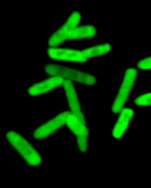
Electrically oxidizing pyocyanin allowed us to control our engineered cells, turning them on or off so they would synthesize (or not) a fluorescent protein. We could rapidly identify what was happening in these cells because the protein emits a green hue.
(a) Device-mediated electronic input consists of applied potential (blue or red step functions) for controlling the oxidation state of redox-mediators (transduced input). Redox mediators intersect with cells to actuate transcription and, depending on actuated gene-of-interest, control biological output.
(b) The electrogenetic device consists of the region encompassing the gene coding for the SoxR protein and the divergent overlapping PsoxR/PsoxS promoters. A gene of interest is placed downstream of the PsoxS promoter. Pyo (O) initiates gene induction and Fcn(R/O), through interactions with respiratory machinery, allows electronic control of induction level. Fcn (R/O), ferro/ferricyanide; Pyo, pyocyanin. The oxidation state of both redox mediators is colorimetrically indicated (Fcn (O) is yellow pentagon; Fcn (R) is white pentagon; Pyo (O) is blue hexagon; Pyo (R) is grey hexagon). Encircled ‘e−‘ and arrows indicate electron movement.

In another example
, we made bacteria that, when switched on, would swim from a stationary position. Bacteria normally swim in starts and stops referred to as a “run” or a “tumble.” The “run” ensures they move in a straight path. When they “tumble,” they essentially remain in a one spot. A protein called CheZ controls the “run” portion of bacteria’s swimming activity. Our electrogenetic switch turned on the synthesis of CheZ, so that the bacteria could move forward.
Bacteria can naturally join forces as biofilms and work together. CDC/Janice Carr, CC BY
We were also able to electrically signal a community of cells to exhibit collective behavior. We made cells with switches controlling the synthesis of a signaling molecule that diffuses to neighboring cells and, in turn, causes changes in their behavior. Electric current turned on cells that, in turn, “programmed” a natural biological signaling process to alter the behavior of nearby cells. We exploited bacterial quorum sensing – a natural process where bacterial cells “talk” to their neighbors and the collection of cells can behave in ways that benefit the entire community.

Perhaps even more interesting, our groups showed that we could both turn on gene expression and turn it off. By reversing the polarity on the electrode, the oxidized pyocyanin becomes reduced – its inactive form. Then, the cells that were turned on were engineered to quickly revert back to their original state. In this way, the group demonstrated the ability to cycle the electrically programmed behavior on and off, repeatedly.

Interestingly, the on and off switch enabled by pyocyanin was fairly weak. By including another redox molecule, ferricyanide, we found a way to amplify the entire system so that the gene expression was very strong, again on and off. The entire system was robust, repeatable and didn’t negatively affect the cells.

SENSING AND RESPONDING ON A CELLULAR LEVEL
Armed with this advance, devices could potentially electrically stimulate bacteria to make therapeutics and deliver them to a site. For example, imagine swallowing a small microelectronic capsule that could record the presence of a pathogen in your GI tract and also contain living bacterial factories that could make an antimicrobial or other therapy – all in a programmable autonomous system.

This current research ties into previous work done here at the University of Maryland where researchers had discovered ways to “record” biological information, by sensing the biological environment, and based on the prevailing conditions, “write” electrons to devices. We and our colleagues “sent out” redox molecules from electrodes, let those molecules interact with the microenvironment near the electrode and then drew them back to the electrode so they could inform the device on what they’d seen. This mode of “molecular communication” is somewhat analogous to sonar, where redox molecules are used instead of sound waves.

These molecular communication efforts were used to identify pathogens, monitor the “stress” in blood levels of individuals with schizophrenia and even determine the differences in melanin from people with red hair. For nearly a decade, the Maryland team has developed methodologies to exploit redox molecules to interrogate biology by directly writing the information to devices with electrochemistry.

Perhaps it is now time to integrate these technologies:

  • Use molecular communication to sense biological function and transfer the information to a device. 
  • Then use the device – maybe a small capsule or perhaps even a cellphone – to program bacteria to make chemicals and other compounds that issue new directions to the biological system. 

It may sound fantastical, many years away from practical uses, but our team is working hard on such valuable applications…stay tuned!

ORIGINAL: Futurism

martes, 28 de febrero de 2017

Microfluidic LEGO bricks put biomedical research in the hands of the masses


3D printed master molds have been used to create microfluidic LEGO bricks that facilitate the study of liquid flow for medical research. The LEGO brick method is being explored by the Department of Biomedical Engineering at the University of California, Irvine, with findings published in the Journal of Micromechanics and Microengineering, January 2017.

What is microfluidics?
Microfluidics is the manipulation and study of sub-microscopic litres of liquid. In a device such as the University of California’s LEGO bricks, liquids are channelled through empty vessels spanning no more than 500 μm (microns, for comparison: a human hair is 50 μm in diameter).

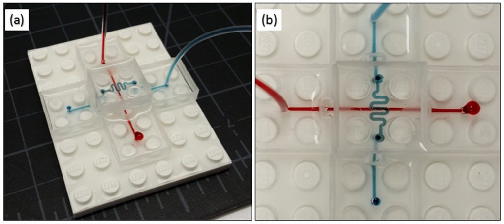
Testing the flow of liquids through the LEGO bricks with colored inks. Image via: Kevin Vittayarukskul and Abraham Phillip Lee

The way a liquid behaves during a flow, and when mixed with other nanoliquids tells researchers certain things about its biological behaviour, Microfludics can also be controlled in a way to produce autonomous movement, as in the example of Harvard University’s soft-robotic Octobot.


Moving .gif shows Harvard’s Octobot that harnesses microfluidic principles to move. Clip via: @NatureNews

In biomedical research microfluidic chips are used to conduct assays that test the reactions between substances, as in lab-on-a-chip technology. Using these devices is preferable to some traditional assay methods as the microscale parts consume less time and resources. California’s LEGO bricks seek to promote these qualities by providing more recognisable devices that are also capable of being mass produced.

Making the microfluidic LEGO bricks
Kevin Vittayarukskul and Professor Abraham Lee’s approach uses Autodesk’s AutoCAD software to first design blocks with an embedded microfluidic channel.

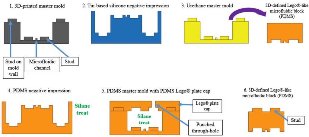
From this a mold is also designed, and then 3D printed on a Perfactory 3 Mini 3D printer by EnvisionTEC that uses the DLP method of vat polymerisation to cure the material. PDMS, (Polydimethylsiloxane) a silicone-based polymer is then used to cast the LEGO bricks.
Process of making the microfluidic LEGO mold. Image via: Kevin Vittayarukskul and Abraham Phillip Lee

The properties of PDMS make it naturally transparent and biocompatible, which is ideal for this kind of research. It is also known for its ability to exactly match the shape of a cast into which it is poured, meaning that none of the DLP 3D printed quality is lost on the final cast.

A microfluidic LEGO kit?
The advantages of being able to stack the microfluidic blocks is that researchers can combine even more channels into a single space. It also allows easy assembly of varying channels, i.e. one straight vessel in to a winding one.

Using a tried and tested building block such as a LEGO brick also means that it has great potential for mass-production. The case with medical research is that it often isn’t accessible by other scholars that could make use of the technology. But a microfluidic LEGO kit could be just the ticket to encourage future biomedical research.

Speaking to EE Times Europe A truly LEGO®-like modular microfluidics platform co-author Professor Abraham Lee explains:
The main goal of this project was to train and educate the next generation of microfluidic developers and researchers. By using actual LEGO’s as the building block and assembly platform, our hope was to attract students as early as young as high schoolers to be interested in the field, learn about microfluidics and stimulate their imagination for new products for applications over a very wide range.
Testing the flow of liquids through the LEGO bricks with colored inks. Image via: Kevin Vittayarukskul and Abraham Phillip Lee


A truly Lego®-like modular microfluidics platform
Kevin Vittayarukskul
and Abraham Phillip Lee
  • Published 24 January 2017 • © 2017 IOP Publishing Ltd
Journal of Micromechanics and Microengineering, Volume 27, Number 3
Author e-mails
Author affiliations
  • Department of Biomedical Engineering, University of California, Irvine, CA, USA
Dates
  • Received 11 October 2016
  • Accepted 15 December 2016
  • Published 24 January 2017
Citation
  • Kevin Vittayarukskul and Abraham Phillip Lee 2017 J. Micromech. Microeng. 27 035004  
DOI: https://doi.org/10.1088/1361-6439/aa53ed

ORIGINAL: 3DPrintingIndustry

Beau Jackson Writer based in London, originally from Yorkshire. Fan of lab-on-a-chip technology, microfluidics, scanning, tech-inspired art and 3D Benchy.
January 25, 2017
 

martes, 24 de enero de 2017

TSRI Scientists Create First Stable Semisynthetic Organism


LA JOLLA, CA – January 23, 2017 – Life’s genetic code has only ever contained four natural bases. These bases pair up to form two “base pairs”—the rungs of the DNA ladder—and they have simply been rearranged to create bacteria and butterflies, penguins and people. Four bases make up all life as we know it.

Until now. Scientists at The Scripps Research Institute (TSRI) have announced the development of the first stable semisynthetic organism. Building on their 2014 study in which they synthesized a DNA base pair, the researchers created a new bacterium that uses the four natural bases (called A, T, C and G), which every living organism possesses, but that also holds as a pair two synthetic bases called X and Y in its genetic code.

TSRI Professor Floyd Romesberg and his colleagues have now shown that their single-celled organism can hold on indefinitely to the synthetic base pair as it divides. Their research was published January 23, 2017, online ahead of print in the journal Proceedings of the National Academy of Sciences.

“We’ve made this semisynthetic organism more life-like,” said Romesberg, senior author of the new study.

While applications for this kind of organism are still far in the future, the researchers say the work could be used to create new functions for single-celled organisms that play important roles in drug discovery and much more.

Building a Unique Organism
When Romesberg and his colleagues announced the development of X and Y in 2014, they also showed that modified E. coli bacteria could hold this synthetic base pair in their genetic code. What these E. coli couldn’t do, however, was keep the base pair in their code indefinitely as they divided. The X and Y base pair was dropped over time, limiting the ways the organism could use the additional information possessed in their DNA.

“Your genome isn’t just stable for a day,” said Romesberg. “Your genome has to be stable for the scale of your lifetime. If the semisynthetic organism is going to really be an organism, it has to be able to stably maintain that information.”

Romesberg compared this flawed organism to an infant. It had some learning to do before it was ready for real life.

In stepped TSRI Graduate Student Yorke Zhang and Brian Lamb, an American Cancer Society postdoctoral fellow in the Romesberg lab at the time of the study. Together, they helped develop the means for the single-celled organism to retain the artificial base pair.

First, Zhang and Lamb, co-first authors of the study, optimized a tool called a nucleotide transporter, which brings the materials necessary for the unnatural base pair to be copied across the cell membrane. “The transporter was used in the 2014 study, but it made the semisynthetic organism very sick,” Zhang explained. The researchers discovered a modification to the transporter that alleviated this problem, making it much easier for the organism to grow and divide while holding on to X and Y.

Next, the researchers optimized their previous version of Y. The new Y was a chemically different molecule that could be better recognized by the enzymes that synthesize DNA molecules during DNA replication. This made it easier for cells to copy the synthetic base pair.

A New Use for CRISPR-Cas9
Finally, the researchers set up a “spell check” system for the organism using CRISPR-Cas9, an increasingly popular tool in human genome editing experiments. But instead of editing a genome, the researchers took advantage of CRISPR-Cas9’s original role in bacteria.

The genetic tools in CRISPR-Cas9 (a DNA segment and an enzyme) originated in bacteria as a kind of immune response. When a bacterium encounters a threat, like a virus, it takes fragments of the invader genome and pastes them into its own genome—a bit like posting a “wanted” poster on the off chance it sees the invader again. Later, it can use those pasted genes to direct an enzyme to attack if the invader returns.

Knowing this, the researchers designed their organism to see a genetic sequence without X and Y as a foreign invader. A cell that dropped X and Y would be marked for destruction, leaving the scientists with an organism that could hold on to the new bases. It was like the organism was immune to unnatural base pair loss.

“We were able to address the problem at a fundamental level,” said Lamb, who now serves as a research scientist at Vertex Pharmaceuticals.

Their semisynthetic organism was thus able to keep X and Y in its genome after dividing 60 times, leading the researchers to believe it can hold on to the base pair indefinitely.

“We can now get the light of life to stay on,” said Romesberg. “That suggests that all of life’s processes can be subject to manipulation.”

A Foundation for Future Research
Romesberg emphasized that this work is only in single cells and is not meant to be used in more complex organisms. He added that the actual applications for this semisynthetic organism are “zero” at this point. So far, scientists can only get the organism to store genetic information.

Next, the researchers plan to study how their new genetic code can be transcribed into RNA, the molecule in cells needed to translate DNA into proteins. “This study lays the foundation for what we want to do going forward,” said Zhang.
Professor Floyd Romesberg (right) and Graduate Student Yorke Zhang led the new study at The Scripps Research Institute, along with Brian Lamb (not pictured).(Photo by Madeline McCurry-Schmidt.)

Additional authors of the study, “A semisynthetic organism engineered for the stable expansion of the genetic alphabet,” were Aaron W. Feldman and Anne Xiaozhou Zhou of TSRI; Thomas Lavergne of the University of Grenoble; and Lingjun Li of Henan Normal University.

The study was supported by the National Institutes of Health (grant GM060005), a National Science Foundation Graduate Research Fellowship (grant DGE-1346837), the National Natural Science Foundation of China (grant 21472036), a Labex ARCANE grant (ANR-11-LABX-0003-01), NanoBio-ICMG platforms (FR 2607) and a postdoctoral fellowship from the American Cancer Society, Illinois Division.

About The Scripps Research Institute
The Scripps Research Institute (TSRI) is one of the world's largest independent, not-for-profit organizations focusing on research in the biomedical sciences. TSRI is internationally recognized for its contributions to science and health, including its role in laying the foundation for new treatments for cancer, rheumatoid arthritis, hemophilia, and other diseases. An institution that evolved from the Scripps Metabolic Clinic founded by philanthropist Ellen Browning Scripps in 1924, the institute now employs more than 2,500 people on its campuses in La Jolla, CA, and Jupiter, FL, where its renowned scientists—including two Nobel laureates and 20 members of the National Academies of Science, Engineering or Medicine—work toward their next discoveries. The institute's graduate program, which awards PhD degrees in biology and chemistry, ranks among the top ten of its kind in the nation. In October 2016, TSRI announced a strategic affiliation with the California Institute for Biomedical Research (Calibr), representing a renewed commitment to the discovery and development of new medicines to address unmet medical needs. For more information, see www.scripps.edu.

# # #

For information:
Office of Communications 
Tel: 858-784-2666 
Fax: 858-784-8118 

martes, 29 de noviembre de 2016

The Robot Revolution Comes to Synthetic Biology

Automation allows thousands of possibilities when building weird new organisms

Photo: Ginkgo Bioworks Robots at Work: Ginkgo Bioworks’ lab automates genetic engineering.
Last month, synthetic biologists at Ginkgo Bioworks raised their glasses—filled with genetically modified beer—to cele­brate the launch of a new automated lab. By applying engineering principles to biology, and with the help of some nifty robotic equipment, Ginkgo has created a factory for churning out exotic life-forms, the likes of which have never before been seen on this planet.

The home brew they were drinking was an example of the potential applications of synthetic biology, a new field that builds on recent progress in genetic assembly methods. Scientists can now manufacture snippets of synthetic DNA and slip them into organisms, giving those critters strange capabilities. For example, the brewer’s yeast used to make the beer for the launch party had genes from an orange tree added to its own DNA. During the fermentation stage of the brewing process, those genes caused the yeast to produce ­valencene, an organic compound with a citrusy flavor. Speaking scientifically, it was delicious.

Ginkgo Bioworks, a hip young company based in Boston, recently raised US $100 million on the promise of finding many such useful applications for synthetic biology. It used some of that cash to build Bioworks2, the company’s vast new lab that uses robotic systems to form an assembly line for organisms.

Ginkgo needs to make microbes on a grand scale in order to find those that can function as tiny biological factories for its customers. Many of the altered organisms will be duds, but through highly organized trial and error, the bioengineers will eventually devise a microbe that turns out a desired substance—like a chemical ingredient used for perfumes, beverages, pesticides, or laundry detergents.

The company’s business model centers on the microbes themselves, not the end products. “We’re not in the business of manufacturing chemicals, flavors, or fragrances,” explains Ginkgo creative director Christina Agapakis. “We specialize in the organisms, and we partner with our customers, who will make the product.” Ginkgo licenses organisms to its customers, she says, and gets royalties if they’re used.

But building an organism to spec is no easy task. Genetics still isn’t well understood; there’s no universal catalog of genes that details each one’s characteristics. Even if researchers know what a particular gene does in an orange tree, for example, when they add it to a yeast cell, it might interact with the native DNA in unexpected ways. If they’re adding several genes from different species to that yeast cell, things get even more complicated.

That’s why Ginkgo takes an engineering approach to biology, applying a rigorous design-build-test cycle to the creation of living organisms. The new lab’s extreme automation is critical to this approach, says Patrick Boyle, ­Ginkgo’s head of organism design. “In grad school, I might have taken my five best ideas and tried them out,” Boyle says. “Here we take our 1,000 best ideas, try them all out, and see which works best.”


Engineering Life
How Ginkgo Bioworks Makes Customized Critters
1: DESIGN Organism designers choose genes from a wide range of animals and plants and combine them in hundreds of different ways. Each mashup is a unique fragment of DNA. Illustration: Erik Vrielink
Design: Ginkgo designers search the scientific literature looking for genes that would cause the yeast to produce useful enzymes. The aim: When the designers feed sugar to the yeast, these enzymes should cause it to carry out chemical reactions that ultimately result in rose oil. But there is a dizzying array of genes and enzymes to consider. “If you have 100 possible enzymes that can serve as a step in a four-step pathway, that’s a lot of design space to explore,” Boyle says.

2: BUILD Robotic instruments take all these fragments and insert them into separate yeast cells. They’re integrated into the cells’ DNA and can change how the yeast function. Illustration: Erik Vrielink
Build: Ginkgo outsources the actual manufacturing of synthetic DNA. When a batch of manufactured DNA arrives at Ginkgo, liquid-handling robots build the new organisms by adding the various snippets to yeast cells. “During my Ph.D., I spent a lot of time moving tiny amounts of fluid around,” says creative director Agapakis. “When we started Ginkgo, a lot of the robots looked like eight-armed grad students—there were a lot of pipettes.” The process has ramped up as these robots have gotten more capable. Ginkgo now has liquid-handling robots that quickly move nanoliters of fluid using targeted pulses of sound.

3: TEST Ginkgo’s customers want the altered yeast to churn out specific products. Spectrometry machines analyze all the yeast cells to see if any are producing the desired output. Illustration: Erik Vrielink
Test: Once the robots have created a thousand yeast variants containing different mashups of genes, it’s time to see if the cells are making their rose oil. Mass spectrometry machines crack open the cells and examine all the molecules inside, checking for the product and also determining whether the yeast is healthy. But success on both counts doesn’t necessarily mean the organism will meet the customer’s needs. Boyle says that in the case of rose oil, Gingko studies each yeast’s overall “fragrance profile.” While a cell may be making certain useful fragrance molecules, it may be making others that are not. “I like the fresh-baked-bread smell, but it’s not great when you’re trying to sell a perfume,” Boyle says.

4: SCALE UP Ginkgo puts its best producers into bioreactors to see how they fare at industrial scale. Lessons learned help the organism designers make choices for the next round of experiments. Illustration: Erik Vrielink
Scale Up: Ginkgo adds one extra step to the typical engineering cycle, since a modified yeast cell that looks like a winner in the lab might not perform as well in the customer’s fermentation vats. In one corner of the lab, robotic systems fill and monitor rows of benchtop bioreactors, using a variety of sensors to watch the processes inside.

To understand how this works in practice, take Ginkgo’s first efforts in the perfume business. The company is working with the French fragrance maker Robertet on a yeast that spits out rose oil, because extracting the substance from rose petals is expensive.

If even their best oil-producing yeast isn’t up to spec, the company’s organism designers go back to the drawing board, using the results of the experiment to inform their next 1,000 best guesses. One of these days, Ginkgo’s bioengineers say, they’ll make the perfect batch—one that comes out smelling like a rose.


ORIGINAL: IEEE Spectrum
Posted 29 Nov 2016

lunes, 10 de octubre de 2016

IChemE Global Awards 2016 Finalist - 'Light-activated synthetic tissues', University of Oxford, UK

The University of Oxford has engineered new high-value synthetic tissues, controllable with an external stimulus. ‘Bottom-up’ approaches in synthetic biology have been used to construct synthetic cells from simple biological components. By using a droplet-based 3D printer synthetic tissues are created, comprising hundreds of communicating synthetic cells, which can perform sophisticated functions such as protein synthesis. In addition, it has shown that 3D-printed synthetic tissues can be controlled externally by light and demonstrate electrical communication, similar to neuronal transmission. Printed synthetic tissues might be used in medicine and could even interface directly with living tissues.

University of Oxford is an IChemE Global Awards finalist for the Biotechnology Award. 

“We are delighted that our project has been chosen as a finalist for the IChemE Global Biotechnology Award. These synthetic tissues, namely materials analogous to biological tissues, can be remotely activated by light and demonstrate rapid electrical signalling through defined pathways, precisely the role of neurons. Our goal is to use these completely new and high value 3D-printed materials in medicinal applications, including drug delivery and tissue replacement.” 
- Michael Booth, Junior Research Fellow, University of Oxford


ORIGINAL: IChemE
Oct 10, 2016