Mostrando entradas con la etiqueta Biocomputing. Mostrar todas las entradas

sábado, 20 de agosto de 2016

Scientists Built a Biological Computer Inside a Cell

Add caption
MIT engineers have developed biological computational circuits capable of both remembering and responding to sequential input data.

The group's work, which is described in this week's issue of Science, represents a critical step in the progression of synthetic biology with the integration of DNA-based memory, in particular, pointing the way toward building large computational systems from biological components—computing devices that are living cells—and, ultimately, programming complex biological functions.

More specifically, Nathaniel Roquet and colleagues at MIT's Synthetic Biology Group were able to implement within a living cell what's known as a state machine: an abstract mathematical model describing computation as a list of of distinct internal states paired with an associated list of operations (or machine inputs) required to transition from state to state. So: a new state is always the result of an old state taken in combination with new inputs (history matters). State machines happen to describe a very large number of different things, from natural language processing algorithms to neurological systems to something as simple as a vending machine.

In a living cell, DNA is the natural candidate for storing state information. After all, that's what DNA does: store information. What Roquet and co. have created is a framework for chemically manipulating DNA such that states are encoded in DNA sequences. As a storage mechanism, this allows for both conveniently reading out a given state via genetic sequencing and also regulating gene expression via state transitions. In other words, the states can be linked to cellular behavior. The DNA serves as the memory for the state machine. The rest is in how, specifically, the DNA is manipulated and what effect that has on cellular behavior.

This could mean integrating biological state machines into tumor models, where they may be used to genetically surveil the activation of genes that may cause cancer

In their experiments, Roquet and co. programmed E. coli cells to react to several substances commonly used in biological laboratory experiments, including an analogue of the antibiotic tetracycline, a sugar called arabinose, and a chemical called DAPG that helps plants protect their roots from pathogens. The cells could be reprogrammed to other inputs as needed, however.

The actual cell behavior being programmed by the researchers was the expression of genes coding for the production of different fluorescent proteins representing different colors. With three different inputs they were able to produce 16 different combinations of colors.

"Synthetic state machines that record and respond to sequences of signaling and gene regulatory events within a cell could be transformative tools in the study and engineering of complex living systems," Roquet writes. In other words, by implementing a state machine (a computer) in a living cell, it's possible to use that state machine to surveil otherwise impossible-to-observe cellular happenings.

For example, progenitor cells (similar to stem cells) develop into differentiated cell types with specific functions thanks to transcription factors, proteins that help regulate gene expression in cells. Transcription factors have allowed researchers to program both progenitor cells to become certain specific types of functional cells—and also to do the opposite, programming functional cells to behave as undifferentiated cells. However, much about the process remains mysterious. A state machine that could record the DNA transitions resulting from TF activation could go a long way toward not only understanding these processes, but manipulating them as well. 

The circuits in the biological state machine are dependent on enzymes called recombinases. These enzymes are activated by various inputs into a cell, such as chemical signals, and act to tweak that cell's DNA. But the tweak that actually occurs depends on the orientation of two DNA sequences known as recognition sites. The important thing is that the effect of changing any two recognition sites (the resulting cellular behavior) depends on how other recognition sites have been altered previously. Hence, memory.

There's really no shortage of potential applications here. The example Roquet gives is in integrating biological state machines into tumor models, where they may be used to genetically surveil the activation of oncogenes (genes that may cause cancer) and deactivation of tumor suppression mechanisms in individual cells.

"This idea that we can record and respond to not just combinations of biological events but also their orders opens up a lot of potential applications," Roquet offers in a statement. "A lot is known about what factors regulate differentiation of specific cell types or lead to the progression of certain diseases, but not much is known about the temporal organization of those factors. That's one of the areas we hope to dive into with our device."

Computers have become "alive," but perhaps not in the way that many of us anticipated. A unicellular organism itself won't ever be packing much computational horsepower, but considered as a building block, the potential is pretty wild.

ORIGINAL: Vice
by MICHAEL BYRNE EDITOR 
July 21, 2016

jueves, 9 de junio de 2016

Physicists confirm (what biologists have already konwn for years) there's a second layer of information hidden in our DNA

Liya Graphics/Shutterstock.com
Theoretical physicists have confirmed that it's not just the information coded into our DNA that shapes who we are - it's also the way DNA folds itself that controls which genes are expressed inside our bodies.

That's something biologists have known for years, and they've even been able to figure out some of the proteins responsible for folding up DNA. But now a group of physicists have been able to demonstrate for the first time through simulations how this hidden information controls our evolution.

Let's back up for a second here, because although it's not necessarily news to many scientists, this second level of DNA information might not be something you're familiar with.

As you probably learnt in high school, Watson and Crick discovered in 1953 that the DNA code that determines who we are is made up of a sequence of the letters G, A, C, and T. 

The order of these letters determines which proteins are made in our cells. So, if you have brown eyes, it's because your DNA contains a particular series of letters that encodes for a protein that makes the dark pigment inside your iris.

But that's not the whole story, because all the cells in your body start out with the exact same DNA code, but every organ has a very different function - your stomach cells don't need to produce the brown eye protein, but they do need to produce digestive enzymes. So how does that work?

Since the '80s, scientists have found that the way DNA is folded up inside our cells actually controls this process. Environmental factors can play a big role in this process too, with things like stress known to turn certain genes on and off through something known as epigenetics.

But the mechanics of the DNA folding is the original control mechanism. That's because every single cell in our body contains around 2 metres of DNA, so to fit inside us, it has to be tightly wrapped up into a bundle called a nucleosome - like a thread around a spool.

And the way the DNA is wrapped up controls which genes are 'read' by the rest of the cell - genes that are all wrapped on the inside won't be expressed as proteins, but those on the outside will. This explains why different cells have the same DNA but different functions.

In recent years, biologists have even started to isolate the mechanical cues that determine the way DNA is folded, by 'grabbing onto' certain parts of the genetic code or changing the shape of the 'spool' the DNA is wrapped around. 

So far, so good, but what do theoretical physicists have to do with all this?

A team from Leiden University in the Netherlands has now been able to step back and look at the process on a whole-genome scale, and confirm through computer simulations that these mechanical cues are actually coded into our DNA. 

The physicists, led by Helmut Schiessel, did this by simulating the genomes of both baker's yeast and fission yeast, and then randomly assigning them a second level of DNA information, complete with mechanical cues.

They were able to show that these cues affected how the DNA was folded and which proteins are expressed - further evidence that the mechanics of DNA are written into our DNA, and they're just as important in our evolution as the code itself.

This means the researchers have shown that there's more than one way that DNA mutations can affect us: by changing the letters in our DNA, or simply by changing the mechanical cues that arrange the way a strand is folded.

"The mechanics of the DNA structure can change, resulting in different packaging and levels of DNA accessibility," they explain, "and therefore differing frequency of production of that protein."

Again, this is confirming what many biologists already knew, but what's really exciting is the fact that the computer simulations open up the possibility for scientists manipulate the mechanical cues that shape DNA - which means they might one day be able to fold DNA to hide unwanted genes, like the ones that trigger disease.

We're a long way off doing that, but the more scientists understand about how our DNA is controlled and folded, the closer we get to being able to improve upon it. The research has been published in PLOS ONE. 


Abstract
Eukaryotic DNA is strongly bent inside fundamental packaging units: the nucleosomes. It is known that their positions are strongly influenced by the mechanical properties of the underlying DNA sequence. Here we discuss the possibility that these mechanical properties and the concomitant nucleosome positions are not just a side product of the given DNA sequence, e.g. that of the genes, but that a mechanical evolution of DNA molecules might have taken place. We first demonstrate the possibility of multiplexing classical and mechanical genetic information using a computational nucleosome model. In a second step we give evidence for genome-wide multiplexing in Saccharomyces cerevisiae and Schizosacharomyces pombe. This suggests that the exact positions of nucleosomes play crucial roles in chromatin function.

Fig 1. Nucleosomal DNA model with bp step dependent mechanical properties.
(A) The rigid base-pair model is forced, using 28 constraints (indicated by red spheres), into a lefthanded superhelical path that mimics the DNA conformation in the nucleosome crystal structure [4].
(B) Fraction of dinucleotides GC and AA/TT/TA at each position along the nucleosome model found in 10 million high affinity sequences produced by MMC at 100 K. The solid and dashed lines indicate minor and major groove bending sites; the nucleosome dyad is at 0 bp. The model recovers the basic nucleosome positioning rules [1, 3].
(C) Same as (B), but on top of 1200 coding sequences (produced by sMMC). The same periodic signals are found albeit with a smaller amplitude.
Fig 2. Mechanical energy landscape along a 500 bp stretch of the YAL002W gene of S. cerevisiae.
(A) Elastic energy of the nucleosome model as a function of position obtained from a Monte Carlo simulation at 50 K.
(B) Effective energy including excluded volume between nucleosomes. In both, (A) and (B), the vertical lines indicate experimentally determined nucleosome positions from the unique nucleosome map [25].
(C) The top graph shows a fraction of the original landscape from (A), the five landscapes below are produced via sMMC with the nucleosome positioned at the corresponding dashed vertical line. The minima can be shifted freely on top of genes, proving that multiplexing is possible. 

Fig 3. Mechanisms underlying multiplexing.
(A) Energy landscape (black dashed curve) of a sequence with highly optimized nucleosome affinity at −5 bp, produced by MMC at very low temperature (15 K). The colored curves are landscapes for three synonymous mutants (three different codon frames) that are optimized via sMMC for high affinity at position 0. The maximum cannot be turned into a minimum in this case, signaling that multiplexing would not be possible on genomes if they were selected for highest nucleosome affinity. 
(B) Distribution of AA, TT and TA dinucleotides around minor groove bending site −25 bp for the shifted nucleosome from Fig 2C bottom. Dashed blue curves: natural preferences (attained through MMC), red curves: distribution obtained from sMMC on that particular stretch of the YAL002W gene; both simulations are performed at 100 K. Though not optimal, sMMC brings in AA at a position close to its preferred position (amplitude almost 1) indicating the plasticity of the mechanical code.
Fig 4. Multiplexing in two eukaryotic genomes.
(A) Normalized Fourier amplitudes for the distribution of the synonymous codons for threonine along nucleosomes on top of genes (purple curve) and for the distribution of the corresponding trinucleotides along nucleosomes outside genes (green curve) [25]. The peaks at 10 bp (indicated by an arrow) are due to nucleosome positioning that appears weaker on top of genes but might signal multiplexing instead.
(B) Same as (A), but for S. pombe [26].
(C) The normalized 10 bp amplitude inside vs. outside genes of all 20 amino acids for the two yeast species. The arrows indicate threonine. All points below the line have smaller amplitudes inside genes, a hallmark of multiplexing.


ORIGINAL: Science Alert
FIONA MACDONALD
9 JUN 2016

jueves, 28 de enero de 2016

Scientists Demonstrate Basics of Nucleic Acid Computing Inside Cells

.
DETAILS: Using strands of nucleic acid, scientists have demonstrated basic computing operations inside a living mammalian cell. Shown examining a cellular “AND” gate are associate professor Philip Santangelo and research scientist Chiara Zurla. (Credit: Rob Felt, Georgia Tech)
Using strands of nucleic acid, scientists have demonstrated basic computing operations inside a living mammalian cell. The research could lead to an artificial sensing system that could control a cell’s behavior in response to such stimuli as the presence of toxins or the development of cancer.

The research uses DNA strand displacement, a technology that has been widely used outside of cells for the design of molecular circuits, motors and sensors. Researchers modified the process to provide both “AND” and “OR” logic gates able to operate inside the living cells and interact with native messenger RNA (mRNA).

The tools they developed could provide a foundation for bio-computers able to sense, analyze and modulate molecular information at the cellular level. Supported by the Defense Advanced Research Projects Agency (DARPA) and the National Science Foundation (NSF), the research was reported December 21 in the journal Nature Nanotechnology.

“The whole idea is to be able to take the logic that is used in computers and port that logic into cells themselves,” said .Philip Santangelo, an associate professor in the .Wallace H. Coulter Department of Biomedical Engineering at Georgia Tech and Emory University. “These devices could sense an aberrant RNA, for instance, and then shut down cellular translation or induce cell death.”

Strand displacement reactions are the biological equivalent of the switches or gates that form the foundation for silicon-based computing. They can be programmed to turn on or off in response to an external stimuli such as a molecule. An “AND” gate, for example, would switch when both conditions were met, while an “OR” gate would switch when either condition was met.

In the switches the researchers used, a fluorophore reporter molecule and its complementary quenching molecule were placed side-by-side to create an “off” mode. Binding of RNA in one of the strands then displaced a portion of nucleic acid, separating the molecules and allowing generation of a signal that created an “on” mode. Two “on” modes on adjacent nucleic acid strands created an “AND” gate.

“Demonstrating individual logic gates is only a first step,” said Georg Seelig, assistant professor of computer science and engineering and electrical engineering at the University of Washington. “In the longer term, we want to expand this technology to create circuits with many inputs, such as those we have constructed in cell-free settings.”

The researchers used ligands designed to bind to specific portions of the nucleic acid strands, which can be created as desired and produced by commercial suppliers.

“We sensed molecules and showed that we could respond to them,” said Santangelo. “We showed that we could utilize native molecules in the cell as part of the circuit, though we haven’t been able to control a cell yet.”

Getting basic computing operations to function inside cells was no easy task, and the research required a number of years to accomplish. Among the challenges were getting the devices into the cells without triggering the switches, providing operation rapid enough to be useful, and not killing the human cell lines that researchers used in the lab.

“We had to chemically change the probes to get them to work inside the cell and to make them stable enough inside the cells,” said Santangelo. “We found that these strand displacement reactions can be slow within the cytosol, so to get them to work faster, we built scaffolding onto the messenger RNA that allowed us to amplify the effects.”

The nucleic acid computers ultimately operated as desired, and the next step is to use their switching to trigger the production of signaling chemicals that would prompt the desired reaction from the cells. Cellular activity is normally controlled by the production of proteins, so the nucleic acid switches will have to be given the ability to produce enough signaling molecules to induce a change.

“We need to generate enough of whatever final signal is needed to get the cell to react,” Santangelo explained. “There are amplification methods used in strand displacement technology, but none of them have been used so far in living cells.”

Even without that final step, the researchers feel they’ve built a foundation that can be used to attain the goal.

“We were able to design some of the basic logical constructs that could be used as building blocks for future work,” Santangelo said. “We know the concentrations of chemicals and the design requirements for individual components, so we can now start putting together a more complicated set of circuits and components.”

Cells, of course, already know how to sense toxic molecules and the development malignant tendencies, and to then take action. But those safeguards can be turned off by viruses or cancer cells that know how to circumvent natural cellular processes.

“Our mechanism would just give cells a hand at doing this,” Santangelo said. “The idea is to add to the existing machinery to give the cells enhanced capabilities.”

Applying an engineering approach to the biological world sets this example apart from other efforts to control cellular machinery.

“What makes DNA strand displacement circuits unique is that all components are fully rationally designed at the level of the DNA sequence,” said Seelig. “This really makes this technology ideal for an engineering approach. In contrast, many other approaches to controlling the cellular machinery rely on components that are borrowed from biology and are not fully understood.”

Beyond those already mentioned, the research team included Benjamin Groves, Yuan-Jyue Chen and Sergii Pochekailov from the University of Washington and Chiara Zurla and Jonathan Kirschman from Georgia Tech and Emory University.

This material is based on work supported by the Defense Advanced Research Projects Agency (DARPA) under contract W911NF-11-2-0068 and by National Science Foundation CAREER award 1253691. The content is solely the responsibility of the authors and does not necessarily represent the official views of DARPA or the NSF.

.
Image shows activation of “AND” gates in cells as observed by fluorescence microscopy.
(Credit: Chiara Zurla, Georgia Tech)
CITATION: Benjamin Groves, et al., “Computing in mammalian cells with nucleic acid strand exchange,” (Nature Nanotechnology, 2015)..http://dx.doi.org/10.1038/nnano.2015.278

Research News
Georgia Institute of Technology
177 North Avenue
Atlanta, Georgia 30332-0181 USA

Media Relations Contact: John Toon (404-894-6986) (.joon@gatech.edu).
Writer: John Toon


ORIGINAL: .Geogia Tech
January 19, 2016

martes, 19 de enero de 2016

Traveling Salesman Applied To DNA Synthesis

One of the fun things about being a programmer is that algorithms that you know can often be applied in unexpected ways in areas that are the domain of other specialists. A good example is the traveling salesman problem being applied to DNA synthesis.

If you want to create a custom strand of DNA, it is fairly automatic these days. You specify the sequence of base pairs you need and a machine can create the DNA for you. Automatic DNA or gene synthesis works by using the standard Polymerase Chain Reaction (PCR) that is used in genetic "fingerprinting". In this process smaller sections of DNA are joined together and reproduced until a usable quantity has been created. This works well, but if the desired DNA contains repeated sections then it fails because there are multiple ways that the DNA fragments can be assembled. 

This seems to be a fundamental problem with the method, but if you are only interested in the protein that the DNA fragment codes for then you have some extra degrees of freedom. The protein that the DNA codes for is a sequence of amino acids and every three letters of the DNA, a codon, specifies a single amino acid, but there are 61 codons producing only 20 amino acids. What this means is that you can change the DNA sequence by substituting equivalent codons and still use it to generate the same protein. 

The redundancy in the codons is the key to solving the repeated sequence problem. All we have to do is change the codons to remove the repeats in the DNA while preserving the repeats in the protein produced. This is what scientists from Duke University have done using an analogy with the traveling salesman problem. The task is to find the least repetitive sequence that still produces the same protein.

Ashutosh Chilkoti, the Theo Pilkington Professor of Biomedical Engineering explains:

"I always thought there was a potential solution, that there must be a way of mathematically figuring it out. I had offered this problem to graduate students before, but nobody wanted to tackle it because it requires a particular combination of high-level math, computer science and molecular biology. But Nicholas Tang was the right guy."

The solution is a version of the "traveling salesman" problem. The classic question is, given a map with a set of cities to visit, what is the shortest route possible that hits every city exactly once before returning to the original city? Once recognized as the traveling salesman problem an exact solution using De Bruijn graphs and a modern mixed integer linear programme solver was found.

After swapping pieces of genetic code in repeating protein recipes, researchers were able to scramble them enough to be commercially synthesized. The dark bands are indicators of 18 repeating polypeptides successfully being fabricated. 

To prove that he had a solution, Tang made non-repetitive DNA for 19 highly repetitive polypeptides and sent the specifications off to a commercial lab. They returned working samples of the DNA with no problems. This would have been impossible using the original DNA specifications. 

Chilkoti and Tang are now working to make the new computer program available online for anybody to use through a simple web form, opening a new area of synthetic biology for all to explore.


ORIGINAL: .I-Programmer
by MIke James 
13 January 2016

martes, 29 de septiembre de 2015

How Your Brain Is Wired Reveals the Real You

The Human Connectome Project finds surprising correlations between brain architecture and behavior

©iStock.com
The brain’s wiring patterns can shed light on a person’s positive and negative traits, researchers report in Nature Neuroscience. The finding, published on September 28, is the first from the Human Connectome Project (HCP), an international effort to map active connections between neurons in different parts of the brain.

The HCP, which launched in 2010 at a cost of US$40 million, seeks to scan the brain networks, or connectomes, of 1,200 adults. Among its goals is to chart the networks that are active when the brain is idle; these are thought to keep the different parts of the brain connected in case they need to perform a task.

In April, a branch of the project led by one of the HCP's co-chairs, biomedical engineer Stephen Smith at the University of Oxford, UK, released a database of resting-state connectomes from about 460 people between 22 and 35 years old. Each brain scan is supplemented by information on approximately 280 traits, such as the person's age, whether they have a history of drug use, their socioeconomic status and personality traits, and their performance on various intelligence tests.

Axis of connectivity
Smith and his colleagues ran a massive computer analysis to look at how these traits varied among the volunteers, and how the traits correlated with different brain connectivity patterns. The team was surprised to find a single, stark difference in the way brains were connected. People with more 'positive' variables, such as more education, better physical endurance and above-average performance on memory tests, shared the same patterns. Their brains seemed to be more strongly connected than those of people with 'negative' traits such as smoking, aggressive behaviour or a family history of alcohol abuse.

Marcus Raichle, a neuroscientist at Washington University in St Louis, Missouri, is impressed that the activity and anatomy of the brains alone were enough to reveal this 'positive-negative' axis. “You can distinguish people with successful traits and successful lives versus those who are not so successful,” he says.

But Raichle says that it is impossible to determine from this study how different traits relate to one another and whether the weakened brain connections are the cause or effect of negative traits. And although the patterns are clear across the large group of HCP volunteers, it might be some time before these connectivity patterns could be used to predict risks and traits in a given individual. Deanna Barch, a psychologist at Washington University who co-authored the latest study, says that once these causal relationships are better understood, it might be possible to push brains toward the 'good' end of the axis.

Van Wedeen, a neuroscientist at Massachusetts General Hospital in Boston, says that the findings could help to prioritize future research. For instance, one of the negative traits that pulled a brain farthest down the negative axis was marijuana use in recent weeks. Wedeen says that the finding emphasizes the importance of projects such as one launched by the US National Institute on Drug Abuse last week, which will follow 10,000 adolescents for 10 years to determine how marijuana and other drugs affect their brains.

Wedeen finds it interesting that the wiring patterns associated with people's general intelligence scores were not exactly the same as the patterns for individual measures of cognition—people with good hand–eye coordination, for instance, fell farther down the negative axis than did those with good verbal memory. This suggests that the biology underlying cognition might be more complex than our current definition of general intelligence, and that it could be influenced by demographic and behavioural factors. “Maybe it will cause us to reconsider what [the test for general intelligence] is measuring,” he says. “We have a new mystery now.”

Much more connectome data should emerge in the next few years. The Harvard Aging Brain Study, for instance, is measuring active brain connections in 284 people aged between 65 and 90, and released its first data earlier this year. And Smith is running the Developing Human Connectome Project in the United Kingdom, which is imaging the brains of 1,200 babies before and after birth. He expects to release its first data in the next few months. Meanwhile, the HCP is analysing genetic data from its participants, which include a large number of identical and fraternal twins, to determine how genetic and environmental factors relate to brain connectivity patterns.

This article is reproduced with permission and was first published on September 28, 2015.

September 28, 2015

viernes, 17 de julio de 2015

The Future of Synthetic Biology: Reading and Writing DNA Using Nanopores

Biology is nothing more than a computational system. Granted, it’s far more sophisticated than any other computer available to us today, but we’re slowly beginning to learn how to read and write DNA as we would with code. Thanks to a group of researcher fellows of the Institute of Electrical and Electronics Engineers (IEEE), we’ve now taken one extra step towards a future of synthetic biology.

Published on IEEE Access, researchers used nanopores – a tiny hole inside of a membrane that allows singular molecules of DNA to pass through – in order to read DNA and proteins, and subsequently write new DNA by inserting mini-genes into mammalian cells.

“In conclusion, the future is brilliant, if you think small and do a bit more research. Nanopores can be used to both READ: detect and sequence DNA and sense proteins, and WRITE DNA into cells. These tools will provide methods to explore areas of biology either impractical to reach, or at least logistically intractable.” – IEEE Study

Photo Credit: IEEE / Genetic Literacy Project
Not only is this breakthrough research helping us better understand our own biology, but is equally bringing everyone else along for the ride. By simplifying the methodological ability to sequence single-cell DNA using nanopores, these researchers have provided molecular and sub-molecular analysis within reach for all bench-top scientists and clinical labs outside of the confines of genomics or spectrometry specialists.

But don’t jump off your seats just yet in celebration, because more research is needed for synthetic biology to make a significant impact.

“Prospects for synthetic biology (and manufacturing) using nanopores to program cells (or micelles) and deliver materials are especially alluring. Chemical processing generally becomes more efficient in a microreactor because mass transport limitations are practically eliminated. However, the synthesis, so far, has been focused at a single cell or few nano-reactor level; it needs to be scaled up.” – IEEE Study

FUTURE IMPLICATIONS
The precision of molecular configuration of ions passing through the pores of membranes points to a future of tiny research making extremely large impacts on the health of society. The future of medicine will largely rely on our ability to read and write DNA like code in order to upgrade our bio-computational systems against fatal diseases. By using nanopores as a means of reading and writing DNA, we are steadily revealing the secrets of our own biology, consequently unlocking future possibilities of enhancing our longevity.


ORIGINAL: Serious Wonder
BY B.J. MURPHY

miércoles, 15 de julio de 2015

Bio-Computing near as MIT Programs Bacteria to Treat Diseases

If you’ve been a reader of Serious Wonder for sometime now, you’ve surely heard of the technology known as nanobots . It’s a technology in which consists of nanoscaled robotics that’ll swim through the bloodstream to deliver drugs and repair any organ damage throughout the body. But what if we can treat diseases, such as colon cancer and immune disorders, by transforming the bacteria in our bodies into bio-cybernetic carriers within our digestive system?

According to a new study published in the journal Cell Systems , researchers at MIT have  achieved just that by using gene expression and genome editing tools, such as CRISPR/Cas9, to modify and enhance gut bacteria, known as Bacteroides thetaiotaomicron, to attain a series of sensors, memory switches, and circuits!

By applying these basic computing elements, MIT has taken one extra step towards a future of bio-computing. Like previous attempts in building genetic circuits inside of organisms such as E. coli, researchers have targeted the gut bacterium B. thetaiotaomicron as a means of figuring out novel ways in delivering drugs throughout the body. However, unlike previous attempts in building genetic circuits, gut bacteria like B. thetaiotaomicron is much more preferable over E. coli, given the bacteria’s abundance in the human gut. For now, researchers are using mice, with long-haul expectations of human deliveries soon after. 

(PHOTO CREDIT:  JANET IWASA )

“We achieve up to 10,000- fold range in constitutive gene expression and 100- fold regulation of gene expression with inducible promoters and use these parts to record DNA-encoded memory in the genome. We use CRISPR interference (CRISPRi) for regulated knockdown of recombinant and endogenous gene expression to alter the metabolic capacity of B. thetaiotaomicronand its resistance to antimicrobial peptides. 

Finally, we show that inducible CRISPRi and recombinase systems can function in B. thetaiotaomicron colonizing the mouse gut. These results provide a blueprint for engineering new chassis and a resource to engineer Bacteroides for surveillance of or therapeutic delivery to the gut microbiome.” –  Cell Systems Study by MIT

FUTURE IMPLICATIONS
The future of margin: 20px;bio-computing may not be fully comprehensible, given the fact that we’re still not entirely sure about the ins and outs of our own biological substrate, but one can surely agree that bio-computing will forever change the way we not only view our own biology, but subsequently how we use it as well. We are on a path towards merging “man” and “machine,” and with MIT’s latest attempts at engineering bio-cybernetic hybrids using our own gut bacteria, one can be rest assured that a future of bio-computing is closer than ever before! What applications can you think of in which we’ll use bio-computing in the next few decades?

Photo Credit: MIT

About the author
B.J. Murphy is the Editor and Social Media Manager of Serious Wonder. He is a futurist, philosopher, activist, author and poet. B.J. is an Advisory Board Member for the NGO nonprofit Lifeboat Foundation and a writer for the Institute for Ethics and Emerging Technologies (IEET).

ORIGIN: Serious Wonder
BY B.J. MURPHY

viernes, 5 de junio de 2015

Computer invents new scientific theory without human help for the first time

The mystery of how flatworms regenerate has been solved independently by a computer(Max Delbrück Center)
One of biology's biggest mysteries - how a sliced up flatworm can regenerate into new organisms - has been solved independently by a computer. The discovery marks the first time that a computer has come up with a new scientific theory without direct human help.
The computer invented an accurate model of the inner-workings of a flatworm(UoM)
Computer scientists from the University of Maryland programmed a computer to randomly predict how a worm's genes formed a regulatory network capable of regeneration, before evaluating these predictions through simulation.

After three days of continuously predicting, simulating and evaluating, the computer was able to come up with a core genetic network that explained how the worm's regeneration took place.
The study by Daniel Lobo and Michael Levin, Inferring Regulatory Networks from Experimental Morphological Phenotypes, was published on Thursday (4 June) in the journal PLOS.

"It's not just statistics or number-crunching," Levin told Popular Mechanics. "The invention of models to explain what nature is doing is the most creative thing scientists do. This is the heart and soul of the scientific enterprise. None of us could have come up with this model; we (as a field) have failed to do so after over a century of effort."

Lobo and Levin are now applying the trial-and-error approach to creating scientific models and theories in different areas, including cancer research.

The pair believe that the approach can be used to better understand the process of metastasis, which causes cancer to spread through the body.

However, despite the computer only taking three days to create the worm model, it took the scientists several years to put together the program.
In order to transfer the computer's abilities to other areas, massive databases of scientific experiments would need to be prepared in order to have enough raw material for discoveries to be made.

"This problem, and our approach, is nearly universal," Levin said. "It can be used with anything, where functional data exist but the underlying mechanism is hard to guess.

"As long as you tweak the formal language, build the database of facts in your field, and provide an appropriate simulator, the whole scheme can be used for many, man applications."

ORIGINAL: International Business Times
June 5, 2015

miércoles, 20 de mayo de 2015

Can We Identify Every Kind of Cell in the Body?

A microscopic quest to find out what we’re really made of.

WHY IT MATTERS
There is still no accurate atlas of human cell types.
A microfluidic device (at center) can carry out experiments on individual cells.
How many types of cells are there in the human body? Textbooks say a couple of hundred. But the true number is undoubtedly far larger.

Aviv Regev. Regev received her M.Sc. from Tel Aviv University, studying biology, computer science, and mathematics in the Interdisciplinary Program for the Fostering of Excellence. She received her Ph.D. in computational biology from Tel Aviv University. Photo: Broad Institute
Piece by piece, a new, more detailed catalogue of cell types is emerging from labs like that of Aviv Regev at the Broad Institute, in Cambridge, Massachusetts, which are applying recent advances in single-cell genomics to study individual cells at a speed and scale previously unthinkable.

The technology applied at the Broad uses fluidic systems to separate cells on microscopic conveyor belts and then submits them to detailed genetic analysis, at the rate of thousands per day. Scientists expect such technologies to find use in medical applications where small differences between cells have big consequences, including cell-based drug screens, stem-cell research, cancer treatment, and basic studies of how tissues develop.

Regev says she has been working with the new methods to classify cells in mouse retinas and human brain tumors, and she is finding cell types never seen before. “We don’t really know what we’re made of,” she says.

Other labs are racing to produce their own surveys and improve the underlying technology. Today a team led by Stephen Quake of Stanford University published its own survey of 466 individual brain cells, calling it “a first step” toward a comprehensive cellular atlas of the human brain.

Such surveys have only recently become possible, scientists say. “A couple of years ago, the challenge was to get any useful data from single cells,” says Sten Linnarsson, a single-cell biologist at the Karolinska Institute in Stockholm, Sweden. In March, Linnarsson’s group used the new techniques to map several thousand cells from a mouse’s brain, identifying 47 kinds, including some subtypes never seen before.

Historically, the best way to study a single cell was to look at it through a microscope. In cancer hospitals, that’s how pathologists decide if cells are cancerous or not: they stain them with dyes, some first introduced in the early 1900s, and consider their location and appearance. Current methods distinguish about 300 different types, says Richard Conroy, a research official at the National Institutes of Health.

Individual cells are captured and separated in bubbles of liquid, readying them for analysis.
The new technology works instead by cataloguing messenger RNA molecules inside a cell. These messages are the genetic material the nucleus sends out to make proteins. Linnarsson’s method attaches a unique molecular bar code to every RNA molecule in each cell. The result is a gene expression profile, amounting to a fingerprint of a cell that reflects its molecular activity rather than what it looks like.

“Previously, cells were defined by one or two markers,” says Linnarsson. “Now we can say what is the full complement of genes expressed in those cells.”

Although researchers determined how to accurately sequence RNA from a single cell a few years ago, it’s only more recently that clever innovations in chemistry and microfluidics have led to an explosion of data. A California company, Cellular Research, showed this year that it could sort cells into micro-wells and then measure the RNA of 3,000 separate cells at once, at the cost of few pennies a cell.

Scientists think the new single-cell methods could overturn previous research findings. That is because previous gene expression studies were based on tissue samples or blood specimens containing thousands, even millions, of cells. Studying such blended mixtures meant researchers were seeing averages, says Eric Lander, head of the Broad Institute.

“Single-cell genomics has come of age in an unbelievable way in just the last 18 months,” Lander told an audience at the National Institutes of Health this year. “And once you realize we are at the point of doing individual cells, how could you ever put up with a fruit smoothie? It is just nuts to be doing genomics on smoothies.”

Lander, one of the leaders of the Human Genome Project, says it may be time to turn pilot projects like those Regev is leading into a wider effort to create a definitive atlas—one cataloguing all human cell types by gene activity and tracking them from the embryo all the way to adulthood.

“It’s a little premature to declare a national or international project until there’s been more piloting, but I think it’s an idea that’s very much in the air,” Lander said in a phone interview. “I think [in two years] we’re going to be in the position where it would be crazy not to have this information. If we had a periodic table of the cells, we would be able to figure out, so to speak, the atomic composition of any given sample.”

Gene profiles might eventually be combined with other efforts to study single cells. Paul Allen, Microsoft’s cofounder, said last December he would be spending $100 million to create a new scientific institute, the Allen Institute for Cell Science It will study stem cells and video their behavior under microscopes as they develop into various cell types, with the ultimate goal of creating a massive animated model. Rick Horwitz, who leads that effort, says that it will serve as a kind of Google Earth for exploring a cell’s life cycle.


The eventual payoff of collecting all this data, says Garry Nolan, an immunologist at Stanford University, won’t be just a catalogue of cell types, but a deeper understanding of how cells work together. “The single-cell approach is a way station that needs to be understood on the way to understanding the greater system,” he says. “In 50 years, we’ll probably be measuring every molecule in the cell dynamically.”

ORIGINAL: MIT Tech Review
May 18, 2015

viernes, 27 de marzo de 2015

This 17-Year-Old Has Discovered DNA Mutations That Could Combat HIV And Meningitis

photo: Gio.tto via Shutterstock]

High schooler Andrew Jin is answering previously unasked questions in biology.


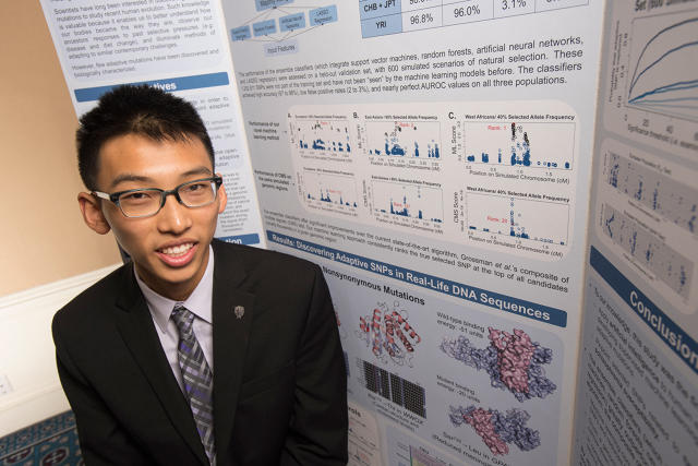
Like plenty of science-oriented high school kids, Andrew Jin is interested in human evolution. But Jin, one of three $150,000 first-place winners in this year's Intel Science Talent Search, took that interest further than most. For his project, the high school senior came up with machine learning algorithms that detect mutations in the human genome—mutations that could one day be used to develop drugs to combat diseases like HIV and schizophrenia.


Initially, Jin wanted to investigate how humans have evolved over the past 10,000 years. "I was doing it out of curiosity," he says. "I started thinking about natural selection and evolution, and that we understand so much about its theory, but we know nothing about reality. I was curious about what mutations help us be sophisticated human beings."

Jin decided to examine 179 human DNA sequences from different parts of the world. Each sequence consisted of 3 million base pairs of DNA—far too much to look at without help from an algorithm. So he set up a machine learning algorithm and found 130 potentially adaptive mutations, related to things like immune response and metabolism, that played a role in human evolution.

Working from a summer program at MIT, Jin refined his research and came up with a handful of mutations, including ones involved in resistance to meningitis and decreased susceptibility to viruses like influenza and HIV, that could potentially be used by pharmaceutical companies in new drug development.

There have been other natural-selection studies in the past looking for adaptive mutations, but Jin says that many of his findings are new. There's still a long way to go before he starts chatting up Big Pharma, however. "There's very, very strong evidence for these mutations playing a role in disease resistance, but in order to confirm, I would have to do biological experiments to study their protective mechanisms. That’s what I’m interested in doing now," says Jin.

Once he gets to college (he's not yet sure where that will be), Jin plans to pursue computer science or biology. But that's not all he's good at: The teen is a talented pianist who has played at Carnegie Hall. "I'm also an avid Boy Scout," he says.

martes, 17 de marzo de 2015

Ido Bachelet DNA nanobots summary with a couple of extra videos

In a brief talk, Bachelet said DNA nanobots will soon be tried in a critically ill leukemia patient. The patient, who has been given roughly six months to live, will receive an injection of DNA nanobots designed to interact with and destroy leukemia cells—while causing virtually zero collateral damage in healthy tissue.

According to Bachelet, his team have successfully tested their method in cell cultures and animals and written two papers on the subject, one in Science and one in Nature.

Contemporary cancer therapies involving invasive surgery and blasts of drugs can be as painful and damaging to the body as the disease itself. If Bachelet's approach proves successful in humans, and is backed by more research in the coming years, the team’s work could signal a transformational moment in cancer treatment.

If this treatment works this will be a medical breakthrough and can be used for many other diseases by delivering drugs more effectively without causing side effects.

2012 Video with answers from George Church, Ido Bachelet and Shawn Douglas on the medical DNA double helix clamshell nanobucket nanobot


George Church indicates the smart DNA nanobot has applications beyond nanomedicine. Applications where there is any need for programmable and targeted release or interaction at the cellular or near molecular scale.

2014 Geek Time Presentation from Ido Bachelet


At the British Friends of Bar-Ilan University's event in Otto Uomo October 2014 Professor Ido Bachelet announced the beginning of the human treatment with nanomedicine. He indicates DNA nanobots can currently identify cells in humans with 12 different types of cancer tumors.

A human patient with late stage leukemia will be given DNA nanobot treatment. Without the DNA nanobot treatment the patient would be expected to die in the summer of 2015. Based upon animal trials they expect to remove the cancer within one month.

Within 1 or 2 years they hope to have spinal cord repair working in animals and then shortly thereafter in humans. This is working in tissue cultures. 

Previously Ido Bachelet and Shawn Douglas have published work on DNA nanobots in the journal Nature and other respected science publications.

One Trillion 50 nanometer nanobots in a syringe will be injected into people to perform cellular surgery.

The DNA nanobots have been tuned to not cause an immune response.

They have been adjusted for different kinds of medical procedures. Procedures can be quick or ones that last many days.


Medicine or treatment released based upon molecular sensing - Only targeted cells are treated


Ido's daughter has a leg disease which requires frequent surgery. He is hoping his DNA nanobots will make the type of surgery she needs relatively trivial - a simple injection at a doctor's office.

We can control powerful drugs that were already developed

Effective drugs that were withdrawn from the market for excessive toxicity can be combined with DNA nanobots for effective delivery. The tiny molecular computers of the DNA nanobots can provide molecular selective control for powerful medicines that were already developed.

Using DNA origami and molecular programming, they are reality. These nanobots can seek and kill cancer cells, mimic social insect behaviors, carry out logical operators like a computer in a living animal, and they can be controlled from an Xbox. Ido Bachelet from the bio-design lab at Bar Ilan University explains this technology and how it will change medicine in the near future.

Ido Bachelet earned his Ph.D. from the Hebrew University in Jerusalem, and was a postdoctoral fellow at M.I.T. and Harvard University. He is currently an assistant professor in the Faculty of Life Sciences and the Nano-Center at Bar Ilan University, Israel, the founder of several biotech companies, and a composer of music for piano and molecules.


Researchers have injected various kinds of DNA nanobots into cockroaches. Because the nanobots are labelled with fluorescent markers, the researchers can follow them and analyse how different robot combinations affect where substances are delivered. The team says the accuracy of delivery and control of the nanobots is equivalent to a computer system.
  • This is the development of the vision of nanomedicine. 
  • This is the realization of the power of DNA nanotechnology. 
  • This is programmable dna nanotechnology.
The DNA nanotechnology cannot perform atomically precise chemistry (yet), but having control of the DNA combined with advanced synthetic biology and control of proteins and nanoparticles is clearly developing into very interesting capabilities.

"This is the first time that biological therapy has been able to match how a computer processor works," says co-author Ido Bachelet of the Institute of Nanotechnology and Advanced Materials at Bar Ilan University.

The team says it should be possible to scale up the computing power in the cockroach to that of an 8-bit computer, equivalent to a Commodore 64 or Atari 800 from the 1980s. Goni-Moreno agrees that this is feasible. "The mechanism seems easy to scale up so the complexity of the computations will soon become higher," he says.

An obvious benefit of this technology would be cancer treatments, because these must be cell-specific and current treatments are not well-targeted. But a treatment like this in mammals must overcome the immune response triggered when a foreign object enters the body.

Bachelet is confident that the team can enhance the robots' stability so that they can survive in mammals. "There is no reason why preliminary trials on humans can't start within five years," he says

Biological systems are collections of discrete molecular objects that move around and collide with each other. Cells carry out elaborate processes by precisely controlling these collisions, but developing artificial machines that can interface with and control such interactions remains a significant challenge. DNA is a natural substrate for computing and has been used to implement a diverse set of mathematical problems, logic circuits and robotics. The molecule also interfaces naturally with living systems, and different forms of DNA-based biocomputing have already been demonstrated. Here, we show that DNA origami can be used to fabricate nanoscale robots that are capable of dynamically interacting with each other in a living animal. The interactions generate logical outputs, which are relayed to switch molecular payloads on or off. As a proof of principle, we use the system to create architectures that emulate various logic gates (AND, OR, XOR, NAND, NOT, CNOT and a half adder). Following an ex vivo prototyping phase, we successfully used the DNA origami robots in living cockroaches (Blaberus discoidalis) to control a molecule that targets their cells.


Ido Bachelet's moonshot to use nanorobotics for surgery has the potential to change lives globally. But who is the man behind the moonshot?

Ido graduated from the Hebrew University of Jerusalem with a PhD in pharmacology and experimental therapeutics. Afterwards he did two postdocs; one in engineering at MIT and one in synthetic biology in the lab of George Church at the Wyss Institute at Harvard. 

Now, his group at Bar-Ilan University designs and studies diverse technologies inspired by nature.

  • They will deliver enzymes that break down cells via programmable nanoparticles.
  • Delivering insulin to tell cells to grow and regenerate tissue at the desired location.
  • Surgery would be performed by putting the programmable nanoparticles into saline and injecting them into the body to seek out remove bad cells and grow new cells and perform other medical work.


Nanoparticles with computational logic has already been done

Load an ensemble of drugs into many particles for programmed release based on situation that is found in the body