Mostrando entradas con la etiqueta Lab On A Chip. Mostrar todas las entradas

miércoles, 21 de diciembre de 2016

Artificial leaf as mini-factory for drugs (for Medicine)

(Nanowerk News) To produce drugs sustainably and cheaply, anywhere you want. Whether in the middle of the jungle or even on Mars. A 'mini-factory' whereby sunlight can be captured to make chemical products. Inspired by the art of nature where leaves are able to collect enough sunlight to produce food, chemical engineers at Eindhoven University of Technology (TU/e) have presented such a scenario.

They describe their prototype reactor - consciously shaped as a leaf -in today's journal Angewandte Chemie ("A leaf-inspired luminescent solar concentrator for energy efficient continuous-flow photochemistry").

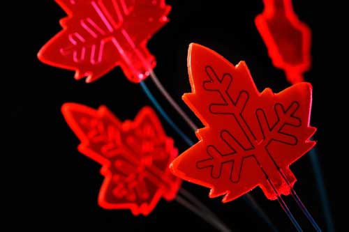
Even with the naked eye the amount of light captured by the 'mini-factories' is visible, lit up bright red. The 'veins' through the leaves are the thin channels through which liquid can be pumped. The start products enter the one channel, light causes the reactions and the end product comes out via the other channels. (Image: Bart van Overbeeke)
Using sunlight to make chemical products has long been a dream of many a chemical engineer. The problem is that the available sunlight generates too little energy to kick off reactions. However, nature is able to do this. Antenna molecules in leaves capture energy from sunlight and collect it in the reaction centers of the leaf where enough solar energy is present for the chemical reactions that give the plant its food (photosynthesis).

Light capture
The researchers came across relatively new materials, known as luminescent solar concentrators (LSC's), which are able to capture sunlight in a similar way. Special light-sensitive molecules in these materials capture a large amount of the incoming light that they then convert into a specific color that is conducted to the edges via light conductivity. These LSC's are often used in practice in combination with solar cells to boost the yield.

Thin channels
The researchers, led by Dr. Timothy Noël, combined the idea of an LSC with their knowledge of microchannels, incorporating very thin channels in a silicon rubber LSC through which a liquid can be pumped. In this way they were able to bring the incoming sunlight into contact with the molecules in the liquid with high enough intensity to generate chemical reactions.

Watch an animation of the artificial leaf.
Surpassed
While the reaction they chose serves as an initial example, the results surpassed all their expectations, and not only in the lab.
"Even an experiment on a cloudy day demonstrated that the chemical production was 40 percent higher than in a similar experiment without LSC material", says research leader Noël. "We still see plenty of possibilities for improvement. We now have a powerful tool at our disposal that enables the sustainable, sunlight-based production of valuable chemical products like drugs or crop protection agents."

Paracetamol on Mars
For the production of drugs there is certainly a lot of potential. The chemical reactions for producing drugs currently require toxic chemicals and a lot of energy in the form of fossil fuels. By using visible light the same reactions become sustainable, cheap and, in theory, countless times faster. But Noël believes it should not have to stop there.

"Using a reactor like this means you can make drugs anywhere, in principle, whether malaria drugs in the jungle or paracetamol on Mars. All you need is sunlight and this mini-factory."

Source: Eindhoven University of Technology


ORIGINAL: NanoWerk
Dec 21, 2016

lunes, 11 de abril de 2016

Scientists have figured out how to make their own molecules from scratch

ETH Zurich/Lucio Isa
And the possibilities are huge.
A new technique for making artificial molecules in the lab has been developed by researchers in Switzerland, and it opens up the possibility of new micro-robots and other microscopic structures being produced for a specific task - such as delivering medicine into targeted areas in the body - in a way that closely mimics the body's natural processes.

"So far, no scientist has succeeded in fully controlling the sequence of individual components when producing artificial molecules on the micro-scale," said lead researcher, Lucio Isa from ETH Zurich.

The process to create these artificial molecules starts with microspheres made of polymer or silica - 1 micrometre in diameter - which are placed side-by-side in tiny indentations and engraved in polymer templates. The indentations set the form of the finished object and heat is then used to bond the spheres together.

The team says this new procedure is more effective than other micro-3D printing technologies because it enables scientists to build single micro-structures from multiple materials. This means they can arrange artificial molecules in the sequence and with the geometry of their choosing.


It also enables them to precisely define magnetic, non-magnetic, and differently charged areas. Small rods, tiny triangles, and basic 3D objects can be created, though the research team wants to expand the capabilities of the process further.

The researchers say the practical benefits of the process could range from
  • self-propelled micro-carriers that move in an external electric field and micro-mixers for lab-on-a-chip applications, to (eventually) 
  • micro-robots for biomedical applications which are able to grab, transport, and release other specific micro-objects.
They also think their new technique could be used to assemble larger 'superstructures' for use in areas such as photonics (light-based signal processing).

Thanks to the level of control the team has managed to develop, the process is extremely versatile. "In principle, our method can be adapted to any material, even metals," said Isa.

"The full programmability of our approach opens up new directions not only for assembling and studying complex materials with single-particle-level control but also for fabricating new microscale devices for sensing, patterning, and delivery applications," concludes the study, which has been published in the journal Science Advances.

ORIGINAL: Science Alert
DAVID NIELD
8 APR 2016

sábado, 21 de noviembre de 2015

'Chemical Laptop' Could Search for Signs of Life Outside Earth


Researchers took the Chemical Laptop to JPL's Mars Yard, where they placed the device on a test rover. This image shows the size comparison between the Chemical Laptop and a regular laptop.
Credits: NASA/JPL-Caltech

If you were looking for the signatures of life on another world, you would want to take something small and portable with you. That's the philosophy behind the "Chemical Laptop" being developed at NASA's Jet Propulsion Laboratory in Pasadena, California: a miniaturized laboratory that analyzes samples for materials associated with life.

"If this instrument were to be sent to space, it would be the most sensitive device of its kind to leave Earth, and the first to be able to look for both amino acids and fatty acids," said Jessica Creamer, a NASA postdoctoral fellow based at JPL.

Like a tricorder from "Star Trek," the Chemical Laptop is a miniaturized on-the-go laboratory, which researchers hope to send one day to another planetary body such as Mars or Europa. It is roughly the size of a regular computing laptop, but much thicker to make room for chemical analysis components inside. But unlike a tricorder, it has to ingest a sample to analyze it. 

"Our device is a chemical analyzer that can be reprogrammed like a laptop to perform different functions," said Fernanda Mora, a JPL technologist who is developing the instrument with JPL's Peter Willis, the project's principal investigator. "As on a regular laptop, we have different apps for different analyses like amino acids and fatty acids."

Amino acids are building blocks of proteins, while fatty acids are key components of cell membranes. Both are essential to life, but can also be found in non-life sources. The Chemical Laptop may be able to tell the difference.
JPL researchers Jessica Creamer, Fernanda Mora and Peter Willis (left to right) pose with the Chemical Laptop, a device designed to detect amino acids and fatty acids. At left is a near-identical copy of the Curiosity rover, which has been on Mars since 2012. Credits: NASA/JPL-Caltech
What it's looking for
Amino acids come in two types: Left-handed and right-handed. Like the left and right hands of a person, these amino acids are mirror images of each other but contain the same components. Some scientists hypothesize that life on Earth evolved to use just left-handed amino acids because that standard was adopted early in life's history, sort of like the way VHS became the standard for video instead of Betamax in the 1980s. It's possible that life on other worlds might use the right-handed kind. 

"If a test found a 50-50 mixture of left-handed and right-handed amino acids, we could conclude that the sample was probably not of biological origin," Creamer said. "But if we were to find an excess of either left or right, that would be the golden ticket. That would be the best evidence so far that life exists on other planets."

The analysis of amino acids is particularly challenging because the left- and right-handed versions are equal in size and electric charge. Even more challenging is developing a method that can look for all the amino acids in a single analysis.

When the laptop is set to look for fatty acids, scientists are most interested in the length of the acids' carbon chain. This is an indication of what organisms are or were present.

How it works
The battery-powered Chemical Laptop needs a liquid sample to analyze, which is more difficult to obtain on a planetary body such as Mars. The group collaborated with JPL's Luther Beegle to incorporate an "espresso machine" technology, in which the sample is put into a tube with liquid water and heated to above 212 degrees Fahrenheit (100 degrees Celsius). The water then comes out carrying the organic molecules with it. The Sample Analysis at Mars (SAM) instrument suite on NASA's Mars Curiosity rover utilizes a similar principle, but it uses heat without water.

Once the water sample is fed into the Chemical Laptop, the device prepares the sample by mixing it with a fluorescent dye, which attaches the dye to the amino acids or fatty acids. The sample then flows into a microchip inside the device, where the amino acids or fatty acids can be separated from one another. At the end of the separation channel is a detection laser. The dye allows researchers see a signal corresponding to the amino acids or fatty acids when they pass the laser.

Inside a "separation channel" of the microchip, there are already chemical additives that mix with the sample. Some of these species will only interact with right-handed amino acids, and some will only interact with the left-handed variety. These additives will change the relative amount of time the left and right-handed amino acids are in the separation channel, allowing scientists to determine the "handedness" of amino acids in the sample.

The Chemical Laptop, developed at JPL, analyzes liquid samples and detects amino acids and fatty acids. These are both chemicals that are essential to life.
Credits: NASA/JPL-Caltech
Testing for future uses
Last year the researchers did a field test at JPL's Mars Yard, where they placed the Chemical Laptop on a test rover.

"This was the first time we showed the instrument works outside of the laboratory setting. This is the first step toward demonstrating a totally portable and automated instrument that can operate in the field," said Mora.

For this test, the laptop analyzed a sample of "green rust," a mineral that absorbs organic molecules in its layers and may be significant in the origin of life, said JPL's Michael Russell, who helped provide the sample.

"One ultimate goal is to put a detector like this on a spacecraft such as a Mars rover, so for our first test outside the lab we literally did that," said Willis.

Since then, Mora has been working to improve the sensitivity of the Chemical Laptop so it can detect even smaller amounts of amino acids or fatty acids. Currently, the instrument can detect concentrations as low as parts per trillion. Mora is currently testing a new laser and detector technology.

Coming up is a test in the Atacama Desert in Chile, with collaboration from NASA's Ames Research Center, Moffett Field, California, through a grant from NASA's Planetary Science & Technology Through Analog Research (PSTAR) program.

"This could also be an especially useful tool for icy-worlds targets such as Enceladus and Europa. All you would need to do is melt a little bit of the ice, and you could sample it and analyze it directly," Creamer said.

The Chemical Laptop technology has applications for Earth, too. It could be used for environmental monitoring -- analyzing samples directly in the field, rather than taking them back to a laboratory. Uses for medicine could include testing whether the contents of drugs are legitimate or counterfeit. 

Creamer recently won an award for her work in this area at JPL's Postdoc Research Day Poster Session.

NASA's PICASSO program, part of the agency's Science Mission Directorate in Washington, supported this research. The California Institute of Technology in Pasadena manages JPL for NASA.


ORIGINAL: NASA
By Elizabeth Landau. NASA's Jet Propulsion Laboratory, Pasadena, Calif.
Nov. 16, 2015

Last Updated: Nov. 16, 2015
818-354-6425

Editor: Martin Perez


martes, 23 de junio de 2015

The Best Design of the Year (Maybe Ever?)

Every year, the Design Museum in London picks a single object and names it the best design of the year. It’s pretty bad sometimes! But this year, the museum picked a winner: A chip that replaces animal test subjects with a complex package of human cells.


It’s called a lung-on-a-chip--a name that is very literally true, lest you think this is simply a computer chip programmed to mimic a lung. It comes from Harvard’s Wyss Institute for Biologically Inspired Engineering, which explains in a great video how it works.


This clear, simple-looking brick of plastic actually contains complex human cells, arranged in a simplified version of the way a lung works: Along the central channels, there’s a lining of human lung cells separated from a lining of capillary blood cells by a porous membrane, just like the air sacs in your lung:


On each side, channels create the flexing movement that an air sac does while you breathe.


In other words, it’s all of the biological complexity of your lungs distilled onto a computer chip.

Scientists can, for example, introduce bacteria to the channels to mimic an infection—and white blood cells in the capillary channel will attack. Or, they can introduce the chemicals you breathe in regularly to mimic air pollution and its affect on your lungs. Or test new medications.

“Bio-inspired micro-devices that mimic whole human organs, such as the lung on a chip, could potentially replace animal testing and bring new therapies to patients faster and at lower cost in the future,” the design team explains in their video. Other labs are working on organs like the heart and even spleen, and Wyss’ ultimate goal is to build ten different organs and link them to create a whole body.

Who do we have to thank for bringing news of the chip to the design world? That would be Paola Antonelli, MoMA’s Senior Curator of Architecture & Design, as Dezeen points out today in its announcement as the award’s media partner. Antonelli not only nominated the chip, she already added it to MoMA’s permanent collection in March, writing on MoMA’s blog:

Esoteric or specialized, perhaps, but universally remarkable in their balance of form, function, and vision, investigations like the Wyss Institute’s Human Organs-on-Chips demonstrate new, radical intersections of synthetic biology and design.

In the past, the Design Museum’s pick have ranged from anodyne at best—a lightbulb, in 2011—to downright tone-deaf, like the jury’s choice of a Zaha Hadid building in Azerbaijan built by a dictatorial regime and named for a president known for his human rights abuses. This year, the jury really turned it around, selecting an object that is not only a brilliant piece of design, but also has the power to end the barbaric practice of animal testing while helping human patients.

Antonelli deserves a lot of credit for caring what’s happening in science, medicine, and technology, and forcing the rest of the design world to broaden insular, myopic field of view to include objects that aren’t just lightbulbs and billion-dollar museums, great though they are. Design—while it won’t save the world—can certainly change it for good.

Contact the author at kelsey@Gizmodo.com.

ORIGINAL: Gizmodo

miércoles, 20 de mayo de 2015

Can We Identify Every Kind of Cell in the Body?

A microscopic quest to find out what we’re really made of.

WHY IT MATTERS
There is still no accurate atlas of human cell types.
A microfluidic device (at center) can carry out experiments on individual cells.
How many types of cells are there in the human body? Textbooks say a couple of hundred. But the true number is undoubtedly far larger.

Aviv Regev. Regev received her M.Sc. from Tel Aviv University, studying biology, computer science, and mathematics in the Interdisciplinary Program for the Fostering of Excellence. She received her Ph.D. in computational biology from Tel Aviv University. Photo: Broad Institute
Piece by piece, a new, more detailed catalogue of cell types is emerging from labs like that of Aviv Regev at the Broad Institute, in Cambridge, Massachusetts, which are applying recent advances in single-cell genomics to study individual cells at a speed and scale previously unthinkable.

The technology applied at the Broad uses fluidic systems to separate cells on microscopic conveyor belts and then submits them to detailed genetic analysis, at the rate of thousands per day. Scientists expect such technologies to find use in medical applications where small differences between cells have big consequences, including cell-based drug screens, stem-cell research, cancer treatment, and basic studies of how tissues develop.

Regev says she has been working with the new methods to classify cells in mouse retinas and human brain tumors, and she is finding cell types never seen before. “We don’t really know what we’re made of,” she says.

Other labs are racing to produce their own surveys and improve the underlying technology. Today a team led by Stephen Quake of Stanford University published its own survey of 466 individual brain cells, calling it “a first step” toward a comprehensive cellular atlas of the human brain.

Such surveys have only recently become possible, scientists say. “A couple of years ago, the challenge was to get any useful data from single cells,” says Sten Linnarsson, a single-cell biologist at the Karolinska Institute in Stockholm, Sweden. In March, Linnarsson’s group used the new techniques to map several thousand cells from a mouse’s brain, identifying 47 kinds, including some subtypes never seen before.

Historically, the best way to study a single cell was to look at it through a microscope. In cancer hospitals, that’s how pathologists decide if cells are cancerous or not: they stain them with dyes, some first introduced in the early 1900s, and consider their location and appearance. Current methods distinguish about 300 different types, says Richard Conroy, a research official at the National Institutes of Health.

Individual cells are captured and separated in bubbles of liquid, readying them for analysis.
The new technology works instead by cataloguing messenger RNA molecules inside a cell. These messages are the genetic material the nucleus sends out to make proteins. Linnarsson’s method attaches a unique molecular bar code to every RNA molecule in each cell. The result is a gene expression profile, amounting to a fingerprint of a cell that reflects its molecular activity rather than what it looks like.

“Previously, cells were defined by one or two markers,” says Linnarsson. “Now we can say what is the full complement of genes expressed in those cells.”

Although researchers determined how to accurately sequence RNA from a single cell a few years ago, it’s only more recently that clever innovations in chemistry and microfluidics have led to an explosion of data. A California company, Cellular Research, showed this year that it could sort cells into micro-wells and then measure the RNA of 3,000 separate cells at once, at the cost of few pennies a cell.

Scientists think the new single-cell methods could overturn previous research findings. That is because previous gene expression studies were based on tissue samples or blood specimens containing thousands, even millions, of cells. Studying such blended mixtures meant researchers were seeing averages, says Eric Lander, head of the Broad Institute.

“Single-cell genomics has come of age in an unbelievable way in just the last 18 months,” Lander told an audience at the National Institutes of Health this year. “And once you realize we are at the point of doing individual cells, how could you ever put up with a fruit smoothie? It is just nuts to be doing genomics on smoothies.”

Lander, one of the leaders of the Human Genome Project, says it may be time to turn pilot projects like those Regev is leading into a wider effort to create a definitive atlas—one cataloguing all human cell types by gene activity and tracking them from the embryo all the way to adulthood.

“It’s a little premature to declare a national or international project until there’s been more piloting, but I think it’s an idea that’s very much in the air,” Lander said in a phone interview. “I think [in two years] we’re going to be in the position where it would be crazy not to have this information. If we had a periodic table of the cells, we would be able to figure out, so to speak, the atomic composition of any given sample.”

Gene profiles might eventually be combined with other efforts to study single cells. Paul Allen, Microsoft’s cofounder, said last December he would be spending $100 million to create a new scientific institute, the Allen Institute for Cell Science It will study stem cells and video their behavior under microscopes as they develop into various cell types, with the ultimate goal of creating a massive animated model. Rick Horwitz, who leads that effort, says that it will serve as a kind of Google Earth for exploring a cell’s life cycle.


The eventual payoff of collecting all this data, says Garry Nolan, an immunologist at Stanford University, won’t be just a catalogue of cell types, but a deeper understanding of how cells work together. “The single-cell approach is a way station that needs to be understood on the way to understanding the greater system,” he says. “In 50 years, we’ll probably be measuring every molecule in the cell dynamically.”

ORIGINAL: MIT Tech Review
May 18, 2015

domingo, 2 de noviembre de 2014

Lego-like modular components make building 3-D 'labs-on-a-chip' a snap


Modular fluidic and instrumentation components developed by researchers at the University of Southern California Viterbi School of Engineering. Credit: USC Viterbi School of Engineering

Thanks to new LEGO-like components developed by researchers at the USC Viterbi School of Engineering, it is now possible to build a 3-D microfluidic system quickly and cheaply by simply snapping together small modules by hand.

Microfluidic systems are used in many fields including engineering, chemistry and biotechnology to precisely manipulate small volumes of fluids for use in applications such as 
  • enzymatic or DNA analysis, 
  • pathogen detection, 
  • clinical diagnostic testing, and 
  • synthetic chemistry. 
Traditionally, microfluidic devices are built in a cleanroom on a two-dimensional surface using the same technology developed to produce integrated circuits for the electronics industry.

Though tiny, designing, assembling and testing a new microfluidics system can take a lot of time and money. Building a single device can often require multiple iterations, each of which can take up to two weeks and several thousand dollars to manufacture. And the more complex the system, the higher the number of iterations needed.

"You test your device and it never works the first time," said Krisna Bhargava, materials science graduate student at the USC Viterbi School of Engineering. "If you've grown up to be an engineer or scientist, you've probably been influenced by LEGO at some point in your childhood. I think every scientist has a secret fantasy that whatever they're building will be as simple to assemble."

Frustrated that reproducing a simple microfluidic circuit could cost him so much time and money, Bhargava set out simplify the construction process. First, he identified the primitive elements commonly used in microfluidic systems, much like how circuitry is broken down in electrical engineering. Basic microfluidic functions would be separated into standardized modular components, not an entirely revolutionary concept. But then, he abandoned the two-dimensional method of building microfluidic devices altogether.

"The founders of the microfluidics field took the same approach as the semiconductor industry: to try to pack in as much integrated structure as possible into a single chip," explained Bhargava. "In electronics, this is important because a high density of transistors has many direct and indirect benefits for computation and signal processing. In microfluidics, our concerns are not with bits and symbolic representations, but rather with the way fluidics are routed, combined, mixed, and analyzed; there's no need to stick with continuing to integrate more and more complex devices."

Borrowing an approach from the electronics industry, which uses prototype boards to build circuits, Bhargava conceived of three-dimensional modular components that encapsulated the common elements of microfluidic systems, as well as a connector that could join the separate components together. Inspired by recent advancements in micron-scale 3D-printing, he and a USC Viterbi research team that included chemical engineering and materials science professor Noah Malmstadt and biomedical engineering graduate student Bryant Thompson, designed computer models for eight modular fluidic and instrumentation components (MFICs, pronounced "em-fix") that would each perform a simple operation. Examples are a "helix" component that can mix two fluid streams and a component that contains an integrated optical sensor for measuring the size of small droplets. The components constructed for this study are approximately 1 cm3, slightly smaller than a standard 6-sided die.

The team's development of these MFICs represents the first attempt to break a device into separate components that can be assembled, disassembled and re-assembled over and over.


viernes, 18 de abril de 2014

Neurology and microfluidic science kits take over for kids in the 21st century

Today kids are not happy enough to play around with the old chemistry kits from yesteryear, they want more, they want real science. Recently the people behind the ISEF science fair, The Society for Science & the Public, launched a competition for children with the aim of letting children make their own modern version of a science kit for kids in the 21st century. Of course in today’s world, children do have access to a great deal more modern technology than what was around in the 60s and 70s.

[Image Courtesy of George Korir]

The results of the competition are in and it was a handmade microfluidic kit that took first prize in the competition. This was made by a graduate student along with the help of his biochemistry professor. For those who don’t know, microfluidic tech is the stuff that is found in lab-on-a-chip devices which are used to analyse small amounts of liquid. These move around and mix chemicals through channels, which are of course very small, on platforms that are very small, such as the size of a computer chip. Scientists have been busy working on devising microfluidic devices that can mimic human organs. First they had to decide what chemical reactions to make in the kit for children.

Holes were then punched out of paper card and these holes corresponded to sequences and were then loaded with the correct chemicals. In order to make the reactions between the chemicals, kids had to then use the cards with a reader that worked with a hand crank. This then released the chemicals slowly, just one single drop for every hole that was punched in the card. Manu Prakash from the Stanford University, who was the professor who helped to make the system, made a statement saying that he could see kids in the future trading the reaction cards and could even become as popular as trading baseball cards were in the past.


In second place came a design of electrodes that when placed onto the body can sense electricity after it sent messages to the brain produced by the body whilst flexing muscles. To do this, electrodes were hooked up to amplifiers and then an electrical device such as a motor or light bulb. This allowed those using it to turn on something, for instance a propeller, simply by squeezing their hand or even just thinking about it.


[Image Courtesy of George Korir]


Chemistry sets were always on the Christmas list of some boys and girls during the 60s and 70s but today they are more than just attaching wires to a battery and small light bulb. The competition could have sparked off a whole new trend when it comes to science kits and they could become the in-thing for budding inventors and scientists for Christmas 2014 and beyond.

For a full list of all winners and their bios head over to the Gordon and Betty Moore Foundation.


April 15th, 2014