Mostrando entradas con la etiqueta GFP. Mostrar todas las entradas

domingo, 26 de agosto de 2018

Test Tube Artificial Neural Network Recognizes "Molecular Handwriting"

Conceptual illustration of a droplet containing an artificial neural network made of DNA that has been designed to recognize complex and noisy molecular information, represented as 'molecular handwriting.' Credit: Olivier Wyart


Test tube chemistry using synthetic DNA molecules can be utilized in complex computing tasks to exhibit artificial intelligence

Researchers at Caltech have developed an artificial neural network made out of DNA that can solve a classic machine learning problem: correctly identifying handwritten numbers. The work is a significant step in demonstrating the capacity to program artificial intelligence into synthetic biomolecular circuits.

The work was done in the laboratory of Lulu Qian, assistant professor of bioengineering. A paper describing the research (paywall) appears online on July 4 and in the July 19 print issue of the journal Nature.

"Though scientists have only just begun to explore creating artificial intelligence in molecular machines, its potential is already undeniable," says Qian. "Similar to how electronic computers and smart phones have made humans more capable than a hundred years ago, artificial molecular machines could make all things made of molecules, perhaps including even paint and bandages, more capable and more responsive to the environment in the hundred years to come."

Artificial neural networks are mathematical models inspired by the human brain. Despite being much simplified compared to their biological counterparts, artificial neural networks function like networks of neurons and are capable of processing complex information. The Qian laboratory's ultimate goal for this work is to program intelligent behaviors (the ability to compute, make choices, and more) with artificial neural networks made out of DNA.

"Humans each have over 80 billion neurons in the brain, with which they make highly sophisticated decisions. Smaller animals such as roundworms can make simpler decisions using just a few hundred neurons. In this work, we have designed and created biochemical circuits that function like a small network of neurons to classify molecular information substantially more complex than previously possible," says Qian.

To illustrate the capability of DNA-based neural networks, Qian laboratory graduate student Kevin Cherry chose a task that is a classic challenge for electronic artificial neural networks: recognizing handwriting.

Human handwriting can vary widely, and so when a person scrutinizes a scribbled sequence of numbers, the brain performs complex computational tasks in order to identify them. Because it can be difficult even for humans to recognize others' sloppy handwriting, identifying handwritten numbers is a common test for programming intelligence into artificial neural networks. These networks must be "taught" how to recognize numbers, account for variations in handwriting, then compare an unknown number to their so-called memories and decide the number's identity.

WHY DNA?
Key to creating biomolecular circuits out of DNA are the strict binding rules between molecules of DNA. A single-stranded DNA molecule is composed of smaller molecules called nucleotides—abbreviated A, T, C, and G—arranged in a string, or sequence. The nucleotides in a single-stranded DNA molecule can bond with those of another single strand to form double-stranded DNA, but the nucleotides bind only in very specific ways: An A nucleotide with a T or a C nucleotide with a G.

Taking advantage of these predictable binding rules, Qian and her colleagues can design short strands of DNA to undergo predictable chemical reactions in a test tube and thereby compute tasks, such as molecular pattern recognition. In 2011, Qian and her colleagues created the first artificial neural network made of DNA molecules that could recognize four simple patterns.

In the work described in the Nature paper, Cherry, who is the first author on the paper, demonstrated that a neural network made out of carefully designed DNA sequences could carry out prescribed chemical reactions to accurately identify "molecular handwriting." Unlike visual handwriting that varies in geometrical shape, each example of molecular handwriting does not actually take the shape of a number. Instead, each molecular number is made up of 20 unique DNA strands chosen from 100 molecules, each assigned to represent an individual pixel in any 10 by 10 pattern. These DNA strands are mixed together in a test tube.

"The lack of geometry is not uncommon in natural molecular signatures yet still requires sophisticated biological neural networks to identify them: for example, a mixture of unique odor molecules comprises a smell," says Qian.

Given a particular example of molecular handwriting, the DNA neural network can classify it into up to nine categories, each representing one of the nine possible handwritten digits from 1 to 9.

First, Cherry built a DNA neural network to distinguish between handwritten 6s and 7s. He tested 36 handwritten numbers and the test tube neural network correctly identified all of them. His system theoretically has the capability of classifying over 12,000 handwritten 6s and 7s—90 percent of those numbers taken from a database of handwritten numbers used widely for machine learning—into the two possibilities.

Crucial to this process was encoding a "winner take all" competitive strategy using DNA molecules, developed by Qian and Cherry. In this strategy, a particular type of DNA molecule dubbed the annihilator was used to select a winner when determining the identity of an unknown number.

"The annihilator forms a complex with one molecule from one competitor and one molecule from a different competitor and reacts to form inert, unreactive species," says Cherry. "The annihilator quickly eats up all of the competitor molecules until only a single competitor species remains. The winning competitor is then restored to a high concentration and produces a fluorescent signal indicating the networks' decision.

Next, Cherry built upon the principles of his first DNA neural network to develop one even more complex, one that could classify single digit numbers 1 through 9. When given an unknown number, this "smart soup" would undergo a series of reactions and output two fluorescent signals, for example, green and yellow to represent a 5, or green and red to represent a 9.

Qian and Cherry plan to develop artificial neural networks that can learn, forming "memories" from examples added to the test tube. This way, Qian says, the same smart soup can be trained to perform different tasks.

"Common medical diagnostics detect the presence of a few biomolecules, for example cholesterol or blood glucose." says Cherry. "Using more sophisticated biomolecular circuits like ours, diagnostic testing could one day include hundreds of biomolecules, with the analysis and response conducted directly in the molecular environment."

The paper is titled "Scaling up molecular pattern recognition with DNA-based winner-take-all neural networks." Funding was provided by the National Science Foundation, the Burroughs Wellcome Fund, and the Shurl and Kay Curci Foundation.

Related:

ORIGINAL: Caltech
by Lori Dajose
07/05/2018

sábado, 21 de octubre de 2017

New Research Points to a Genetic Switch That Can Let Our Bodies Talk to Electronics


Shutterstock
IN BRIEF
Our bodies are biologically based and therefore are not equipped to communicate with electronics efficiently. New research could make it possible to genetically engineer our cells to be able to communicate with electronics.

The development has the potential to allow us to eventually build apps that autonomously detect and treat disease.

Microelectronics has transformed our lives. Cellphones, earbuds, pacemakers, defibrillators – all these and more rely on microelectronics’ very small electronic designs and components. Microelectronics has changed the way we collect, process and transmit information.

Such devices, however, rarely provide access to our biological world; there are technical gaps. We can’t simply connect our cellphones to our skin and expect to gain health information. For instance, is there an infection? What type of bacteria or virus is involved? We also can’t program the cellphone to make and deliver an antibiotic, even if we knew whether the pathogen was Staph or Strep. There’s a translation problem when you want the world of biology to communicate with the world of electronics.

The research we’ve just published with colleagues in Nature Communications brings us one step closer to closing that communication gap.
Electronic control of gene expression and cell behaviour in Escherichia coli through redox signalling

ABSTRACT:
The ability to interconvert information between electronic and ionic modalities has transformed our ability to record and actuate biological function. Synthetic biology offers the potential to expand communication ‘bandwidth’ by using biomolecules and providing electrochemical access to redox-based cell signals and behaviours. While engineered cells have transmitted molecular information to electronic devices, the potential for bidirectional communication stands largely untapped. Here we present a simple electrogenetic device that uses redox biomolecules to carry electronic information to engineered bacterial cells in order to control transcription from a simple synthetic gene circuit. Electronic actuation of the native transcriptional regulator SoxR and transcription from the PsoxS promoter allows cell response that is quick, reversible and dependent on the amplitude and frequency of the imposed electronic signals. Further, induction of bacterial motility and population based cell-to-cell communication demonstrates the versatility of our approach and potential to drive intricate biological behaviours.

Source: NATURE COMMS
Rather than relying on the usual molecular signals, like hormones or nutrients, that control a cell’s gene expression, we created a synthetic “switching” system in bacterial cells that recognizes electrons instead. This new technology – a link between electrons and biology – may ultimately allow us to program our phones or other microelectronic devices to autonomously detect and treat disease.

COMMUNICATING WITH ELECTRONS, NOT MOLECULES
One of the barriers scientists have encountered when trying to link microelectronic devices with biological systems has to do with information flow. In biology, almost all activity is made possible by the transfer of molecules like
  • glucose, 
  • epinephrine, 
  • cholesterol and 
  • insulin 
signaling between cells and tissues. Infecting bacteria secrete molecular toxins and attach to our skin using molecular receptors. To treat an infection, we need to detect these molecules to identify the bacteria, discern their activities and determine how to best respond.

Microelectronic devices don’t process information with molecules. A microelectronic device typically has silicon, gold, chemicals like boron or phosphorus and an energy source that provides electrons. By themselves, they’re poorly suited to engage in molecular communication with living cells.

Free electrons don’t exist in biological systems so there’s almost no way to connect with microelectronics. There is, however, a small class of molecules that stably shuttle electrons. These are called “redox” molecules; they can transport electrons, sort of like wire does. The difference is that in wire, the electrons can flow freely to any location within; redox molecules must undergo chemical reactions – oxidation or reduction reactions – to “hand off” electrons.
Bacteria are engineered to respond to a redox molecule activated by an electrode by creating an electrogenetic switch. Bentley and Payne, CC BY-ND

TURNING CELLS ON AND OFF
Capitalizing on the electronic nature of redox molecules, we genetically engineered bacteria to respond to them. We focused on redox molecules that could be “programmed” by the electrode of a microelectronic device. The device toggles the molecule’s oxidation state – it’s either 
  • oxidized (loses an electron) or 
  • reduced (gains an electron). 
The electron is supplied by a typical energy source in electronics like a battery.

We wanted our bacteria cells to turn “on” and “off” due to the applied voltage – voltage that oxidized a naturally occurring redox molecule, pyocyanin.

Electrically oxidizing pyocyanin allowed us to control our engineered cells, turning them on or off so they would synthesize (or not) a fluorescent protein. We could rapidly identify what was happening in these cells because the protein emits a green hue.
(a) Device-mediated electronic input consists of applied potential (blue or red step functions) for controlling the oxidation state of redox-mediators (transduced input). Redox mediators intersect with cells to actuate transcription and, depending on actuated gene-of-interest, control biological output.
(b) The electrogenetic device consists of the region encompassing the gene coding for the SoxR protein and the divergent overlapping PsoxR/PsoxS promoters. A gene of interest is placed downstream of the PsoxS promoter. Pyo (O) initiates gene induction and Fcn(R/O), through interactions with respiratory machinery, allows electronic control of induction level. Fcn (R/O), ferro/ferricyanide; Pyo, pyocyanin. The oxidation state of both redox mediators is colorimetrically indicated (Fcn (O) is yellow pentagon; Fcn (R) is white pentagon; Pyo (O) is blue hexagon; Pyo (R) is grey hexagon). Encircled ‘e−‘ and arrows indicate electron movement.

In another example
, we made bacteria that, when switched on, would swim from a stationary position. Bacteria normally swim in starts and stops referred to as a “run” or a “tumble.” The “run” ensures they move in a straight path. When they “tumble,” they essentially remain in a one spot. A protein called CheZ controls the “run” portion of bacteria’s swimming activity. Our electrogenetic switch turned on the synthesis of CheZ, so that the bacteria could move forward.
Bacteria can naturally join forces as biofilms and work together. CDC/Janice Carr, CC BY
We were also able to electrically signal a community of cells to exhibit collective behavior. We made cells with switches controlling the synthesis of a signaling molecule that diffuses to neighboring cells and, in turn, causes changes in their behavior. Electric current turned on cells that, in turn, “programmed” a natural biological signaling process to alter the behavior of nearby cells. We exploited bacterial quorum sensing – a natural process where bacterial cells “talk” to their neighbors and the collection of cells can behave in ways that benefit the entire community.

Perhaps even more interesting, our groups showed that we could both turn on gene expression and turn it off. By reversing the polarity on the electrode, the oxidized pyocyanin becomes reduced – its inactive form. Then, the cells that were turned on were engineered to quickly revert back to their original state. In this way, the group demonstrated the ability to cycle the electrically programmed behavior on and off, repeatedly.

Interestingly, the on and off switch enabled by pyocyanin was fairly weak. By including another redox molecule, ferricyanide, we found a way to amplify the entire system so that the gene expression was very strong, again on and off. The entire system was robust, repeatable and didn’t negatively affect the cells.

SENSING AND RESPONDING ON A CELLULAR LEVEL
Armed with this advance, devices could potentially electrically stimulate bacteria to make therapeutics and deliver them to a site. For example, imagine swallowing a small microelectronic capsule that could record the presence of a pathogen in your GI tract and also contain living bacterial factories that could make an antimicrobial or other therapyall in a programmable autonomous system.

This current research ties into previous work done here at the University of Maryland where researchers had discovered ways to “record” biological information, by sensing the biological environment, and based on the prevailing conditions, “write” electrons to devices. We and our colleagues “sent out” redox molecules from electrodes, let those molecules interact with the microenvironment near the electrode and then drew them back to the electrode so they could inform the device on what they’d seen. This mode of “molecular communication” is somewhat analogous to sonar, where redox molecules are used instead of sound waves.

These molecular communication efforts were used to identify pathogens, monitor the “stress” in blood levels of individuals with schizophrenia and even determine the differences in melanin from people with red hair. For nearly a decade, the Maryland team has developed methodologies to exploit redox molecules to interrogate biology by directly writing the information to devices with electrochemistry.

Perhaps it is now time to integrate these technologies:

  • Use molecular communication to sense biological function and transfer the information to a device. 
  • Then use the device – maybe a small capsule or perhaps even a cellphone – to program bacteria to make chemicals and other compounds that issue new directions to the biological system. 

It may sound fantastical, many years away from practical uses, but our team is working hard on such valuable applications…stay tuned!

ORIGINAL: Futurism

miércoles, 1 de marzo de 2017

A Giant Neuron Has Been Found Wrapped Around the Entire Circumference of the Brain

Allen Institute for Brain Science
This could be where consciousness forms. For the first time, scientists have detected a giant neuron wrapped around the entire circumference of a mouse's brain, and it's so densely connected across both hemispheres, it could finally explain the origins of consciousness.

Using a new imaging technique, the team detected the giant neuron emanating from one of the best-connected regions in the brain, and say it could be coordinating signals from different areas to create conscious thought.

This recently discovered neuron is one of three that have been detected for the first time in a mammal's brain, and the new imaging technique could help us figure out if similar structures have gone undetected in our own brains for centuries.

At a recent meeting of the Brain Research through Advancing Innovative Neurotechnologies initiative in Maryland, a team from the Allen Institute for Brain Science described how all three neurons stretch across both hemispheres of the brain, but the largest one wraps around the organ's circumference like a "crown of thorns".
You can see them highlighted in the image at the top of the page.

Lead researcher Christof Koch told Sara Reardon at Nature that they've never seen neurons extend so far across both regions of the brain before.
Oddly enough, all three giant neurons happen to emanate from a part of the brain that's shown intriguing connections to human consciousness in the past - the claustrum, a thin sheet of grey matter that could be the most connected structure in the entire brain, based on volume.

This relatively small region is hidden between the inner surface of the neocortex in the centre of the brain, and communicates with almost all regions of cortex to achieve many higher cognitive functions such as 
  • language, 
  • long-term planning, and 
  • advanced sensory tasks such as 
  • seeing and 
  • hearing.
"Advanced brain-imaging techniques that look at the white matter fibres coursing to and from the claustrum reveal that it is a neural Grand Central Station," Koch wrote for Scientific American back in 2014. "Almost every region of the cortex sends fibres to the claustrum."

The claustrum is so densely connected to several crucial areas in the brain that Francis Crick of DNA double helix fame referred to it a "conductor of consciousness" in a 2005 paper co-written with Koch.

They suggested that it connects all of our external and internal perceptions together into a single unifying experience, like a conductor synchronises an orchestra, and strange medical cases in the past few years have only made their case stronger.

Back in 2014, a 54-year-old woman checked into the George Washington University Medical Faculty Associates in Washington, DC, for epilepsy treatment.

This involved gently probing various regions of her brain with electrodes to narrow down the potential source of her epileptic seizures, but when the team started stimulating the woman's claustrum, they found they could effectively 'switch' her consciousness off and on again.

Helen Thomson reported for New Scientist at the time:
"When the team zapped the area with high frequency electrical impulses, the woman lost consciousness. She stopped reading and stared blankly into space, she didn't respond to auditory or visual commands and her breathing slowed.

As soon as the stimulation stopped, she immediately regained consciousness with no memory of the event. The same thing happened every time the area was stimulated during two days of experiments.
"

According to Koch, who was not involved in the study, this kind of abrupt and specific 'stopping and starting' of consciousness had never been seen before.

Another experiment in 2015 examined the effects of claustrum lesions on the consciousness of 171 combat veterans with traumatic brain injuries.

They found that claustrum damage was associated with the duration, but not frequency, of loss of consciousness, suggesting that it could play an important role in the switching on and off of conscious thought, but another region could be involved in maintaining it.

And now Koch and his team have discovered extensive neurons in mouse brains emanating from this mysterious region.

In order to map neurons, researchers usually have to inject individual nerve cells with a dye, cut the brain into thin sections, and then trace the neuron's path by hand.

It's a surprisingly rudimentary technique for a neuroscientist to have to perform, and given that they have to destroy the brain in the process, it's not one that can be done regularly on human organs.

Koch and his team wanted to come up with a technique that was less invasive, and engineered mice that could have specific genes in their claustrum neurons activated by a specific drug.

"When the researchers fed the mice a small amount of the drug, only a handful of neurons received enough of it to switch on these genes," Reardon reports for Nature.

"That resulted in production of a green fluorescent protein that spread throughout the entire neuron. The team then took 10,000 cross-sectional images of the mouse brain, and used a computer program to create a 3D reconstruction of just three glowing cells."

We should keep in mind that just because these new giant neurons are connected to the claustrum doesn't mean that Koch's hypothesis about consciousness is correct - we're a long way from proving that yet.

It's also important to note that these neurons have only been detected in mice so far, and the research has yet to be published in a peer-reviewed journal, so we need to wait for further confirmation before we can really delve into what this discovery could mean for humans.

But the discovery is an intriguing piece of the puzzle that could help up make sense of this crucial, but enigmatic region of the brain, and how it could relate to the human experience of conscious thought.

The research was presented at the 15 February meeting of the Brain Research through Advancing Innovative Neurotechnologies initiative in Bethesda, Maryland.

ORIGINAL: ScienceAlert
BEC CREW
28 FEB 2017

miércoles, 8 de abril de 2015

Fluorescent proteins light up science by making the invisible visible

Multiple fluorescent proteins illuminate the cells in a human brainstem. Jeff Lichtman/Harvard University, CC BY-NC-ND

When you look up at the blue sky, where are the stars that you see at night? They’re there but we can’t see them. A firefly flitting across a field is invisible to us during the day, but at night we can easily spot its flashes. Similarly, proteins, viruses, parasites and bacteria inside living cells can’t be seen by the naked eye under normal conditions. But a technique using a fluorescent protein can light up cells' molecular machinations like a microscopic flashlight.

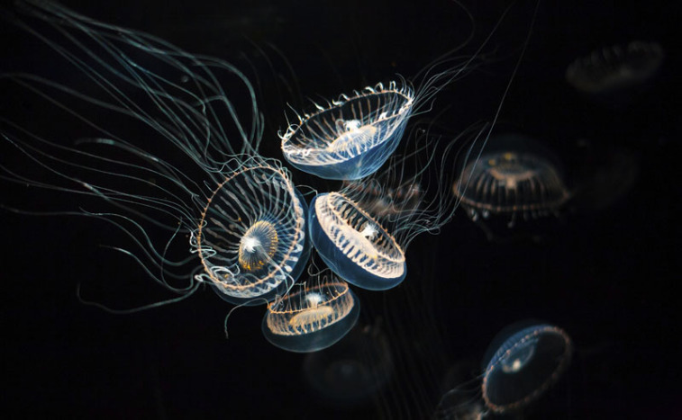
The crystal jellyfish has about 300 photo organs on the bottom edge of the jellyfish’s umbrella.Courtesy Steven Haddock – http://biolum.eemb.ucsb.edu, Author provided
The first fluorescent protein found in nature comes from the crystal jellyfish, Aequorea victoria, where it is responsible for the green light emitted by its photo organs. It’s called green fluorescent protein (GFP). We don’t know why these jellyfish have this lit-up feature.

Fluorescent proteins absorb light with short wavelengths, such as blue light, and immediately return it with a different color light that has a longer wavelength, such as green. In Aequorea victoria, a protein named aequorin produces blue light which GFP converts into the green light emitted by the jellyfish’s photo organs. This visibility under standard conditions is extremely rare; most other organisms have fluorescent proteins that are only visible if they are illuminated by external blue light sources.

Close up of a few of the photo organs. Courtesy Steven Haddock – http://biolum.eemb.ucsb.edu,Author provided
After the green fluorescent jellyfish protein, many other fluorescent proteins have been both found in nature and created in the lab. We now have a spectrum of fluorescent colors available to us that make previously invisible biological structures and processes visible in blazing fluorescent glory. Many new applications reliant on these colors are being published on a regular basis.
Petri dish with bacterial colonies expressing differently colored fluorescent proteins. These fluorescent proteins developed by Roger Tsien’s group are called the mFruits and have names like mHoneydew, mTomato, mCherry, mRaspberry, and mPlum. Paul Steinbach and Roger Y. Tsien, University of California, San DiegoCC BY-SA

Shining a light on imaging
Fluorescent protein technology has led to many other interesting developments designed to improve imaging with these glowing molecules.

CaMPARI is one new technique, short for calcium-modulated photoactivatable ratiometric integrator. By exploiting the fact that calcium concentrations change when nerve cells send signals, CaMPARI is able to light up all the neurons that have fired in a living organism. The technique is based on a fluorescent protein called EOS, which changes its fluorescence from green to red. In fruit flies, zebrafish and mice, CaMPARI-genetically-modified neurons fluoresce red if they are active and green if they are less active.

CaMPARI fluorescence in a larval zebrafish brain showing active neurons (magenta) that were marked while the fish was swimming freely. Looger Lab (HHMI/Janelia), Science, VOL 347, ISSUE 6223.

Before CaMPARI, all the fluorescent calcium indicators available temporarily lit up when the neuron fired. They couldn’t record the firing history of neurons or indicate whether a neuron had fired in the past. According to Loren Looger, one of the researchers who worked on the development of CaMPARI, “The most enabling thing about this technology may be that you don’t have to have your organism under a microscope during your experiment. So we can now visualize neural activity in fly larvae crawling on a plate or fish swimming in a dish.

The CLARITY technique removes opaque parts and makes the whole brain transparent.

Expanding and transparent brains
Even with the help of light emitted by fluorescent proteins, it’s difficult to image neurons tangled deep within the brain. Ed Boyden, a neuroscientist from MIT, has created a method to expand brains to make fluorescent neurons deep within the brain more visible. He uses acrylate, which forms a dense mesh to hold the brain in place and expand in the presence of water thereby inflating the brain equally by about 4.5 times in each direction. It’s a lot like a diaper expanding when it gets wet. Boyden thinks that this “expansion microscopy may provide a key tool for comprehensive, precise, circuit-wide, brain mapping.

Intact adult mouse brain before and after the CLARITY process. The Deisseroth Lab
One of the reasons expansion microscopy is so useful is that the brain can be made see-through before it is blown up several sizes larger. In 2013 Karl Deisseroth and Viviana Gradinaru at Stanford published a method called CLARITY that removes opaque molecules such as fats and makes the brain transparent without changing its shape. According to Thomas Insel, director of the US National Institute of Mental Health, “This is probably one of the most important advances for doing neuroanatomy in decades.” Since developing CLARITY for brains, Gradinaru has extended the method to all other organs including an entire mouse.

Both of these methods can be applied to brains that have been genetically modified with fluorescent proteins, therefore allowing for the visualization of neurons deep within the brain.

Mouse neurons labeled by GFPs. Wellcome ImagesCC BY-NC-ND
In 2008, the three scientists responsible for taking GFP from the jellyfish and making it a common tool used in over a million experiments all over the world were awarded the 100th Nobel Prize in chemistry. And in 2014 three other scientists were awarded the Nobel Prize for using fluorescent protein to increase the resolution of light microscopes.

E. coli with GFPs glowing in their petri dishes. Carlos de PazCC BY-NC-SA

Revolutionary and resilient
I’ve been researching the photochemistry and photophysics of fluorescent proteins since they were first used in imaging technology in 1994, I’ve written two books on them, and still I’m stunned by the many different ways in which this fairly simple protein can be used. Perhaps I shouldn’t be surprised that plasmid DNA molecules coding for GFP have survived space flight – not inside the rocket, but on the outside where they were exposed to 1800F (1000C) temperatures and mad friction. 53% of the DNA intentionally placed inside the screw heads in the TEXUS-49 rocket mission expressed fully fluorescent GFP when inserted into cells upon return to earth.

Like stars at night, fluorescent proteins have been lighting up science for the last 20 years. And it won’t be long before they’re guiding surgeons to tumorous growths during surgery and allowing researchers to switch on and off selected biomolecular processes.

ORIGINAL: The Conversation
By Marc Zimmer. Professor of Chemistry and Dean of Studies at Connecticut College
April 7 2015, 6.16am EDT

DISCLOSURE STATEMENT. Marc Zimmer receives funding from NIH.
The Conversation is funded by Gordon and Betty Moore Foundation, Howard Hughes Medical Institute, Robert Wood Johnson Foundation, Alfred P Sloan Foundation and William and Flora Hewlett Foundation. Our global publishing platform is funded by Commonwealth Bank of Australia.

martes, 31 de marzo de 2015

Bioluminescence: Nature’s Light Show [Photo Gallery]

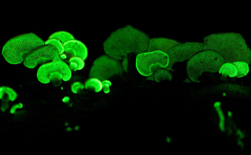
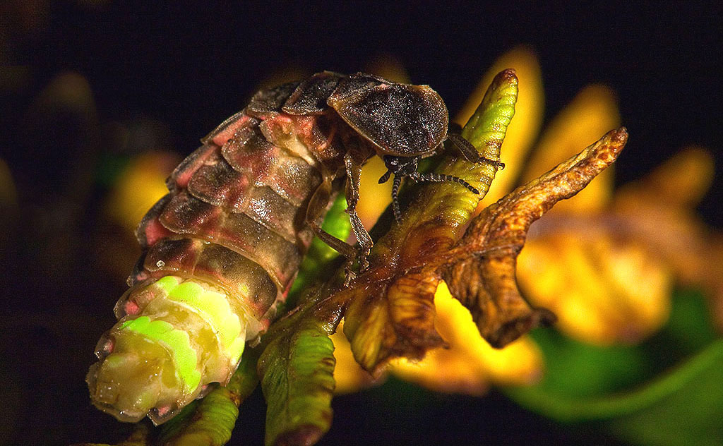
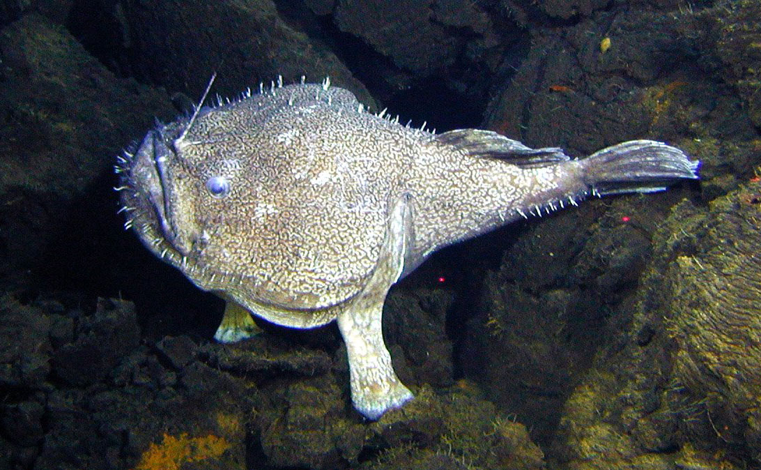
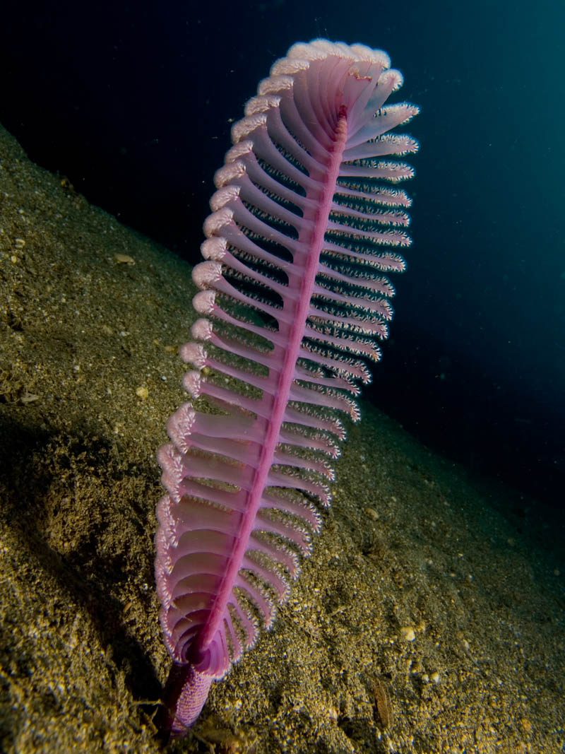
Long before humans figured out that sporting glowy lights could make you look really cool at dance parties, animals large and small had already figured out that light could be an advantage. Organisms that make their own light through bioluminescence use their flashy accessories to 
  • lure prey, 
  • attract mates, 
  • warn off predators, and 
  • perform a host of other functions.

Most bioluminescent organisms reside in the ocean, but landlubbers find ways to make good use of light too. Just recently, scientists discovered that bioluminescent mushrooms glow green in order to attract insects that help them disperse their spores, and while we’ve known of fireflies’ flashing abilities for a long time, late last year scientists learned how they actually add oxygen to their light-producing cells.

Science has taken advantage of natural glowing for its own ends—the Aequoria victoria jellyfish shown above was the source for the isolation of green fluorescent protein (GFP), which is used throughout biological experiments to measure gene expression. GFP, which earned its discoverers the 2008 Nobel Prize in Chemistry, lights up green when exposed to ultraviolet or blue light—the latter being produced by the jellyfish using the protein aequorin. Researchers have even isolated bioluminescent proteins from bacteria to create glow-in-the-dark plants. But equally beautiful specimens can be found out in nature. Here are some glowing examples.


Image Credit: istock / GaryKavanagh


ORIGINAL:
World Science Festival
03/31/15 

martes, 17 de marzo de 2015

Ido Bachelet DNA nanobots summary with a couple of extra videos

In a brief talk, Bachelet said DNA nanobots will soon be tried in a critically ill leukemia patient. The patient, who has been given roughly six months to live, will receive an injection of DNA nanobots designed to interact with and destroy leukemia cells—while causing virtually zero collateral damage in healthy tissue.

According to Bachelet, his team have successfully tested their method in cell cultures and animals and written two papers on the subject, one in Science and one in Nature.

Contemporary cancer therapies involving invasive surgery and blasts of drugs can be as painful and damaging to the body as the disease itself. If Bachelet's approach proves successful in humans, and is backed by more research in the coming years, the team’s work could signal a transformational moment in cancer treatment.

If this treatment works this will be a medical breakthrough and can be used for many other diseases by delivering drugs more effectively without causing side effects.

2012 Video with answers from George Church, Ido Bachelet and Shawn Douglas on the medical DNA double helix clamshell nanobucket nanobot


George Church indicates the smart DNA nanobot has applications beyond nanomedicine. Applications where there is any need for programmable and targeted release or interaction at the cellular or near molecular scale.

2014 Geek Time Presentation from Ido Bachelet


At the British Friends of Bar-Ilan University's event in Otto Uomo October 2014 Professor Ido Bachelet announced the beginning of the human treatment with nanomedicine. He indicates DNA nanobots can currently identify cells in humans with 12 different types of cancer tumors.

A human patient with late stage leukemia will be given DNA nanobot treatment. Without the DNA nanobot treatment the patient would be expected to die in the summer of 2015. Based upon animal trials they expect to remove the cancer within one month.

Within 1 or 2 years they hope to have spinal cord repair working in animals and then shortly thereafter in humans. This is working in tissue cultures. 

Previously Ido Bachelet and Shawn Douglas have published work on DNA nanobots in the journal Nature and other respected science publications.

One Trillion 50 nanometer nanobots in a syringe will be injected into people to perform cellular surgery.

The DNA nanobots have been tuned to not cause an immune response.

They have been adjusted for different kinds of medical procedures. Procedures can be quick or ones that last many days.


Medicine or treatment released based upon molecular sensing - Only targeted cells are treated


Ido's daughter has a leg disease which requires frequent surgery. He is hoping his DNA nanobots will make the type of surgery she needs relatively trivial - a simple injection at a doctor's office.

We can control powerful drugs that were already developed

Effective drugs that were withdrawn from the market for excessive toxicity can be combined with DNA nanobots for effective delivery. The tiny molecular computers of the DNA nanobots can provide molecular selective control for powerful medicines that were already developed.

Using DNA origami and molecular programming, they are reality. These nanobots can seek and kill cancer cells, mimic social insect behaviors, carry out logical operators like a computer in a living animal, and they can be controlled from an Xbox. Ido Bachelet from the bio-design lab at Bar Ilan University explains this technology and how it will change medicine in the near future.

Ido Bachelet earned his Ph.D. from the Hebrew University in Jerusalem, and was a postdoctoral fellow at M.I.T. and Harvard University. He is currently an assistant professor in the Faculty of Life Sciences and the Nano-Center at Bar Ilan University, Israel, the founder of several biotech companies, and a composer of music for piano and molecules.


Researchers have injected various kinds of DNA nanobots into cockroaches. Because the nanobots are labelled with fluorescent markers, the researchers can follow them and analyse how different robot combinations affect where substances are delivered. The team says the accuracy of delivery and control of the nanobots is equivalent to a computer system.
  • This is the development of the vision of nanomedicine. 
  • This is the realization of the power of DNA nanotechnology. 
  • This is programmable dna nanotechnology.
The DNA nanotechnology cannot perform atomically precise chemistry (yet), but having control of the DNA combined with advanced synthetic biology and control of proteins and nanoparticles is clearly developing into very interesting capabilities.

"This is the first time that biological therapy has been able to match how a computer processor works," says co-author Ido Bachelet of the Institute of Nanotechnology and Advanced Materials at Bar Ilan University.

The team says it should be possible to scale up the computing power in the cockroach to that of an 8-bit computer, equivalent to a Commodore 64 or Atari 800 from the 1980s. Goni-Moreno agrees that this is feasible. "The mechanism seems easy to scale up so the complexity of the computations will soon become higher," he says.

An obvious benefit of this technology would be cancer treatments, because these must be cell-specific and current treatments are not well-targeted. But a treatment like this in mammals must overcome the immune response triggered when a foreign object enters the body.

Bachelet is confident that the team can enhance the robots' stability so that they can survive in mammals. "There is no reason why preliminary trials on humans can't start within five years," he says

Biological systems are collections of discrete molecular objects that move around and collide with each other. Cells carry out elaborate processes by precisely controlling these collisions, but developing artificial machines that can interface with and control such interactions remains a significant challenge. DNA is a natural substrate for computing and has been used to implement a diverse set of mathematical problems, logic circuits and robotics. The molecule also interfaces naturally with living systems, and different forms of DNA-based biocomputing have already been demonstrated. Here, we show that DNA origami can be used to fabricate nanoscale robots that are capable of dynamically interacting with each other in a living animal. The interactions generate logical outputs, which are relayed to switch molecular payloads on or off. As a proof of principle, we use the system to create architectures that emulate various logic gates (AND, OR, XOR, NAND, NOT, CNOT and a half adder). Following an ex vivo prototyping phase, we successfully used the DNA origami robots in living cockroaches (Blaberus discoidalis) to control a molecule that targets their cells.


Ido Bachelet's moonshot to use nanorobotics for surgery has the potential to change lives globally. But who is the man behind the moonshot?

Ido graduated from the Hebrew University of Jerusalem with a PhD in pharmacology and experimental therapeutics. Afterwards he did two postdocs; one in engineering at MIT and one in synthetic biology in the lab of George Church at the Wyss Institute at Harvard. 

Now, his group at Bar-Ilan University designs and studies diverse technologies inspired by nature.

  • They will deliver enzymes that break down cells via programmable nanoparticles.
  • Delivering insulin to tell cells to grow and regenerate tissue at the desired location.
  • Surgery would be performed by putting the programmable nanoparticles into saline and injecting them into the body to seek out remove bad cells and grow new cells and perform other medical work.


Nanoparticles with computational logic has already been done

Load an ensemble of drugs into many particles for programmed release based on situation that is found in the body


viernes, 28 de noviembre de 2014

DNA Survives Re-Entry Into Earth's Atmosphere

photo credit: Adrian Mettauer. When the TEXUS-49 sounding rocket launched it carried DNA coatings on the outside of its payload

Double-stranded DNA molecules applied to the outside of a rocket payload have survived being blasted into space: they briefly entered near total vacuum and returned through the atmosphere. At the end of this, the molecules could still transfer genetic information.

Contrary to some misunderstandings, the Rosetta mission did not find DNA on Comet Churyumov-Gerasimenko 67P. It did however find organic molecules. This revived discussion of the possibility, known as panspermia” that life is distributed around the galaxy through molecules on comets or asteroids.

One of the challenges to this idea has been the question of whether something as complex as DNA could survive the extreme heat generated when entering the atmosphere. Exposure to cosmic rays or solar radiation while unprotected by the atmosphere represents another challenge to the theory.

However, the concept is looking more credible after a team from the University of Zurich pipetted DNA onto the outside of the TEXUS-49 sounding rocket, and collected it again after re-entry. They found the DNA could be inserted into bacteria and connective tissue cells and still function.

Dr. Cora Thiel came up with the idea of the experiment while planning to use TEXUS-49's payload to study how gene expression changes in human cells in zero gravity. She started thinking about the biosignatures, which she describes as “molecules that can prove the existence of past or present extraterrestrial life.

The outside of the same rocket looked like a particularly tough test – DNA cocooned inside an asteroid might have a better chance of surviving than something on a rocket's metallic surface.

Reporting in PLOS ONE, Thiel used DNA carrying a green fluorescent protein and an antibiotic resistance cassette and applied the material both on the front surface of the payload and in more protected spots at the bottom and in grooves where screws were inserted.

Gas temperatures at the front of the craft reached over 1000° C. Even inside, it got to 130° C. Some of the DNA had burned off, but 53% was recovered from the payload bottom and as much as 35% was intact enough to produce both fluorescent proteins and antibiotic resistance when inserted into E. coli. Control areas had no detectable DNA, eliminating the possibility of contamination after the return to Earth. Mutation rates were low.

Thiel and her co-authors note that the findings are important for future missions to other planets or moons. “For these missions, it is essential to know whether the detected biomarkers definitely originate from the analysed site or if they could be potential contamination from “stowaways” which traveled as hitchhikers on the spacecraft or analytical equipment,” they argue. Knowing just what DNA can survive will help future projects decide what scrubbing needs to be done before launch.

The flight was only 13 minutes long, so there was no time to see how the DNA stood up to the radiation in space, but the capacity to survive the heat of re-entry could reshape thinking.

ORIGINAL: IFLScience
by Stephen Luntz
November 27, 2014

lunes, 27 de enero de 2014

Scientists film how the brain makes memories for the first time ever


For the first time in history, scientists at Albert Einstein College of Medicine of Yeshiva University have captured how our brain makes memories in video, watching how molecules morph into the structures that, at the end of the day, make who we are. If there's a soul, this how it gets made.

Before this, Japanese scientists observed how a thought was formed, which was an entirely different process. According to the scientists, the has been a "technological tour de force:"

These insights into the molecular basis of memory were made possible by a technological tour de force never before achieved in animals: a mouse model developed at Einstein in which molecules crucial to making memories were given fluorescent "tags" so they could be observed traveling in real time in living brain cells.


The process, which has been documented in two Science papers, required researchers to "stimulate neurons from the mouse's hippocampus, where memories are made and stored, and then watched fluorescently glowing beta-actin mRNA molecules form in the nuclei of neurons and travel within dendrites, the neuron's branched projections."

What they have found is fascinating: "mRNA in neurons is regulated through a novel process described as "masking" and "unmasking," which allows beta-actin protein to be synthesized at specific times and places and in specific amounts."

This is precisely how scientists imagined things worked, according to Doctor Robert Singer, the research papers' senior author:

This observation that neurons selectively activate protein synthesis and then shut it off fits perfectly with how we think memories are made. Frequent stimulation of the neuron would make mRNA available in frequent, controlled bursts, causing beta-actin protein to accumulate precisely where it's needed to strengthen the synapse.

It kind of sounds like a computer storing data bits into a hard drive!


ORIGINAL: Sploid