Mostrando entradas con la etiqueta Image Processing. Mostrar todas las entradas

jueves, 2 de junio de 2016

See The Difference One Year Makes In Artificial Intelligence Research

AN IMPROVED WAY OF LEARNING ABOUT NEURAL NETWORKS

Google/ Geometric IntelligenceThe difference between Google's generated images of 2015, and the images generated in 2016.


Last June, Google wrote that it was teaching its artificial intelligence algorithms to generate images of objects, or "dream." The A.I. tried to generate pictures of things it had seen before, like dumbbells. But it ran into a few problems. It was able to successfully make objects shaped like dumbbells, but each had disembodied arms sticking out from the handles, because arms and dumbbells were closely associated. Over the course of a year, this process has become incredibly refined, meaning these algorithms are learning much more complete ideas about the world.

New research shows that even when trained on a standardized set of images,, A.I. can generate increasingly realistic images of objects that it's seen before. Through this, the researchers were also able to sequence the images and make low-resolution videos of actions like skydiving and playing violin. The paper, from the University of Wyoming, Albert Ludwigs University of Freiburg, and Geometric Intelligence, focuses on deep generator networks, which not only create these images but are able to show how each neuron in the network affects the entire system's understanding.

Looking at generated images from a model is important because it gives researchers a better idea about how their models process data. It's a way to take a look under the hood of algorithms that usually act independent of human intervention as they work. By seeing what computation each neuron in the network does, they can tweak the structure to be faster or more accurate.

"With real images, it is unclear which of their features a neuron has learned," the team wrote. "For example, if a neuron is activated by a picture of a lawn mower on grass, it is unclear if it ‘cares about’ the grass, but if an image...contains grass, we can be more confident the neuron has learned to pay attention to that context."

They're researching their research—and this gives a valuable tool to continue doing so.

Screenshot
Take a look at some other examples of images the A.I. was able to produce.

ORIGINAL: Popular Science
May 31, 2016

domingo, 17 de mayo de 2015

Silicon Chips That See Are Going to Make Your Smartphone Brilliant

Many gadgets will be able to understand images and video thanks to chips designed to run powerful artificial-intelligence algorithms.

WHY IT MATTERS
Many applications for mobile computers could be more powerful with advanced image recognition.

Many of the devices around us may soon acquire powerful new abilities to understand images and video, thanks to hardware designed for the machine-learning technique called deep learning.

Companies like Google have made breakthroughs in image and face recognition through deep learning, using giant data sets and powerful computers (see “10 Breakthrough Technologies 2013: Deep Learning”). Now two leading chip companies and the Chinese search giant Baidu say hardware is coming that will bring the technique to phones, cars, and more.

Chip manufacturers don’t typically disclose their new features in advance. But at a conference on computer vision Tuesday, Synopsys, a company that licenses software and intellectual property to the biggest names in chip making, showed off a new image-processor core tailored for deep learning. It is expected to be added to chips that power smartphones, cameras, and cars. The core would occupy about one square millimeter of space on a chip made with one of the most commonly used manufacturing technologies.

Pierre Paulin, a director of R&D at Synopsys, told MIT Technology Review that the new processor design will be made available to his company’s customers this summer. Many have expressed strong interest in getting hold of hardware to help deploy deep learning, he said.

Synopsys showed a demo in which the new design recognized speed-limit signs in footage from a car. Paulin also presented results from using the chip to run a deep-learning network trained to recognize faces. It didn’t hit the accuracy levels of the best research results, which have been achieved on powerful computers, but it came pretty close, he said. “For applications like video surveillance it performs very well,” he said. The specialized core uses significantly less power than a conventional chip would need to do the same task.

The new core could add a degree of visual intelligence to many kinds of devices, from phones to cheap security cameras. It wouldn’t allow devices to recognize tens of thousands of objects on their own, but Paulin said they might be able to recognize dozens.

That might lead to novel kinds of camera or photo apps. Paulin said the technology could also enhance car, traffic, and surveillance cameras. For example, a home security camera could start sending data over the Internet only when a human entered the frame. “You can do fancier things like detecting if someone has fallen on the subway,” he said.

Jeff Gehlhaar, vice president of technology at Qualcomm Research, spoke at the event about his company’s work on getting deep learning running on apps for existing phone hardware. He declined to discuss whether the company is planning to build support for deep learning into its chips. But speaking about the industry in general, he said that such chips are surely coming. Being able to use deep learning on mobile chips will be vital to helping robots navigate and interact with the world, he said, and to efforts to develop autonomous cars.

I think you will see custom hardware emerge to solve these problems,” he said. “Our traditional approaches to silicon are going to run out of gas, and we’ll have to roll up our sleeves and do things differently.” Gehlhaar didn’t indicate how soon that might be. Qualcomm has said that its coming generation of mobile chips will include software designed to bring deep learning to camera and other apps (see “Smartphones Will Soon Learn to Recognize Faces and More”).

Ren Wu, a researcher at Chinese search company Baidu, also said chips that support deep learning are needed for powerful research computers in daily use. “You need to deploy that intelligence everywhere, at any place or any time,” he said.

Being able to do things like analyze images on a device without connecting to the Internet can make apps faster and more energy-efficient because it isn’t necessary to send data to and fro, said Wu. He and Qualcomm’s Gehlhaar both said that making mobile devices more intelligent could temper the privacy implications of some apps by reducing the volume of personal data such as photos transmitted off a device.

You want the intelligence to filter out the raw data and only send the important information, the metadata, to the cloud,” said Wu.


ORIGINAL: Tech Review
May 14, 2015

miércoles, 8 de abril de 2015

Fluorescent proteins light up science by making the invisible visible

Multiple fluorescent proteins illuminate the cells in a human brainstem. Jeff Lichtman/Harvard University, CC BY-NC-ND

When you look up at the blue sky, where are the stars that you see at night? They’re there but we can’t see them. A firefly flitting across a field is invisible to us during the day, but at night we can easily spot its flashes. Similarly, proteins, viruses, parasites and bacteria inside living cells can’t be seen by the naked eye under normal conditions. But a technique using a fluorescent protein can light up cells' molecular machinations like a microscopic flashlight.

The crystal jellyfish has about 300 photo organs on the bottom edge of the jellyfish’s umbrella.Courtesy Steven Haddock – http://biolum.eemb.ucsb.edu, Author provided
The first fluorescent protein found in nature comes from the crystal jellyfish, Aequorea victoria, where it is responsible for the green light emitted by its photo organs. It’s called green fluorescent protein (GFP). We don’t know why these jellyfish have this lit-up feature.

Fluorescent proteins absorb light with short wavelengths, such as blue light, and immediately return it with a different color light that has a longer wavelength, such as green. In Aequorea victoria, a protein named aequorin produces blue light which GFP converts into the green light emitted by the jellyfish’s photo organs. This visibility under standard conditions is extremely rare; most other organisms have fluorescent proteins that are only visible if they are illuminated by external blue light sources.

Close up of a few of the photo organs. Courtesy Steven Haddock – http://biolum.eemb.ucsb.edu,Author provided
After the green fluorescent jellyfish protein, many other fluorescent proteins have been both found in nature and created in the lab. We now have a spectrum of fluorescent colors available to us that make previously invisible biological structures and processes visible in blazing fluorescent glory. Many new applications reliant on these colors are being published on a regular basis.
Petri dish with bacterial colonies expressing differently colored fluorescent proteins. These fluorescent proteins developed by Roger Tsien’s group are called the mFruits and have names like mHoneydew, mTomato, mCherry, mRaspberry, and mPlum. Paul Steinbach and Roger Y. Tsien, University of California, San DiegoCC BY-SA

Shining a light on imaging
Fluorescent protein technology has led to many other interesting developments designed to improve imaging with these glowing molecules.

CaMPARI is one new technique, short for calcium-modulated photoactivatable ratiometric integrator. By exploiting the fact that calcium concentrations change when nerve cells send signals, CaMPARI is able to light up all the neurons that have fired in a living organism. The technique is based on a fluorescent protein called EOS, which changes its fluorescence from green to red. In fruit flies, zebrafish and mice, CaMPARI-genetically-modified neurons fluoresce red if they are active and green if they are less active.

CaMPARI fluorescence in a larval zebrafish brain showing active neurons (magenta) that were marked while the fish was swimming freely. Looger Lab (HHMI/Janelia), Science, VOL 347, ISSUE 6223.

Before CaMPARI, all the fluorescent calcium indicators available temporarily lit up when the neuron fired. They couldn’t record the firing history of neurons or indicate whether a neuron had fired in the past. According to Loren Looger, one of the researchers who worked on the development of CaMPARI, “The most enabling thing about this technology may be that you don’t have to have your organism under a microscope during your experiment. So we can now visualize neural activity in fly larvae crawling on a plate or fish swimming in a dish.

The CLARITY technique removes opaque parts and makes the whole brain transparent.

Expanding and transparent brains
Even with the help of light emitted by fluorescent proteins, it’s difficult to image neurons tangled deep within the brain. Ed Boyden, a neuroscientist from MIT, has created a method to expand brains to make fluorescent neurons deep within the brain more visible. He uses acrylate, which forms a dense mesh to hold the brain in place and expand in the presence of water thereby inflating the brain equally by about 4.5 times in each direction. It’s a lot like a diaper expanding when it gets wet. Boyden thinks that this “expansion microscopy may provide a key tool for comprehensive, precise, circuit-wide, brain mapping.

Intact adult mouse brain before and after the CLARITY process. The Deisseroth Lab
One of the reasons expansion microscopy is so useful is that the brain can be made see-through before it is blown up several sizes larger. In 2013 Karl Deisseroth and Viviana Gradinaru at Stanford published a method called CLARITY that removes opaque molecules such as fats and makes the brain transparent without changing its shape. According to Thomas Insel, director of the US National Institute of Mental Health, “This is probably one of the most important advances for doing neuroanatomy in decades.” Since developing CLARITY for brains, Gradinaru has extended the method to all other organs including an entire mouse.

Both of these methods can be applied to brains that have been genetically modified with fluorescent proteins, therefore allowing for the visualization of neurons deep within the brain.

Mouse neurons labeled by GFPs. Wellcome ImagesCC BY-NC-ND
In 2008, the three scientists responsible for taking GFP from the jellyfish and making it a common tool used in over a million experiments all over the world were awarded the 100th Nobel Prize in chemistry. And in 2014 three other scientists were awarded the Nobel Prize for using fluorescent protein to increase the resolution of light microscopes.

E. coli with GFPs glowing in their petri dishes. Carlos de PazCC BY-NC-SA

Revolutionary and resilient
I’ve been researching the photochemistry and photophysics of fluorescent proteins since they were first used in imaging technology in 1994, I’ve written two books on them, and still I’m stunned by the many different ways in which this fairly simple protein can be used. Perhaps I shouldn’t be surprised that plasmid DNA molecules coding for GFP have survived space flight – not inside the rocket, but on the outside where they were exposed to 1800F (1000C) temperatures and mad friction. 53% of the DNA intentionally placed inside the screw heads in the TEXUS-49 rocket mission expressed fully fluorescent GFP when inserted into cells upon return to earth.

Like stars at night, fluorescent proteins have been lighting up science for the last 20 years. And it won’t be long before they’re guiding surgeons to tumorous growths during surgery and allowing researchers to switch on and off selected biomolecular processes.

ORIGINAL: The Conversation
By Marc Zimmer. Professor of Chemistry and Dean of Studies at Connecticut College
April 7 2015, 6.16am EDT

DISCLOSURE STATEMENT. Marc Zimmer receives funding from NIH.
The Conversation is funded by Gordon and Betty Moore Foundation, Howard Hughes Medical Institute, Robert Wood Johnson Foundation, Alfred P Sloan Foundation and William and Flora Hewlett Foundation. Our global publishing platform is funded by Commonwealth Bank of Australia.

domingo, 11 de enero de 2015

The Algorithm That Unscrambles Fractured Images

The ongoing revolution in image processing has produced yet another way to extract images from a complex environment.


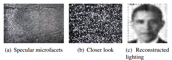
Take a hammer to a mirror and you will fracture the image it produces as well as the glass. Keep smashing and the image becomes more broken. When the pieces of glass are the size of glitter, the reflections will be random and the image unrecognisable.

It’s easy to imagine that reconstructing this image would be close to impossible. Not so, say Zhengdong Zhang and pals at the Massachusetts Institute of Technology in Cambridge. Today, these guys unveil SparkleVision, an image processing algorithm that reassembles the smashed imaged.

The problem that Zhang and co attack is to work out the contents of a picture reflected off a screen covered in glitter. The approach is to photograph the glitter and then process the resulting image in a way that unscrambles the picture.

It turns out that there is a straightforward way to approach this. Zhang and co consider each piece of glitter to be a randomly oriented micromirror. So light from the picture hits a micromirror and is reflected to a sensor inside the camera.

That means there is a simple mapping from each pixel in the original picture to a sensor in the camera. The task is to determine that mapping for every pixel. “There exists a forward scrambling matrix, and in principle we can find its inverse and unscramble the image,” they say.

To find this unscrambling matrix, Zhang and co shine a set of test images at the glitter screen and record where the pixels in the original image end up in the camera.

From this, they can create an algorithm that unscrambles any other image placed in exactly the same spot as the test images. They call this algorithm SparkleVision.


That’s a handy piece of software that could have interesting applications in retrieving images reflected off glitter-like surfaces such as certain types of foliage, wet surfaces, metals and so on.

And Zhang and co hope to make the software more useful. In its current incarnation, the software can only unscramble images placed in the exact location of the test images. But in theory, the test images should provide enough data to unscramble images from any part of the light field. “Thus, our system could be naturally extended to work as a lightfield camera,” they say.

The work is part of a growing body that is currently revolutionising photography and image processing, Other researchers have worked out how to unscramble images from all kinds of distorted reflections and surfaces, sometimes even without using lenses.

These so-called “random cameras” are dramatically widening the capability of optics specialists. And SparkleVision looks set to take its place among them.

Ref: http://arxiv.org/abs/1412.7884 : SparkleVision: Seeing the World through Random Specular Microfacets

ORIGINAL: Technology Review