Mostrando entradas con la etiqueta Ratón. Mostrar todas las entradas

martes, 26 de marzo de 2013

Mice get brain boost from transplanted human tissue

ORIGINAL: Science News
March 7, 2013 

Study suggests support cells may enhance people’s thinking prowess
A CHANGE OF VENUE. Human astrocytes (large yellow-green cells with white nuclei) grow in the brain of a 10-month-old mouse. Mouse cell nuclei are stained blue. Mice with human astrocytes implanted in their brains perform better on learning and memory tests than normal mice do, suggesting that human astrocytes are better than their rodent counterparts at modulating learning. X. Han et al/Cell Stem Cell 2013

Transplanting human brain cells into mice makes the mice smarter, a new study shows.

But the smart-making brain cells are not the nerve cells most people think of as controlling thoughts. Instead, they are part of the supporting cast of brain cells known as glia (Greek for “glue”).

Scientists have long seen glia, including a subset known as astrocytes, as support cells that feed neurons, mop up excess neurotransmitters and generally help hold the brain together. The new study, published March 7 in Cell Stem Cell, shows that glial cells also influence memory formation and could change how scientists think the brain works, says R. Douglas Fields of the National Institute of Child Health and Human Development. “It’s a paradigm-shifting paper,” says Fields, who was not involved in the work.

In the new study, researchers led by neurologist and stem cell biologist Steven Goldman and neurobiologist Maiken Nedergaard of the University of Rochester Medical Center in New York implanted human cells called glial progenitor cells into the brains of newborn mice. Glial progenitor cells are a type of stem cell that is poised to make several varieties of glia, including astrocytes. Previously, the researchers had transplanted human glial progenitor cells into the brains of mice that had a genetic disorder mimicking multiple sclerosis. The glial progenitor cells healed the mice, allowing them to live a normal life span. That result held promise that such cell transplants might help people with neurological disorders.
WELL-CONNECTED. A human astrocyte (green) sends out more tentacles and monitors many more neuron-to-neuron connections than mouse astrocytes (red) do. Mice with human astrocytes implanted in their brains do better on learning and memory tests than normal mice do, suggesting that human astrocytes are better than their rodent counterparts at modulating learning.
X. Han et al/Cell Stem Cell 2013

The researchers also noticed something curious in the brains of mice that had received human cell transplants. “The shocker was that all the glial progenitors were human and had completely taken over the mouse progenitors,” Goldman says. The finding made the researchers wonder what effect human cells might have on otherwise normal mice.

Although many neuroscientists essentially ignore glia, it is becoming clear that the cells — which make up about 90 percent of the brain — are more important than some people believe. Astrocytes are required for nerve cells, or neurons, to make connections, called synapses, with each other. While neurons pretty much look and behave the same from species to species, human astrocytes are much larger and more complex than those from other species, leading some scientists to wonder whether the cells are at least partly responsible for the human brain’s computing power.

To find out, the Rochester researchers tested human glial progenitor cells in the brains of normal mice. By the time the mice were 6 months old, the human cells had pushed out the mouse progenitor cells and replaced many of the mouse astrocytes with human astrocytes. Some mice got a transplant of mouse glial progenitors instead of human cells to make sure any effect was due to the action of human cells and not to having extra brain cells.

Astrocytes use calcium to communicate. In lab dish tests, human astrocytes passed calcium signals three times faster than mouse astrocytes did. And the human astrocytes helped forge stronger synapses between mouse neurons than the mouse’s own astrocytes did.

The researchers also put mice through a battery of tests, probing the animals’ ability to learn mazes, distinguish new objects from old ones, and learn that a certain sound portends a mild electric shock. It took normal mice and mice with mouse cell transplants several tries to pick up on the association between the sound and the shock. Mice with human astrocytes “pretty much picked up the association immediately and got more fearful,” Goldman says.

Since the mice have their own neurons, the memory boost must have come from the human cells, the researchers conclude. While evidence points to the astrocytes as the source of the enhancement, the researchers can’t rule out that undeveloped progenitor cells might also contribute.

In any case, the results indicate that human cells not only aid in learning and memory, but do it better than their rodent counterparts do.“It’s a stunning result. It provides the first unequivocal evidence that astrocytes may well have been one of the evolutionary drivers of human capabilities,” says Bruce Ransom, a neuroscientist at the University of Washington. “As completely outrageous as it sounds, I think the evidence is such now that we have to take that very seriously.

CITATIONS
X. Han et al. Forebrain engraftment by human glial progenitor cells enhances synaptic plasticity and learning in adult mice. Cell Stem Cell. Vol. 12, March 7, 2013, p. 342. doi: 10.1016/j.stem.2012.12.015. [Go to]


SUGGESTED READING

T. Hesman Saey. Astrocytes are rising stars. Science News. Vol. 174, August 2, 2008, p. 5. Available online: [Go to]

J. Netting. Gray matters. Science News. Vol. 159, April 7, 2001, p. 222. Available online: [Go to]

J. Travis. Protein triggers nerve connections. Science News. Vol. 164, November 29, 2003, p. 350. Available online: [Go to]

domingo, 27 de enero de 2013

Mice With Firefly Genes Glow in Response to Tumor Growth

ORIGINAL: MedGadget
Jan 24, 2013
The progression of P16 increases in mice as they age visible from the younger mice (left) to the older mice (right).
Researchers at University of North Carolina have engineered laboratory mice which exhibit a firefly gene that could help scientists study cancer development. The p16INK4a (p16) gene is known to play a role in tumor suppression, so the team introduced the firefly gene so that it would be activated whenever the p16 gene is.

Add caption
Over time the mice with induced tumors were tracked and the glow was used to follow the activity of the p16 gene as it reacted to tumor progression. Some findings were that older mice glowed brighter, as expected, and the sites where cancer seemed to originate were particularly luminescent.

The researchers used these mice to make several unexpected discoveries. First, the group was able to track the accumulation of senescent cells in aging mice by assessing how brightly each mouse glowed. Surprisingly, the brightest animals were no more likely to die from spontaneous cancer than dimmer animals of the same age. That is, the number of senescent cells in the mouse did not predict its risk of dying.

Another surprise came from the disparities in p16 levels among the mice. The authors studied a large group of genetically identical animals that were all housed in the same way and fed the same diet. However, despite identical genetic and environmental conditions, the brightness of individual mice at any given age was highly variable, suggesting that factors beyond genetics and diet influence aging.

The glowing mice also provide a window into the formation of cancers. Expression of p16 is activated in the earliest stages of cancer formation to suppress cancer. Usually activation of p16 prevents cancer, but rarely this tumor suppressor mechanism fails and tumors develop, while still activating the p16 gene. As such, all tumors forming in these mice strongly glowed, allowing researchers to monitor early tumor formation in a wide variety of cancer types. In contrast to expectations, the researchers also found that p16 was activated not only in the tumor cells themselves, but also in normal, neighboring cells.

martes, 14 de agosto de 2012

Chemical makes blind mice see

ORIGINAL: Berkeley
By Robert Sanders, Media Relations | July 25, 2012
BERKELEY —

Mice with a genetic disease that causes blindness regained some sight after injection with a chemical “photoswitch.” The eye of the untreated mouse on the left shows no response to light, while the pupil of the mouse on the right, which was injected with the chemical, contracts in light.

Mice with a genetic disease that causes blindness regained some sight after injection with a chemical “photoswitch. The eye of the untreated mouse on the left shows no response to light, while the pupil of the mouse on the right, which was injected with the chemical, contracts in light.

A team of University of California, Berkeley, scientists in collaboration with researchers at the University of Munich and University of Washington, in Seattle, has discovered a chemical that temporarily restores some vision to blind mice, and is working on an improved compound that may someday allow people with degenerative blindness to see again.

The approach could eventually help those with retinitis pigmentosa, a genetic disease that is the most common inherited form of blindness, as well as age-related macular degeneration, the most common cause of acquired blindness in the developed world. In both diseases, the light sensitive cells in the retina — the rods and cones — die, leaving the eye without functional photoreceptors.

The chemical, called AAQ, acts by making the remaining, normally “blind” cells in the retina sensitive to light, said lead researcher Richard Kramer, UC Berkeley professor of molecular and cell biology. AAQ is a photoswitch that binds to protein ion channels on the surface of retinal cells. When switched on by light, AAQ alters the flow of ions through the channels and activates these neurons much the way rods and cones are activated by light.

This is similar to the way local anesthetics work: they embed themselves in ion channels and stick around for a long time, so that you stay numb for a long time,” Kramer said. “Our molecule is different in that it’s light sensitive, so you can turn it on and off and turn on or off neural activity.

Because the chemical eventually wears off, it may offer a safer alternative to other experimental approaches for restoring sight, such as gene or stem cell therapies, which permanently change the retina. It is also less invasive than implanting light-sensitive electronic chips in the eye.

The advantage of this approach is that it is a simple chemical, which means that you can change the dosage, you can use it in combination with other therapies, or you can discontinue the therapy if you don’t like the results. As improved chemicals become available, you could offer them to patients. You can’t do that when you surgically implant a chip or after you genetically modify somebody,” Kramer said.

This is a major advance in the field of vision restoration,” said co-author Dr. Russell Van Gelder, an ophthalmologist and chair of the Department of Ophthalmology at the University of Washington, Seattle.

Kramer, Van Gelder, chemist Dirk Trauner and their colleagues at UC Berkeley, the University of Washington, Seattle, and the University of Munich will publish their findings Thursday, July 26, in the journal Neuron.

The blind mice in the experiment had genetic mutations that made their rods and cones die within months of birth and inactivated other photopigments in the eye. After injecting very small amounts of AAQ into the eyes of the blind mice, Kramer and his colleagues confirmed that they had restored light sensitivity because the mice’s pupils contracted in bright light, and the mice showed light avoidance, a typical rodent behavior impossible without the animals being able to see some light. Kramer is hoping to conduct more sophisticated vision tests in rodents injected with the next generation of the compound.

The photoswitch approach offers real hope to patients with retinal degeneration,” Van Gelder said. “We still need to show that these compounds are safe and will work in people the way they work in mice, but these results demonstrate that this class of compound restores light sensitivity to retinas blind from genetic disease.

From optogenetics to implanted chips

The current technologies being evaluated for restoring sight to people whose rods and cones have died include injection of stem cells to regenerate the rods and cones; “optogenetics,” that is, gene therapy to insert a photoreceptor gene into blind neurons to make them sensitive to light; and installation of electronic prosthetic devices, such as a small light-sensitive retinal chip with electrodes that stimulate blind neurons. Several dozen people already have retinal implants and have had rudimentary, low vision restored, Kramer said.

Eight years ago, Kramer, Trauner, a former UC Berkeley chemist now at the University of Munich, and their colleagues developed an optogenetic technique to chemically alter potassium ion channels in blind neurons so that a photoswitch could latch on. Potassium channels normally open to turn a cell off, but with the attached photoswitch, they were opened when hit by ultraviolet light and closed when hit by green light, thereby activating and deactivating the neurons.

Subsequently, Trauner synthesized AAQ (acrylamide-azobenzene-quaternary ammonium), a photoswitch that attaches to potassium channels without the need to genetically modify the channel. Tests of this compound are reported in the current Neuron paper.

New versions of AAQ now being tested are better, Kramer said. They activate neurons for days rather than hours using blue-green light of moderate intensity, and these photoswitches naturally deactivate in darkness, so that a second color of light is not needed to switch them off.

This is what we are really excited about,” he said.

Coauthors with Kramer, Van Gelder and Trauner are UC Berkeley current or former post-docs or graduate students Aleksandra Polosukhina, Jeffrey Litt, Ivan Tochitsky, Ivan De Kouchkovsky, Tracy Huang and Katharine Borges; and post-doctoral fellow Joseph Nemargut and ophthalmology resident Yivgeny Sychev at the University of Washington.

The work was supported by the National Eye Institute of the National Institutes of Health (EY018957 & EY003176) and Research to Prevent Blindness.

RELATED INFORMATION
Richard Kramer laboratory Web site

miércoles, 6 de junio de 2012

Neuroscientists reach major milestone in whole-brain circuit mapping project

May 31, 2012

Projections from a motor cortex AAV injection (credit: CSHL)
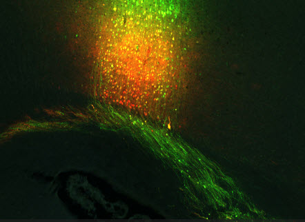
Cold Spring Harbor, NY – Neuroscientists at Cold Spring Harbor Laboratory (CSHL) reached an important milestone today, publicly releasing the first installment of data from the 500 terabytes so far collected in their pathbreaking project to construct the first whole-brain wiring diagram of a vertebrate brain, that of the mouse

Add caption
The data consist of gigapixel images (each close to 1 billion pixels) of whole-brain sections that can be zoomed to show individual neurons and their processes, providing a “virtual microscope.” The images are integrated with other data sources from the web, and are being made fully accessible to neuroscientists as well as interested members of the general public (http://mouse.brainarchitecture.org). The data are being released pre-publication in the spirit of open science initiatives that have become familiar in digital astronomy (e.g., Sloan Digital Sky Survey) but are not yet as widespread in neurobiology. 


Each sampled brain is represented in about 500 images, each image showing an optical section through a 20 micron-thick slice of brain tissue. A multi-resolution viewer permits users to journey through each brain from “front” to “back,” and thus enables them to follow the pathways taken through three-dimensional brain space by tracer-labeled neuronal pathways. The tracers were picked to follow neuronal inputs and outputs of given brain regions.

We’re executing a grid-based “shotgun” strategy for neuronal tract tracing that we first proposed a few years ago, and which I am pleased to note has gained acceptance elsewhere within the neuroscience community,” says Partha P. Mitra, Ph.D., the Crick-Clay Professor of Biomathematics at CSHL and director of the Mouse Brain Architecture (MBA) Project. After the initial June 1 release, project data will be made public continuously on a monthly basis, Mitra says.

Project addresses a large gap in knowledge

Our project seeks to address a remarkable gap in our knowledge of the brain,” Mitra explains. “Our knowledge of how the brain is wired remains piecemeal and partial after a century of intense activity. Francis Crick and Ted Jones emphasized this in an article published in Nature nearly 20 years ago. Yet to understand how the brain works (or fails to work in neurological or neuropsychiatric disease), it is critical that we understand this wiring diagram more fully. Further, there remain fundamental questions about brain evolution that cannot be addressed without obtaining such wiring diagrams for the brains of different species.


martes, 1 de mayo de 2012

Compañía de cosméticos combate la experimentación con animales mediante "La tortura" de seres humanos

ORIGINAL: Ecouterre

Cosméticos Lush ha asociado con la Sociedad Humanitaria de los Estados Unidos y la Humane Society International para poner en marcha la campaña mundial más grande de la historia para poner fin a las pruebas de cosméticos en animales. Sin embargo, el proveedor de productos para el cuidado de la piel no se detuvo allí. Para subrayar su punto, LUSH convirtió la vitrina de su tienda de Regent Street en Londres en un cuadro de la crueldad, donde la artista de performance Traides Jacqueline "sufrió" 10 horas de alimentación forzada, inyecciones de líquidos, y otros procedimientos de tortura empleados en forma regular por la industria de la belleza

No era un espectáculo agradable, ni era la intención de cosméticos LUSH que lo fuera.

Aunque los ensayos con animales se prohibieron en toda Europa hace casi dos décadas, la venta de los animales los productos ensayados todavía se permite en el Reino Unido y otros países.
En los Estados Unidos, la ley federal exige a las empresas asegurar que sus productos son seguros, pero no requiere la experimentación con animales.


"La industria de la belleza continúa la experimentación con animales innecesaria y es hora de que deje de hacerlo" dice Kate Willett, director de toxicología regulatoria, evaluación de riesgos y alternativas de HSUS. "Los consumidores no tienen mucho conocimiento de que algunos champús y lápices de labios todavía implican envenenamiento químico de animales como conejos y ratones. La ciencia nos ha ido más allá de esto y no hay lugar para las pruebas de cosméticos en animales en la sociedad moderna."


"La lucha contra la experimentación con animales" es la campaña de parte de la conciencia, la petición de parte. A medida que la iniciativa se lanza en 48 países y más de 700 tiendas de LUSH en los Estados Unidos, Canadá, Europa, India, Australia, Nueva Zelanda, Corea del Sur, y Rusia, los miembros del público se les insta a firmar peticiones nacionales en las tiendas de LUSH y en línea para exigir el fin a la práctica anticuada.

"Los animales no deberían tener que depender de los códigos voluntarios de conducta, sino que debe ser protegida por las leyes que obligan a todos los sólidos que las empresas adopten métodos humanitarios para llevar sus productos al mercado", dice Brandi Salas, director de la campaña del Norte de América de Lush.


miércoles, 25 de enero de 2012

Reglamentación sobre Animales de Laboratorio: ¿Cuánto espacio necesitan?

ORIGINAL: Huffington Post
De Russell McLendon y la Red de la Madre Naturaleza:


Fuente: http://www.sciencedaily.com/releases/2007/12/071219122855.htm
Los animales de laboratorio han hecho mucho por la humanidad en los últimos años, nos han ayudado a encontrar tratamientos y curas para una variedad de enfermedades humanas. Los EE.UU. está tratando de retribuír a los animales de laboratorio su servicio - incluso si se trata de servicios involuntarios - por la promulgación de la primera actualización de su "Guía para el Cuidado y Uso de Animales de Laboratorio" desde 1996.

Esa actualización, de acuerdo con la Oficina de Bienestar de Animales de Laboratorio de los EE.UU. "faculta continuo avance en el cuidado humano y el uso de animales vertebrados en la investigación, la formación en investigación y pruebas biológicas".

La última edición de la guía, que fue presentada el año pasado y apenas entró en vigor este mes, incluye nuevos requisitos de espacio para una amplia gama de animales de laboratorio, desde las ratas y conejos a pollos y chimpancés. Los activistas por los Derechos de los animales, han pasado años presionando por esos cambios, y que en gran medida dan bienvenida a la nueva normativa.

Pero como informa NPR, algunos investigadores están erizados en la idea de comprar nuevas jaulas para los miles de ratas.

"El efecto sería, tendríamos que comprar más de estas jaula, y nuestra estimación fue de alrededor $ 300.000 dólares en jaulas, por lo menos", informó Bob Adams, el jefe del principal  complejo de ratas de laboratorio en Universidad Johns Hopkins, dice NPR. "En resumidas cuentas, hay más trabajo, hay más costos para todos, y para nuestra operación."

fuente: http://www.medicalnewstoday.com/articles/119170.php
No es que los científicos se muestran ambivalentes sobre el bienestar animal, añade José Thulin del Colegio Médico de Wisconsin. "Yo no quiero que nadie piense que la comunidad científica no quiere poner en práctica las nuevas directrices, y que no se preocupan por sus animales", dice a NPR. "Ese no es el caso en lo absoluto." No es simplemente una falta de investigación sobre la vivienda de los roedores, dice, creando dudas acerca "del impacto positivo y medible en los animales resultado de la ampliación de los tamaños des jaulas."

Las instituciones de investigación deben completar al menos una auto-evaluación en el marco de la nueva normativa antes del 31 de diciembre 2012, y hay una cierta confusión sobre la cantidad de espacio de maniobra que tendrán. Un consultor que ayudó a redactar las reglas de las que describe simplemente como "recomendaciones", por ejemplo, sin embargo, el sitio web OLAW (Oficina para el Bienestar de los Animales de Laboratorio) advierte de que "está totalmente prohibida, toda  desviaciones del programa de la Guía por razones de conveniencia, costo u otras consideraciones de bienestar no relacionadas con los animales, no son aceptables".

Y mientras que los críticos censuran la "incertidumbre" que esto crea, algunos defensores de los derechos de los animales expresan su preocupación que los EE.UU. puedan incumplir las nuevas reglas. "Si se va a utilizar millones o decenas de millones de roedores en este país, tenemos una obligación con el bienestar de estos animales" dijo a NPR, Kathleen Conlee de la Sociedad Protectora de Animales de Estados Unidos. Los grupos de HSUS y similares "tiene la esperanza de que el Instituto Nacionales de Salud (NIH) hará cumplir estrictamente las recomendaciones de la guía", añade.

La Oficina de Bienestar de Animales de Laboratorio recibirá comentarios del público sobre las nuevas normas hasta el final de este mes.

Consulte la guía completa (PDF) para un vistazo a algunas de las necesidades actualizadas.