Mostrando entradas con la etiqueta humanos. Mostrar todas las entradas

miércoles, 4 de diciembre de 2013

Scientists Just Sequenced the DNA From A 400,000-Year-Old Early Human

DNA from a group of ancient human fossils found in Spain (above), has been mysteriously related to an ancient lineage called the Denisovans, previously found only in Siberia. Photo by Javier Trueba, Madrid Scientific Films
Since its discovery in 1990, La Sima de los Huesos, an underground cave in Northern Spain’s Atapuerca Mountains, has yielded more than 6,000 fossils from 28 individual ancient human ancestors, making it Europe’s most significant site for the study of ancient humans. But despite years of analysis, the exact age and even the species to which these individuals belonged has been in doubt.

Now, though, an international group of scientists has extracted and sequenced DNA from the fossilized femur of one of these individuals for the first time. The resulting data—which represent the oldest genetic material ever sequenced from a hominin, or ancient human ancestor—finally give us an idea of the age and lineage of these mysterious individuals, and it’s not what many scientists expected.

The fossilized bone tested, a femur, is roughly 400,000 years old. But the big surprise is that, although scientists had previously believed the fossils belonged to Neanderthals because of their anatomical appearance, the DNA analysis actually shows they’re more closely related to Denisovans, a recently-discovered third lineage of human ancestors known only from DNA isolated from a few fossils found in Siberia in 2010. The findings, published today in Nature, will force anthropologists to further reconsider how the Denisovans, Neanderthals and the direct ancestors of modern-day humans fit together in a complicated family tree.
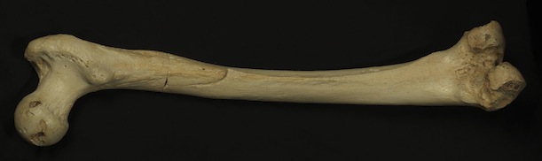
The femur from which DNA was extracted for analysis. Photo by Javier Trueba, Madrid Scientific Films
The analysis was enabled by recent advances in methods for recovering ancient DNA fragments developed at the Max Planck Institute for Evolutionary Anthropology in Germany, previously used to analyze the DNA of a cave bear fossil found in the same cave. “This wouldn’t have been possible just two years ago,” says Juan Luis Arsuaga, a paleontologist at the University of Madrid who led the initial excavations of the cave and collaborated on the new study. “And even given these new methods, we still didn’t expect these bones to preserve DNA, because they’re so old—ten times older than some of the oldest Neanderthals from whom we’ve taken DNA.

After extracting a two grams of crushed bone from the femur, a group of scientists led by Matthias Meyer isolated the mitochondrial DNA (mtDNA), a pool of genetic material that’s distinct from the DNA in the chromosomes located in our cells’ nuclei. Instead, this mtDNA lives in our cells’ mitochondria—microscopic organelles responsible for cellular respiration—and is much shorter in length than nuclear DNA.

There’s another quirk of mtDNA that makes it especially valuable as a means of studying the evolution of ancient humans: Unlike your nuclear DNA, which is a mix of DNA from both your parents, your mtDNA comes solely from your mother, because most of a sperm’s mitochondria are found in its tail, which it sheds after fertilization. As a result, mtDNA is nearly identical from generation to generation, and a limited number of distinct sequences of mtDNA (called haplogroups) have been observed in both modern humans and ancient human ancestors. Unlike anatomical characteristics and nuclear DNA, which can vary within a group and make it difficult to confidently distinguish one from another, mtDNA is generally consistent, making it easier to link a particular specimen with a lineage.

Which is why, when the researchers compared the femur’s mtDNA to previously sequenced samples from Neanderthals, from a Denisovan finger bone and tooth found in Siberia and from many different modern humans, they found it so surprising that it more closely resembled the Denisovans. “This was really unexpected,” Arsuaga says. “We had to think really hard to come up with a few scenarios that could potentially explain this.

Anthropologists had already known that all three lineages (humans, Neanderthals and Denisovans) shared a common ancestor, but it’s far from clear how all three groups fit together, and the picture is further clouded by the fact that interbreeding may have occurred between them after they diverged. Helpfully, comparing the femur’s mtDNA to the Neanderthal, Denisovan and modern human samples allowed the researchers to estimate its age—based upon known rates of mtDNA mutation, the previously established ages of the other samples, and the degree of difference between them—leading to the 400,000 year figure.

To explain how a Neanderthal-looking individual could come to have Denisovan mtDNA during this time period, the scientists present several different hypothetical scenarios. It’s possible, for instance, that
  • the fossil in question belongs to a lineage that served as ancestors of both Neanderthals and Denisovans, or more likely, one that came after the split between the two groups (estimated to be around 1 million years ago) and was closely related to the latter but not the former. 
  • It’s also a possibility that the femur belongs to a third, different group, and that its similarities to Denisovan mtDNA are explained by either interbreeding with the Denisovans or the existence of yet another hominin lineage that bred with both Denisovans and the La Sima de los Huesos population and introduced the same mtDNA to both groups.
If this sounds like a complicated family tree to you, you’re not alone. This analysis, along with earlier work, adds further mystery to an already puzzling situation. Initial testing on the Denisovan finger bone found in Siberia, for instance, found that it shared mtDNA with modern humans living in New Guinea, but nowhere else. Meanwhile, it was previously thought that Neanderthals had settled in Europe and Denisovans further east, on the other side of the Ural Mountains. The new analysis complicates that idea.

For now, the researchers believe the most plausible scenario (illustrated below) is the femur belongs to a lineage that split off from Denisovans sometime after they diverged from the common ancestor of both Neanderthals and modern humans. But perhaps the most exciting conclusion to come out of this work is that it proves that genetic material can survive for at least 400,000 years, and can be analyzed even after that amount of degradation. Armed with this knowledge and the new techniques, anthropologists can now attempt to genetically survey many other ancient specimens in hopes of better understanding our family tree.
Image via Nature/Meyer et. al.
December 4, 2013

miércoles, 28 de agosto de 2013

Researcher controls colleague's motions in first human brain-to-brain interface (w/ Video)

ORIGINAL: MedicalXPress
by Doree Armstrong & Michelle Ma

University of Washington researcher Rajesh Rao, left, plays a computer game with his mind. Across campus, researcher Andrea Stocco, right, wears a magnetic stimulation coil over the left motor cortex region of his brain. Stocco's right index
 (Medical Xpress)—University of Washington researchers have performed what they believe is the first noninvasive human-to-human brain interface, with one researcher able to send a brain signal via the Internet to control the hand motions of a fellow researcher.

Using electrical brain recordings and a form of magnetic stimulation, Rajesh Rao sent a brain signal to Andrea Stocco on the other side of the UW campus, causing Stocco's finger to move on a keyboard.

While researchers at Duke University have demonstrated brain-to-brain communication between two rats, and Harvard researchers have demonstrated it between a human and a rat, Rao and Stocco believe this is the first demonstration of human-to-human brain interfacing.

"The Internet was a way to connect computers, and now it can be a way to connect brains," Stocco said. "We want to take the knowledge of a brain and transmit it directly from brain to brain."

The researchers captured the full demonstration on video recorded in both labs. The version available at the end of this release has been edited for length.

Rao, a UW professor of computer science and engineering, has been working on brain-computer interfacing (BCI) in his lab for more than 10 years and just published a textbook on the subject. In 2011, spurred by the rapid advances in BCI technology, he believed he could demonstrate the concept of human brain-to-brain interfacing. So he partnered with Stocco, a UW research assistant professor in psychology at the UW's Institute for Learning & Brain Sciences.

On Aug. 12, Rao sat in his lab wearing a cap with electrodes hooked up to an electroencephalography machine, which reads electrical activity in the brain. Stocco was in his lab across campus wearing a purple swim cap marked with the stimulation site for the transcranial magnetic stimulation coil that was placed directly over his left motor cortex, which controls hand movement.

This image shows the cycle of the experiment. Brain signals from the "Sender" are recorded. When the computer detects imagined hand movements, a "fire" command is transmitted over the Internet to the TMS machine, which causes an upward 
The team had a Skype connection set up so the two labs could coordinate, though neither Rao nor Stocco could see the Skype screens.

Rao looked at a computer screen and played a simple video game with his mind. When he was supposed to fire a cannon at a target, he imagined moving his right hand (being careful not to actually move his hand), causing a cursor to hit the "fire" button. Almost instantaneously, Stocco, who wore noise-canceling earbuds and wasn't looking at a computer screen, involuntarily moved his right index finger to push the space bar on the keyboard in front of him, as if firing the cannon. Stocco compared the feeling of his hand moving involuntarily to that of a nervous tic.

"It was both exciting and eerie to watch an imagined action from my brain get translated into actual action by another brain," Rao said. "This was basically a one-way flow of information from my brain to his. The next step is having a more equitable two-way conversation directly between the two brains."


The technologies used by the researchers for recording and stimulating the brain are both well-known. Electroencephalography, or EEG, is routinely used by clinicians and researchers to record brain activity noninvasively from the scalp. Transcranial magnetic stimulation, or TMS, is a noninvasive way of delivering stimulation to the brain to elicit a response. Its effect depends on where the coil is placed; in this case, it was placed directly over the brain region that controls a person's right hand. By activating these neurons, the stimulation convinced the brain that it needed to move the right hand.

Computer science and engineering undergraduates Matthew Bryan, Bryan Djunaedi, Joseph Wu and Alex Dadgar, along with bioengineering graduate student Dev Sarma, wrote the computer code for the project, translating Rao's brain signals into a command for Stocco's brain.

"Brain-computer interface is something people have been talking about for a long, long time," said Chantel Prat, assistant professor in psychology at the UW's Institute for Learning & Brain Sciences, and Stocco's wife and research partner who helped conduct the experiment. "We plugged a brain into the most complex computer anyone has ever studied, and that is another brain."

At first blush, this breakthrough brings to mind all kinds of science fiction scenarios. Stocco jokingly referred to it as a "Vulcan mind meld." But Rao cautioned this technology only reads certain kinds of simple brain signals, not a person's thoughts. And it doesn't give anyone the ability to control your actions against your will.

Both researchers were in the lab wearing highly specialized equipment and under ideal conditions. They also had to obtain and follow a stringent set of international human-subject testing rules to conduct the demonstration.

"I think some people will be unnerved by this because they will overestimate the technology," Prat said. "There's no possible way the technology that we have could be used on a person unknowingly or without their willing participation."

Stocco said years from now the technology could be used, for example, by someone on the ground to help a flight attendant or passenger land an airplane if the pilot becomes incapacitated. Or a person with disabilities could communicate his or her wish, say, for food or water. The brain signals from one person to another would work even if they didn't speak the same language.

Rao and Stocco next plan to conduct an experiment that would transmit more complex information from one brain to the other. If that works, they then will conduct the experiment on a larger pool of subjects.


Explore further: Artifact suppression and analysis of brain activities with EEG signals

More information: homes.cs.washington.edu/~rao/brain2brain/
Provided by University of Washington

domingo, 31 de marzo de 2013

Dolphin discovery-Bubble Rings!

ORIGINAL: Ryan Burke

Dolphins and a human producing unbelievable bubble rings that defy explanation - simply amazing must see vid

lunes, 12 de noviembre de 2012

Sheril Kirshenbaum: La ciencia de besar. (La Ciudad de Las Ideas)

Sheril Kirshenbaum, autora de “La ciencia de besar”, estudió cualidades químicas, sociales e históricas del beso, el cual es más que una simple demostración de cariño.

“The Kiss by the Hôtel de Ville,” taken in Paris in 1950. (Photo: Robert Doisneau)
Kirshenbaum, científica de la Universidad de Texas, en una entrevista con BBC Mundo, definió al beso como "presionar los labios contra otra persona, otras partes del cuerpo o hasta objetos" y explicita que existen razones variadas por las que la gente besa, pero “nuestros labios están repletos de terminaciones nerviosas, así que el contacto más suave enviará una gran gama de señales al cerebro que usualmente hacen que se sienta bien”.

El beso tiene el poder de generar fenómenos químicos en los cuerpos, que son responsables del enamoramiento. Mientras que la hormona oxitocinanos da una fuerte sensación de apego a otra persona, es responsable de atar y mantener nuestras relaciones por un periodo largo de tiempo”, la dopamina se eleva cuando besamos, y es “responsable de esos sentimientos de deseo ardiente y el no poder esperar para estar con esa persona”, expresó Kirshenbaum.

La científica también identificó la actuación de un neurotransmisor, la serotonina, “que es responsable de los pensamientos incesantes hacia alguien, especialmente cuando besamos a alguien nuevo”.

Según la autora, un beso es capaz de brindarnos pistas sobre cuán compatibles somos con la persona con la cual nos besamos; “comenzamos a usar nuestro sentido del olfato, gusto y tacto, y todo ello nos provee de una serie de pistas sobre” la compatibilidad.

Kirshenbaum identificó que las mujeres se sienten más atraídas a hombres con diferentes genes a los suyos, por lo cual les atrae inicialmente el aroma de esos hombres, desconociendo de manera consciente por qué se sienten de esa manera. Según manifestó ante la BBC, experimentos científicos han revelado que las mujeres parecen descifrar inmediatamente si alguien es un buen complemento genético dado que sería bueno para sus hijos. 

No sólo se limitó a analizar el beso romántico, sino también el beso social, aquel que se da en la mejilla como saludo y que tiene un “significado evolutivo, porque refuerza lazos, acicala o refleja una jerarquía social”. No obstante, en algunos países de América Latina o de Europa, Francia, Italia, es más probable ver a gente besándose en espacios abiertos que en China, Japón o Medio Oriente, donde no se ven manifestaciones de afecto públicas.

Kirshenbaum también estudió la cualidad histórica del beso, preguntándose qué conductas desplegaban los individuos antes de descubrir el beso. “Se lamerían, soplarían, chuparían, pellizcarían, hasta existió en una cultura donde de hecho se mordían las pestañas cuando tenían encuentros íntimos”, expresa la científica.

Asimismo, reivindicó las capacidades positivas del beso, el cual puede minimizar el stress, disminuir la presión arterial o fortalecer los lazos sociales.

(Foto: flickr.com/richardmasoner)


viernes, 6 de julio de 2012

Spaceflight May Extend the Lifespan of Microscopic Worm

ORIGINAL: Science Daily

ScienceDaily (July 6, 2012) — The effect of spaceflight on a microscopic worm --Caenorhabditis elegans (C. elegans) -- could help it to live longer.

Image of worms post flight. (Credit: Image courtesy of University of Nottingham) 

The discovery was made by an international group of scientists studying the loss of bone and muscle mass experienced by astronauts after extended flights in space. The results of this research have been published July 5 2012, in the online journal Scientific Reports.

Dr Nathaniel Szewczyk, from The University of Nottingham, was part of the ICE-FIRST project which involved scientists from Japan, France, the US, and Canada. They discovered that spaceflight suppressed accumulation of toxic proteins that normally accumulate within aging muscle. They also discovered a group of genes that are expressed at lower levels during spaceflight. When the expression of these same genes were lowered in worms back on Earth the worms lived longer.

Dr Szewczyk, an expert in muscle metabolism, said: "We identified seven genes, which were down-regulated in space and whose inactivation extended lifespan under laboratory conditions."

How do these genes play a role in longevity control? Dr. Szewczyk said: "We are not entirely certain, but it would appear that these genes are involved in how the worm senses the environment and signals changes in metabolism in order to adapt to the environment. For example, one of the genes we have identified encodes insulin which, because of diabetes, is well known to be associated with metabolic control. In worms, flies, and mice insulin is also associated with modulation of lifespan."

What could this mean for space travellers? He said: "Well, most of us know that muscle tends to shrink in space. These latest results suggest that this is almost certainly an adaptive response rather than a pathological one. Counter-intuitively, muscle in space may age better than on Earth. It may also be that spaceflight slows the process of aging."

Dr Szewczyk's role was to provide expertise in the culturing of worms in CeMM -- a special liquid food for worms. Dr Szewczyk transported the samples to and from the Russian launch site and ran a series of 'health' checks to ensure that the tiny astronauts were fit for flying. On their return he helped with the analysis of the data.

Nottingham's space biology lab

Dr Szewczyk studies the signals that control muscle protein degradation in the human body. C. elegans is the perfect substitute for studying long-term changes in human physiology because they suffer from muscle atrophy -- muscle loss -- under many of the same conditions that people do.

C. elegans was the first multi-cellular organism to have its genetic structure completely mapped and many of its 20,000 genes perform the same functions as those in humans. Two thousand of these genes have a role in promoting muscle function and 50 to 60 per cent of these have very obvious human counterparts.

When the research began Dr Szewczyk was working at NASA. He is now based at The University of Nottingham's MRC and Arthritis Research UK Centre for Musculoskeletal Ageing Research

The experiment in 2004 involved a consignment of live worms being despatched to the International Space Station (ISS) onboard the Dutch DELTA mission.

He uses worms which originate from a garbage dump in Bristol. C. elegans often feed on decaying fruit and vegetable matter.

They have since taken part in five spaceflights to the ISS with the aim of learning more about the effect of microgravity on the physiology of the human body.

Notably, in 2003 Dr Szewczyk's C. elegans made the news when they survived the Space Shuttle Columbia disaster. Living in petri dishes and enclosed in aluminium canisters the worms survived re-entry and impact on the ground and were recovered weeks after the disaster.

This spaceflight work teaches us things about the body that we couldn't learn on Earth. They have led to the publication of research into how to block muscle degradation using a form of gene therapy in PLoS ONE and publication of a muscle repair mechanism in PLoS Genetics. The work on C. elegans has also established that worms can live and reproduce for at least six months in space. This makes it an ideal and cost-effective experimental system to investigate the effects of long duration and distance space exploration as recently reported in Interface, a journal of The Royal Society. Together these missions have established that the team is not only better able to understand how muscle works on Earth but they are also in a position to send worms to other planets and experiment on them along the way.

Astronaut now being studied

Another member of the Centre's team is currently examining the effects of spaceflight upon the muscles of the current European record holder for time spent in space.

Andre Kuipers, the Dutch astronaut who flew the mission in 2004, has just returned from ISS with yet another worm experiment from space for the team at Nottingham and is also, himself, being studied.

That experiment, led by Professor Marco Narici, is to study the effects of long-duration spaceflight on human muscle.

Story Source:
The above story is reprinted from materials provided by University of Nottingham .

Note: Materials may be edited for content and length. For further information, please contact the source cited above.


Journal Reference:
Yoko Honda, Akira Higashibata, Yohei Matsunaga, Yukiko Yonezawa, Tsuyoshi Kawano, Atsushi Higashitani, Kana Kuriyama, Toru Shimazu, Masashi Tanaka, Nathaniel J. Szewczyk, Noriaki Ishioka, Shuji Honda. Genes down-regulated in spaceflight are involved in the control of longevity in Caenorhabditis elegans. Scientific Reports, 2012; 2 DOI: 10.1038/srep00487


viernes, 1 de junio de 2012

A Disease Spread By Parasitic Bugs Is Being Called 'The New AIDS'

ORIGINAL: BusinessInsider
 06 01, 2012

A disease spread by parasitic bugs is being dubbed the "new AIDS of the Americas" by researchers because its initial symptoms are hard to detect. Terry Feuerborn/Flickr
According to a lengthy editorial in the journal PLoS Neglected Tropical Diseases, Chagas disease is slowly — and surreptitiously — spreading to the U.S. from Latin America. Here, a concise guide to the stealthy illness:

What is Chagas?
Chagas disease has infected more than 300,000 people living in the United States and up to 8 million worldwide. It was once largely limited to Latin America, but now the illness is spreading north because of travel and immigration. Chagas is caused by a black wingless beetle called the Triatoma bug, which feeds on human blood and releases a parasite called Trypanosoma cruzi. The tiny 0.78 inch (20 mm) insect typically feeds near the lips of sleepers, earning the nickname "kissing bug." When the beetle is done feeding, it defecates, "pooping out copies of the parasite," says Maryn McKenna at Wired. A sleeping person scratches the itch and unknowingly smears the feces into the wound. "Voila, Chagas infection."

Original: Wired
What are the symptoms?
The parasite has a long incubation period similar to HIV/AIDS, causing the disease to come in two phases: acute and severe. During the acute phase, victims can experience fever, general sickness, or swelling in one eye. Afterward the disease goes into remission — sometimes for several years — until victims one day begin experiencing constipation and digestive problems. This is the severe stage, and eventually causes a quarter of the Chagas disease sufferers to develop enlarged hearts or intestines, which can burst and cause sudden death

Can it spread between humans?
Yes. The disease can spread human-to-human, especially through blood transfusions, if the blood isn't tested for the parasite.

Is Chagas disease curable?
If caught early enough, yes — though a typical treatment takes three months of harsh medication. The main problem, say researchers, is that the long incubation period makes symptoms difficult to detect. There's also a stigma attached that makes sufferers reluctant to seek medical help. It's commonly known in Latin America as "a disease of the poor," says Cassie Murdoch at Jezebel, "so there hasn't been much invested in finding new treatments."


This story was originally published by The Week.


VERSIÓN EN ESPAÑOL
Una enfermedad transmitida por insectos parásitos se está apodado el "El nuevo SIDA de Estados Unidos" por los investigadores debido a que sus síntomas iniciales son difíciles de detectar. Terry Feuerborn / Flickr

De acuerdo con un extenso editorial en la revista PLoS Neglected Tropical Diseases, la enfermedad de Chagas está poco a poco - y subrepticiamente - extiéndose a los EE.UU. desde América Latina. A continuación, una guía concisa para la enfermedad sigilosa:

¿Qué es la enfermedad de Chagas?
La enfermedad de Chagas ha infectado a más de 300.000 personas que viven en los Estados Unidos y en todo el mundo hasta 8 millones de dólares. Alguna vez fue en gran medida limitado a América Latina, pero ahora la enfermedad se está extendiendo hacia el norte debido a los viajes y la inmigración. Chagas es causada por un escarabajo negro sin alas llamado insecto Triatoma, que se alimenta de sangre humana y libera un parásito llamado Trypanosoma cruzi. El pequeño 0,78 pulgadas (20 mm) por lo general se alimenta de insectos cerca de los labios de la personas mientraas duermen, ganándose el apodo de "insecto del beso". Cuando el escarabajo termina de alimentarse, defeca, "dejando copias del parásito en las heces", dice Maryn McKenna en Wired. Una persona dormida se rasca la picazón y sin saberlo, frota las las heces en la herida "Voila, la infección de Chagas".

¿Cuáles son los síntomas?
El parásito tiene un largo período de incubación similares al VIH / SIDA, causante de la enfermedad que se presentan en dos fases: aguda y severa. Durante la fase aguda, las víctimas pueden experimentar fiebre, enfermedad general o hinchazón en un ojo. Después de la enfermedad entra en remisión - a veces durante varios años - hasta que las víctimas un día comienzan a experimentar el estreñimiento y problemas digestivos. Esta es la etapa severa, con el tiempo produce en una cuarta parte de quienes sufren la enfermedad de Chagas que desarrollen agrandamiento del corazón o los intestinos, que pueden romperse y causar la muerte súbita.

¿Puede propagarse entre los humanos?
Sí. La enfermedad puede propagarse de humano a humano, especialmente a través de transfusiones de sangre, si la sangre no se ha probado para el parásito.

¿Es la enfermedad de Chagas se puede curar?
Si se detecta a tiempo, sí - a través de un tratamiento típico tiene tres meses de medicación dura. El principal problema, dicen los investigadores, es que el largo período de incubación hace que los síntomas sean difíciles de detectar. También hay un estigma que hace que los enfermos reacios a buscar ayuda médica. Es comúnmente conocido en América Latina como "una enfermedad de los pobres", dice Cassie Murdoch en Jezabel,  "por lo que no se ha invertido mucho en la búsqueda de nuevos tratamientos."

lunes, 7 de mayo de 2012

La selección darwiniana sigue influyendo en la evolución humana

ORIGINAL: Europapress.es

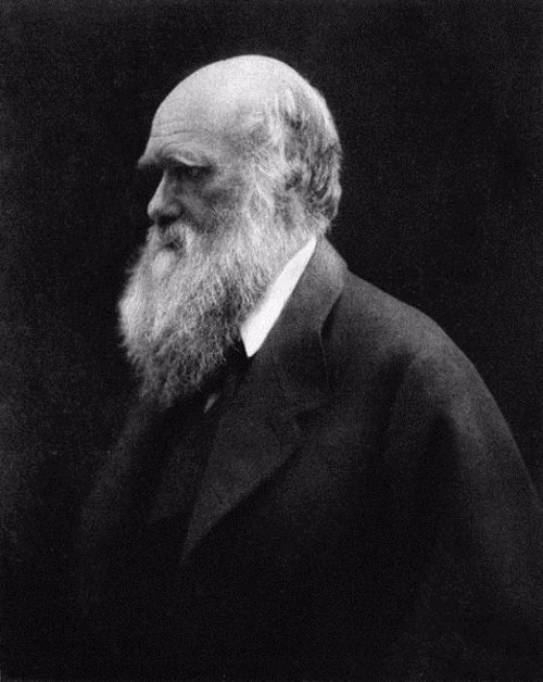
Foto: Julia Margaret Cameron
/ Wikimedia Commons
 
MADRID, 30 Abr. (EUROPA PRESS) -

Una nueva evidencia demuestra que los seres humanos están en constante evolución, y que la selección natural y sexual sigue teniendo lugar en nuestra especie. A pesar de los avances en la medicina y la tecnología, la reciente investigación ha revelado que los seres humanos siguen evolucionando, al igual que otras especies.

Para el estudio, un grupo internacional de científicos, que incluye a expertos de la Universidad de Sheffield, analizó los registros de la iglesia de unas 6.000 personas finlandesas, nacidas entre 1760 y 1849, para determinar si los cambios demográficos, culturales y tecnológicos de la revolución agrícola afectaron la selección natural y sexual en nuestra especie.

El líder del proyecto, el doctor Virpi Lummaa, declara que "los nuevos avances no han frenado la evolución de nuestra especie. Hemos demostrado que una selección significativa ha tenido lugar en poblaciones muy recientes, y es probable que siga teniendo lugar, por lo cual, los seres humanos continúan siendo afectados, tanto por la selección natural, como por la sexual". Los autores también observaron que los hombres y las mujeres no son iguales respecto a la selección darwiniana.

El investigador principal, el doctor Alexandre Courtiol, añade que "las características que aumentan el éxito de apareamiento de los hombres tienden a evolucionar más rápido que las de las mujeres. Esto se debe a que aparearse con un mayor número de individuos aumenta el éxito reproductivo, más en hombres que en mujeres. Sorprendentemente, sin embargo, la selección afecta a las personas ricas y pobres en la misma medida".

Los expertos necesitaban información detallada sobre un gran número de sujetos para poder estudiar la selección durante el ciclo de vida de los individuos: la supervivencia hasta la edad adulta, el éxito de apareamiento, y la fertilidad.

La genealogía es muy popular en Finlandia, y el país cuenta con algunos de los mejores datos disponibles para la investigación, gracias a los registros detallados de la iglesia, como nacimientos, defunciones, matrimonios y el estado de la riqueza. Según afirma Lummaa, "estos datos imparciales -ya que la selección natural y sexual actúa de manera diferente en diferentes clases de personas- permitieron estudiar cómo evoluciona la especie humana".