Mostrando entradas con la etiqueta Johns Hopkins U. Mostrar todas las entradas

viernes, 18 de septiembre de 2015

3-D printed guide helps regrow complex nerves after injury

University of Minnesota. Scientists have developed a first-of-its-kind, 3-D printed guide that helps regrow both the sensory and motor functions of complex nerves after injury. The groundbreaking research has the potential to help more than 200,000 people annually who experience nerve injuries or disease.
This is a 3-D printed nerve regeneration pathway implanted in a rat helped to improve walking in 10 to 12 weeks after implantation. Credit: University of Minnesota College of Science and Engineering

A national team of researchers has developed a first-of-its-kind, 3D-printed guide that helps regrow both the sensory and motor functions of complex nerves after injury. The groundbreaking research has the potential to help more than 200,000 people annually who experience nerve injuries or disease.

Collaborators on the project are from the University of Minnesota, Virginia Tech, University of Maryland, Princeton University, and Johns Hopkins University.

Nerve regeneration is a complex process. Because of this complexity, regrowth of nerves after injury or disease is very rare, according to the Mayo Clinic. Nerve damage is often permanent. Advanced 3D printing methods may now be the solution.

In a new study, published today in the journal Advanced Functional Materials, researchers used a combination of 3D imaging and 3D printing techniques to create a custom silicone guide implanted with biochemical cues to help nerve regeneration. The guide's effectiveness was tested in the lab using rats.

To achieve their results, researchers used a 3D scanner to reverse engineer the structure of a rat's sciatic nerve. They then used a specialized, custom-built 3D printer to print a guide for regeneration. Incorporated into the guide were 3D-printed chemical cues to promote both motor and sensory nerve regeneration. The guide was then implanted into the rat by surgically grafting it to the cut ends of the nerve. Within about 10 to 12 weeks, the rat's ability to walk again was improved.

"This represents an important proof of concept of the 3D printing of custom nerve guides for the regeneration of complex nerve injuries," said University of Minnesota mechanical engineering professor Michael McAlpine, the study's lead researcher. "Someday we hope that we could have a 3D scanner and printer right at the hospital to create custom nerve guides right on site to restore nerve function."

Scanning and printing takes about an hour, but the body needs several weeks to regrow the nerves. McAlpine said previous studies have shown regrowth of linear nerves, but this is the first time a study has shown the creation of a custom guide for regrowth of a complex nerve like the Y-shaped sciatic nerve that has both sensory and motor branches.

"The exciting next step would be to implant these guides in humans rather than rats," McAlpine said. In cases where a nerve is unavailable for scanning, McAlpine said there could someday be a "library" of scanned nerves from other people or cadavers that hospitals could use to create closely matched 3D-printed guides for patients.

In addition to McAlpine, major contributors to the research team include Blake N. Johnson, Virginia Tech; Xiaofeng Jia, University of Maryland and Johns Hopkins University; and Karen Z. Lancaster, Esteban Engel, and Lynn W. Enquist, Princeton University.

This research was funded by grants from the National Institutes of Health, the Defense Advanced Research Projects Agency, the Maryland Stem Cell Research Fund, and the Grand Challenges Program at Princeton University.

To read more about the study entitled "3D Printed Anatomical Nerve Regeneration Pathways," visit the Advanced Functional Materials website.

Story Source:
The above post is reprinted from materials provided by University of Minnesota. 
Note: Materials may be edited for content and length.

Journal Reference:
Blake N. Johnson, Karen Z. Lancaster, Gehua Zhen, Junyun He, Maneesh K. Gupta, Yong Lin Kong, Esteban A. Engel, Kellin D. Krick, Alex Ju, Fanben Meng, Lynn W. Enquist, Xiaofeng Jia, Michael C. McAlpine. 3D Printed Anatomical Nerve Regeneration Pathways. Advanced Functional Materials, 2015; 


ORIGINAL: ScienceDaily
September 18, 2015

lunes, 14 de septiembre de 2015

Neurotechnology Provides Near-Natural Sense of Touch

Revolutionizing Prosthetics program achieves goal of restoring sensation


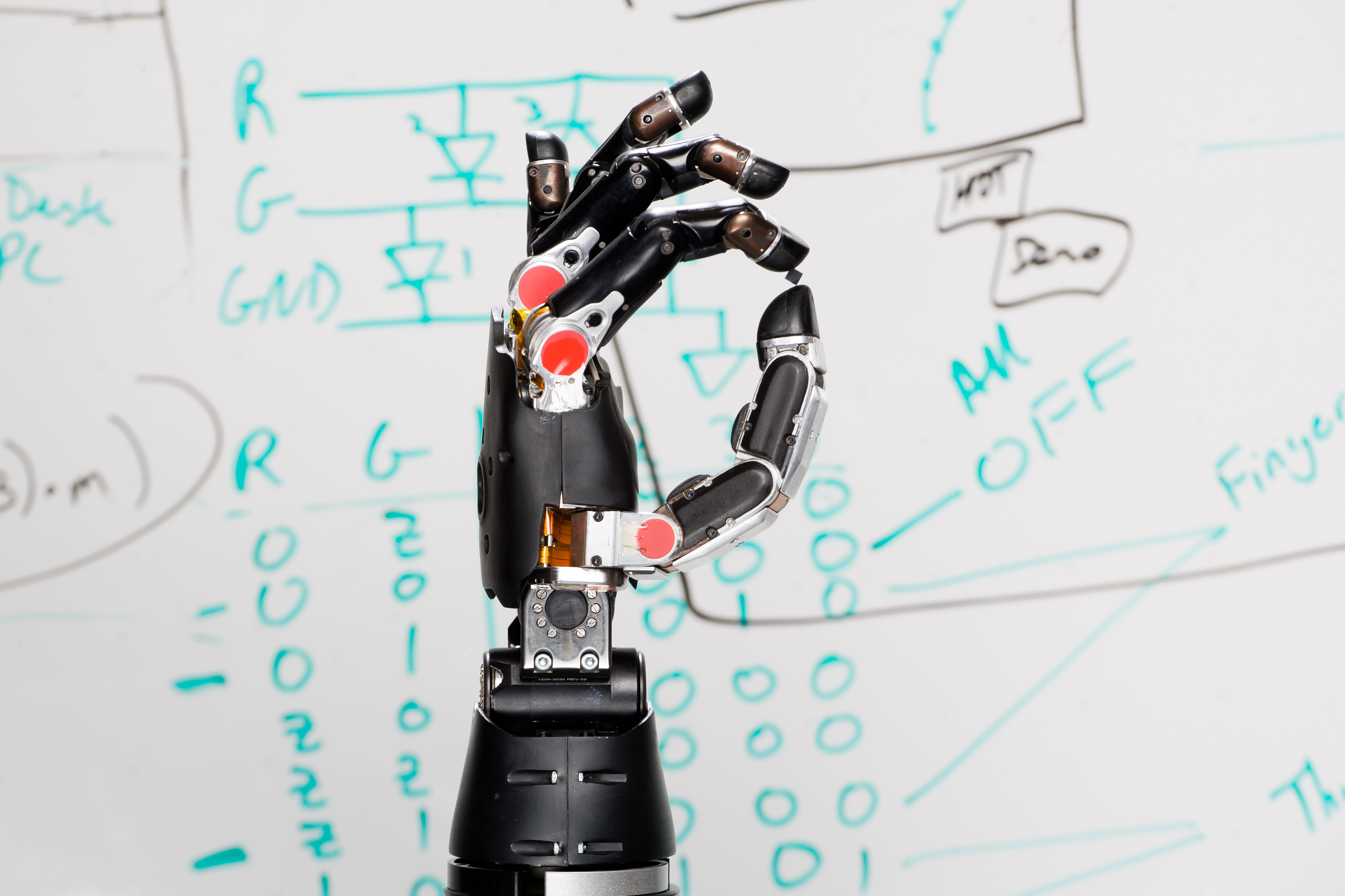
Modular Prosthetic Limb courtesy of the Johns Hopkins University 

A 28-year-old who has been paralyzed for more than a decade as a result of a spinal cord injury has become the first person to be able to “feel” physical sensations through a prosthetic hand directly connected to his brain, and even identify which mechanical finger is being gently touched.

The advance, made possible by sophisticated neural technologies developed under DARPA’s Revolutionizing Prosthetics points to a future in which people living with paralyzed or missing limbs will not only be able to manipulate objects by sending signals from their brain to robotic devices, but also be able to sense precisely what those devices are touching.

“We’ve completed the circuit,” said DARPA program manager Justin Sanchez. “Prosthetic limbs that can be controlled by thoughts are showing great promise, but without feedback from signals traveling back to the brain it can be difficult to achieve the level of control needed to perform precise movements. By wiring a sense of touch from a mechanical hand directly into the brain, this work shows the potential for seamless bio-technological restoration of near-natural function.”

The clinical work involved the placement of electrode arrays onto the paralyzed volunteer’s sensory cortex—the brain region responsible for identifying tactile sensations such as pressure. In addition, the team placed arrays on the volunteer’s motor cortex, the part of the brain that directs body movements.

Wires were run from the arrays on the motor cortex to a mechanical hand developed by the Applied Physics Laboratory (APL) at Johns Hopkins University. That gave the volunteer—whose identity is being withheld to protect his privacy—the capacity to control the hand’s movements with his thoughts, a feat previously accomplished under the DARPA program by another person with similar injuries.


Then, breaking new neurotechnological ground, the researchers went on to provide the volunteer a sense of touch. The APL hand contains sophisticated torque sensors that can detect when pressure is being applied to any of its fingers, and can convert those physical “sensations” into electrical signals. The team used wires to route those signals to the arrays on the volunteer’s brain.

In the very first set of tests, in which researchers gently touched each of the prosthetic hand’s fingers while the volunteer was blindfolded, he was able to report with nearly 100 percent accuracy which mechanical finger was being touched. The feeling, he reported, was as if his own hand were being touched.

“At one point, instead of pressing one finger, the team decided to press two without telling him,” said Sanchez, who oversees the Revolutionizing Prosthetics program. “He responded in jest asking whether somebody was trying to play a trick on him. That is when we knew that the feelings he was perceiving through the robotic hand were near-natural.”

Sanchez described the basic findings on Thursday at Wait, What? A Future Technology Forum, hosted by DARPA in St. Louis. Further details about the work are being withheld pending peer review and acceptance for publication in a scientific journal.

The restoration of sensation with implanted neural arrays is one of several neurotechnology-based advances emerging from DARPA’s 18-month-old Biological Technologies Office, Sanchez said. “DARPA’s investments in neurotechnologies are helping to open entirely new worlds of function and experience for individuals living with paralysis and have the potential to benefit people with similarly debilitating brain injuries or diseases,” he said.

In addition to the Revolutionizing Prosthetics program that focuses on restoring movement and sensation, DARPA’s portfolio of neurotechnology programs includes the
which seek to develop closed-loop direct interfaces to the brain to restore function to individuals living with memory loss from traumatic brain injury or complex neuropsychiatric illness.

For more information about Wait, What? please visit: www.darpawaitwhat.com (!!)


ORIGINAL: DARPA
OUTREACH@DARPA.MIL
9/11/2015

miércoles, 17 de julio de 2013

Physicists, biologists unite to expose how cancer spreads

ORIGINAL: Princeton
by Catherine Zandonella
April 26, 2013; 01:00 p.m.

Cancer cells that can break out of a tumor and invade other organs are more aggressive and nimble than nonmalignant cells, according to a new multi-institutional nationwide study. These cells exert greater force on their environment and can more easily maneuver small spaces.

The researchers report in the journal Scientific Reports that a systematic comparison of metastatic breast-cancer cells to healthy breast cells revealed dramatic differences between the two cell lines in their mechanics, migration, oxygen response, protein production and ability to stick to surfaces. The researchers discovered new insights into how cells make the transition from nonmalignant to metastatic, a process that is not well understood.

The resulting catalogue of differences could someday help researchers detect cancerous cells earlier and someday prevent or treat metastatic cancer, which is responsible for 90 percent of all cancer deaths, according to the study. It was conducted by a network of 12 federally funded Physical Sciences-Oncology Centers (PS-OC) sponsored by the National Cancer Institute. PS-OC is a collaboration of researchers in the physical and biological sciences seeking a better understanding of the physical and chemical forces that shape the emergence and behavior of cancer.
(Image by Guillaume Lambert)


A multi-institutional study including researchers from Princeton University's Physical Sciences-Oncology Center found that metastatic cancer cells are more aggressive and nimble than nonmalignant cells. The Princeton group used silicon-etched microchannels (above) to study the behavior and physical properties of cancer cells. In this device, metastatic cancer cells enter the narrow channels at one end and accelerate as they rapidly move down the channel. Such high motility is a hallmark of metastasis and also indicative of high glucose metabolism, another hallmark of cancer.

"By bringing together different types of experimental expertise to systematically compare metastatic and nonmetastatic cells, we have advanced our knowledge of how metastasis occurs," said Robert Austin, professor of physics and leader of the Princeton PS-OC, along with senior co-investigator Thea Tlsty of the University of California-San Francisco.

Researchers with the Princeton PS-OC, for instance, determined that metastatic cells, in spite of moving more slowly than nonmalignant cells, move farther and in a straighter line, Austin said. The investigators studied the cells' behavior in tiny cell-sized chambers and channels etched out of silicon and designed to mimic the natural environment of the body's interior.

"The mobility of these metastatic cells is an essential feature of their ability to break through the tough membrane [the extracellular matrix] that the body uses to wall off the tumor from the rest of the body," Austin said. "These cells are essentially jail-breakers."

The tiny silicon chambers were built using Princeton's expertise in microfabrication technology — typically used to create small technologies such as integrated circuits and solar cells — and are an example of the type of expertise that physicists and engineers can bring to cancer research, Austin said. For the current study, the Princeton team included physics graduate students David Liao and Guillaume Lambert, and postdoctoral researchers Liyu Liu and Saurabh Vyawahare. They worked closely with a research group led by James Sturm, Princeton's William and Edna Macaleer Professor of Engineering and Applied Science and director of the Princeton Institute for the Science and Technology of Materials (PRISM) where the microfabrication was done.

The Princeton PS-OC also includes collaborators at the Johns Hopkins University School of Medicine, the Salk Institute for Biological Studies and the University of California-Santa Cruz.

The nationwide PS-OC program aims to crack the difficulty of understanding and treating cancer by bringing in researchers from physics, engineering, computer science and chemistry, said Nastaran Zahir Kuhn, program manager for the PS-OC at the National Cancer Institute.

Other notable findings from the paper include that metastatic cells recover more rapidly from the stress of a low-oxygen environment than nonmetastatic cells, which is consistent with previous studies. Although the low-oxygen environment did kill many of the metastatic cells, the survivors rebounded vigorously, underscoring the likely role of individual cells in the spread of cancer. The study also looked at total protein production and detected proteins in the metastatic cells that are consistent with the physical properties such as mobility that malignant cells need to invade the extracellular matrix.

"The PS-OC program aims to bring physical sciences tools and perspectives into cancer research," Kuhn said. "The results of this study demonstrate the utility of such an approach, particularly when studies are conducted in a standardized manner from the beginning."

For the nationwide project, nearly 100 investigators from 20 institutions and laboratories conducted their experiments using the same two cell lines, reagents and protocols to assure that results could be compared. The experimental methods ranged from physical measurements of how the cells push on surrounding cells to measurements of gene and protein expression.

"Roughly 20 techniques were used to study the cell lines, enabling identification of a number of unique relationships between observations," Kuhn said.

For example, a technique known as atomic force microscopy indicated that metastatic cells are softer than nonmalignant cells whereas a different technique, traction force microscopy, suggested that metastatic cells exert more force on their surroundings, Kuhn said. Together these two findings may indicate that metastatic cells can exert force to stick to, migrate on and remodel the tough extracellular matrix that surrounds the tumor, while remaining flexible enough to squeeze through small spaces in that membrane.