Mostrando entradas con la etiqueta Bioquímica Celular. Mostrar todas las entradas

lunes, 28 de abril de 2014

At the origin of cell division


SISSA

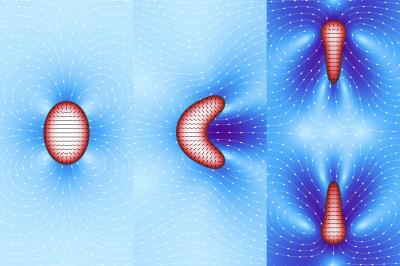
Droplets of filamentous material enclosed in a lipid membrane: these are the models of a "simplified" cell used by the SISSA physicists Luca Giomi and Antonio DeSimone, who simulated the spontaneous emergence of cell motility and division -- that is, features of living material -- in inanimate "objects." The research is one of the cover stories of the April 10th online issue of the journal Physical Review Letters. Giomi and DeSimone's artificial cells are in fact computer models that mimic some of the physical properties of the materials making up the inner content and outer membrane of cells.

The two researchers varied some of the parameters of the materials, recording what happened: "our 'cells' are a 'bare bones' representation of a biological cell, which normally contains microtubules, elongated proteins enclosed in an essentially lipid cell membrane," explains Giomi, first author of the study. "The filaments contained in the 'cytoplasm' of our cells slide over one another exerting a force that we can control."

The force exerted by the filaments is the variable that competes with another force, the surface tension that keeps the membrane surrounding the droplet from collapsing. The generates a flow in the fluid surrounding the droplet, which in turn is propelled by such self-generated flow. When the flow becomes very strong, the droplet deforms to the point of dividing. "When the force of the flow prevails over the force that keeps the membrane together we have cellular division," explains DeSimone, director of the SISSA mathLab, SISSA's mathematical modelling and scientific computing laboratory.

"We showed that by acting on a single physical parameter in a very simple model we can reproduce similar effects to those obtained with experimental observations," continues DeSimone. Empirical observations on microtubule specimens have shown that these also move outside the cell environment, in a manner proportional to the energy they have (derived from ATP, the cell "fuel"). "Similarly, our droplets, fuelled by their 'inner' energy alone -- without forces acting from the outside -- are able to move and even divide."

"Acquiring motility and the ability to divide is a fundamental step for life and, according to our simulations, the laws governing these phenomena could be very simple. Observations like ours can prepare the way for the creation of functioning artificial cells, and not only," comments Giomi. "Our work is also useful for understanding the transition from non-living to living matter on our planet. The development of the early forms of life, in other words."

Chemists and biologists who study the origin of life don't have access to cells that are sufficiently simple to be observed directly. "Even the simplest organism existing today has undergone billions of years of evolution," explains Giomi, "and will always contain fairly complex structures. Starting from schematic organisms as we do is like turning the clock back to when the first rudimentary living beings made their first appearance. We are currently starting studies to understand how cell metabolism emerged."

VIDEO: Artificial cell simulation (courtesy of Physical Review Letters):http://goo.gl/vLDcbB
Source: Sissa Medialab


ORIGINAL: eScienceNews
April 16, 2014 - 20:28 in Physics & Chemistry

domingo, 6 de abril de 2014

At the origin of cell division


SISSA

Droplets of filamentous material enclosed in a lipid membrane: these are the models of a "simplified" cell used by the SISSA physicists Luca Giomi and Antonio DeSimone, who simulated the spontaneous emergence of cell motility and division -- that is, features of living material -- in inanimate "objects." The research is one of the cover stories of the April 10th online issue of the journal Physical Review Letters. Giomi and DeSimone's artificial cells are in fact computer models that mimic some of the physical properties of the materials making up the inner content and outer membrane of cells.

The two researchers varied some of the parameters of the materials, recording what happened: "our 'cells' are a 'bare bones' representation of a biological cell, which normally contains microtubules, elongated proteins enclosed in an essentially lipid cell membrane," explains Giomi, first author of the study. "The filaments contained in the 'cytoplasm' of our cells slide over one another exerting a force that we can control."

The force exerted by the filaments is the variable that competes with another force, the surface tension that keeps the membrane surrounding the droplet from collapsing. The generates a flow in the fluid surrounding the droplet, which in turn is propelled by such self-generated flow. When the flow becomes very strong, the droplet deforms to the point of dividing. "When the force of the flow prevails over the force that keeps the membrane together we have cellular division," explains DeSimone, director of the SISSA mathLab, SISSA's mathematical modelling and scientific computing laboratory.

"We showed that by acting on a single physical parameter in a very simple model we can reproduce similar effects to those obtained with experimental observations," continues DeSimone. Empirical observations on microtubule specimens have shown that these also move outside the cell environment, in a manner proportional to the energy they have (derived from ATP, the cell "fuel"). "Similarly, our droplets, fuelled by their 'inner' energy alone -- without forces acting from the outside -- are able to move and even divide."

"Acquiring motility and the ability to divide is a fundamental step for life and, according to our simulations, the laws governing these phenomena could be very simple. Observations like ours can prepare the way for the creation of functioning artificial cells, and not only," comments Giomi. "Our work is also useful for understanding the transition from non-living to living matter on our planet. The development of the early forms of life, in other words."

Chemists and biologists who study the origin of life don't have access to cells that are sufficiently simple to be observed directly. "Even the simplest organism existing today has undergone billions of years of evolution," explains Giomi, "and will always contain fairly complex structures. Starting from schematic organisms as we do is like turning the clock back to when the first rudimentary living beings made their first appearance. We are currently starting studies to understand how cell metabolism emerged."

VIDEO: Artificial cell simulation (courtesy of Physical Review Letters):
http://goo.gl/vLDcbB
Source: Sissa Medialab


ORIGINAL: eScienceNews
April 16, 2014 - 20:28 in Physics & Chemistry

miércoles, 15 de mayo de 2013

Cells as living calculators

ORIGINAL: MIT
Anne Trafton, MIT News Office
May 15, 2013

Using analog computation circuits, MIT engineers design cells that can compute logarithms, divide and take square roots.

MIT engineers have created synthetic biology circuits that can perform analog computations such as taking logarithms and square roots in living cells. ILLUSTRATION COURTESY OF THE RESEARCHERS

MIT engineers have transformed bacterial cells into living calculators that can compute logarithms, divide, and take square roots, using three or fewer genetic parts.

Inspired by how analog electronic circuits function, the researchers created synthetic computation circuits by combining existing genetic “parts,” or engineered genes, in novel ways.

The circuits perform those calculations in an analog fashion by exploiting natural biochemical functions that are already present in the cell rather than by reinventing them with digital logic, thus making them more efficient than the digital circuits pursued by most synthetic biologists, according to Rahul Sarpeshkar and Timothy Lu, the two senior authors on the paper, describing the circuits in the May 15 online edition of Nature.

In analog you compute on a continuous set of numbers, which means it’s not just black and white, it’s gray as well,” says Sarpeshkar, an associate professor of electrical engineering and computer science and the head of the Analog Circuits and Biological Systems group at MIT

Analog computation would be particularly useful for designing cellular sensors for pathogens or other molecules, the researchers say. Analog sensing could also be combined with digital circuits to create cells that can take a specific action triggered by a threshold concentration of certain molecules.

You could do a lot of upfront sensing with the analog circuits because they’re very rich and a relatively small amount of parts can give you a lot of complexity, and have that output go into a circuit that makes a decision — is this true or not?” says Lu, an assistant professor of electrical engineering and computer science and biological engineering. 

Lead author of the Nature paper is MIT postdoc Ramiz DanielJacob Rubens, a graduate student in microbiology, is also an author of the paper.

Analog advantages 
Sarpeshkar has previously identified thermodynamic similarities between analog transistor circuits and the chemical circuits that take place inside cells. In 2011, he took advantage of those similarities to model biological interactions between DNA and proteins in an electronic circuit, using only eight transistors

In the new Nature paper, Sarpeshkar, Lu and colleagues have done the reverse — mapping analog electronic circuits onto cells. Sarpeshkar has long advocated analog computing as a more efficient alternative to digital computation at the moderate precision of computation seen in biology. These analog circuits are efficient because they can take in a continuous range of inputs, and they exploit the natural continuous computing functions that are already present in cells. In the case of cells, that continuous input might be the amount of glucose present. In transistors, it’s a range of continuous input currents or voltages.

Digital circuits, meanwhile, represent every value as zero or one, ignoring the range of possibilities in between. This can be useful for creating circuits that perform logic functions such as AND, NOT and OR inside cells, which many synthetic biologists have done. These circuits can reveal whether or not a threshold level of a certain molecule is present, but not the exact amount of it.

Digital circuits also require many more parts, which can drain the energy of the cell hosting them. “If you build too many parts to make some function, the cell is not going to have the energy to keep making those proteins,” Sarpeshkar says.

Doing the math
To create an analog adding or multiplying circuit that can calculate the total quantity of two or more compounds in a cell, the researchers combined two circuits, each of which responds to a different input. In one circuit, a sugar called arabinose turns on a transcription factor that activates the gene that codes for green fluorescent protein (GFP). In the second, a signaling molecule known as AHL also turns on a gene that produces GFP. By measuring the total amount of GFP, the total amount of both inputs can be calculated. 

To subtract or divide, the researchers swapped one of the activator transcription factors with a repressor, which turns off production of GFP when the input molecule is present. The team also built an analog square root circuit that requires just two parts, while a recently reported digital synthetic circuit for performing square roots had more than 100.

Analog computation is very efficient,” Sarpeshkar says. “To create digital circuits at a comparable level of precision would take many more genetic parts.

Another of the team’s circuits can perform division by calculating the ratio of two different molecules. Cells often perform this kind of computation on their own, which is critical for monitoring the relative concentrations of molecules such as NAD and NADH, which are frequently converted from one to the other as they help other cellular reactions take place.

That ratio is important for controlling a lot of cellular processes, and the cell naturally has enzymes that can recognize those ratios,” Lu says. “Cells can already do a lot of these things on their own, but for them to do it over a useful range requires extra engineering.

That extra engineering included modifying the circuits so that they can compute with inputs over a range of 1 to 10,000 — much wider than the range of a naturally occurring cell circuit. 

It’s nice to see that frameworks from electrical engineering can be concisely and elegantly mapped into synthetic biology,” says Eric Klavins, an associate professor of electrical engineering and adjunct associate professor of biological engineering at the University of Washington who was not part of the research team.

The researchers are now trying to create analog circuits in nonbacterial cells, including mammalian cells. They are also working on expanding the library of genetic parts that can be incorporated into the circuits. “Right now we’re using three of the most commonly used transcription factors in biology, but we’d like to do this with additional parts and make this a generalizable platform so everyone else can use it,” Lu says.

We have just scratched the surface of what sophisticated analog feedback circuits can do in living cells,” says Sarpeshkar, whose lab is working on building further new analog circuits in cells. He believes the new approach of what he terms “analog synthetic biology” will create a new set of fundamental and applied circuits that can dramatically improve the fine control of gene expression, molecular sensing, computation and actuation.

The research was funded by MIT Lincoln Laboratory, the Office of Naval Research and the National Science Foundation.

martes, 5 de febrero de 2013

Una bacteria convierte toxinas en oro

ORIGINAL: MadrI+D
05/02/2013

Una especie de bacteria puede convertirse en el 'rey Midas' de la naturaleza, ya que es capaz de crear diminutas pepitas de oro para ayudarse a crecer en las soluciones tóxicas del metal precioso. Investigadores creen que la molécula con la que las bacterias crean estas partículas podría ser utilizada en el futuro para recoger oro de los desechos mineros.

Los microbios pueden utilizar algunos metales para desarrollarse, como el hierro, pero otros les resultan letales, como es el caso del oro y la plata. El oro soluble es tóxico para la mayoría de las microbios, pero resulta que se han encontrado biopelículas -ecosistemas microbianos con diferentes microorganismos- sobre la superficie de las pepitas de oro. Y esas bacterias pueden ser, precisamente, las culpables de la acumulación del oro sólido.

Frank Reith, un microbiólogo ambiental en la Universidad de Adelaida (Australia), encontró ya hace diez años algunas de las primeras evidencias de que las bacterias prosperan en partículas de oro. En varios sitios, a miles de kilómetros de distancia, encontró una bacteria, la Cupriavidus metallidurans, que desintoxica el oro disuelto acumulando pequeñas nanopartículas de oro en el interior de sus células. 

EN LAS MINAS
Ahora, un estudio firmado por un equipo de científicos canadienses de la Universidad McMaster de Hamilton(Ontario) en la revista Nature Chemical Biology se ha preguntado si otra bacteria, la Delftia acidovorans, actúa de forma similar. Descubrieron que esta bacteria no metaboliza el oro soluble como su congénere, si no que lo solidifica en el exterior, bajo una forma no tóxica. Utiliza una molécula para crear estructuras sólidas complejas, similares a las que se encuentran en las pepitas de oro. El proceso se desarrolla en unos segundos, a temperatura ambiente y en condiciones de acidez neutra. Según los científicos, la bacteria es aún más eficaz que los productos utilizados actualmente por la industria para producir nanopartículas de oro.

Source:  Nature Chemical Biology. (a) Gold resident bacteria D. acidovorans (wild type, i), the NRPS-null D. acidovorans mutant strain (ΔdelG, ii) and C. metallidurans (iii) were grown for 3 d and overlaid with soft agarose containing 10 mM AuCl3 for 2 h.
Los investigadores creen que se podría utilizar esta bacteria para crear oro de las aguas residuales producidas en las minas. Sería como sacar un tesoro de la basura.