Mostrando entradas con la etiqueta División Celular. Mostrar todas las entradas

domingo, 6 de abril de 2014

At the origin of cell division


SISSA

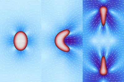
Droplets of filamentous material enclosed in a lipid membrane: these are the models of a "simplified" cell used by the SISSA physicists Luca Giomi and Antonio DeSimone, who simulated the spontaneous emergence of cell motility and division -- that is, features of living material -- in inanimate "objects." The research is one of the cover stories of the April 10th online issue of the journal Physical Review Letters. Giomi and DeSimone's artificial cells are in fact computer models that mimic some of the physical properties of the materials making up the inner content and outer membrane of cells.

The two researchers varied some of the parameters of the materials, recording what happened: "our 'cells' are a 'bare bones' representation of a biological cell, which normally contains microtubules, elongated proteins enclosed in an essentially lipid cell membrane," explains Giomi, first author of the study. "The filaments contained in the 'cytoplasm' of our cells slide over one another exerting a force that we can control."

The force exerted by the filaments is the variable that competes with another force, the surface tension that keeps the membrane surrounding the droplet from collapsing. The generates a flow in the fluid surrounding the droplet, which in turn is propelled by such self-generated flow. When the flow becomes very strong, the droplet deforms to the point of dividing. "When the force of the flow prevails over the force that keeps the membrane together we have cellular division," explains DeSimone, director of the SISSA mathLab, SISSA's mathematical modelling and scientific computing laboratory.

"We showed that by acting on a single physical parameter in a very simple model we can reproduce similar effects to those obtained with experimental observations," continues DeSimone. Empirical observations on microtubule specimens have shown that these also move outside the cell environment, in a manner proportional to the energy they have (derived from ATP, the cell "fuel"). "Similarly, our droplets, fuelled by their 'inner' energy alone -- without forces acting from the outside -- are able to move and even divide."

"Acquiring motility and the ability to divide is a fundamental step for life and, according to our simulations, the laws governing these phenomena could be very simple. Observations like ours can prepare the way for the creation of functioning artificial cells, and not only," comments Giomi. "Our work is also useful for understanding the transition from non-living to living matter on our planet. The development of the early forms of life, in other words."

Chemists and biologists who study the origin of life don't have access to cells that are sufficiently simple to be observed directly. "Even the simplest organism existing today has undergone billions of years of evolution," explains Giomi, "and will always contain fairly complex structures. Starting from schematic organisms as we do is like turning the clock back to when the first rudimentary living beings made their first appearance. We are currently starting studies to understand how cell metabolism emerged."

VIDEO: Artificial cell simulation (courtesy of Physical Review Letters):
http://goo.gl/vLDcbB
Source: Sissa Medialab


ORIGINAL: eScienceNews
April 16, 2014 - 20:28 in Physics & Chemistry

domingo, 28 de julio de 2013

Floppy Cells

ORIGINAL: The Scientist
By Kate Yandell
July 24, 2013

Cell division in L-forms—bacterial variants that have no cell walls—could shed light on how primitive life forms replicated.

PINCH HITTING: When a walled Bacillus subtilis cell divides, complicated cellular machinery segregates its contents and builds a new peptidoglycan wall across its center (1) before the bacterium splits into two daughter cells (2). L-form bacteria, which don’t have cell walls, dispense with the normal replication methods, at least in some cases. Instead, L-forms produce extra cell membrane and extra chromosomes and become large and irregularly shaped (3). Biomechanical forces cause smaller cells to break off through blebbing (4) or tubulation (5).
LUCY READING-IKKANDA
The paper
R. Mercier et al., “Excess membrane synthesis drives a primitive mode of cell proliferation,” Cell, 152:997-1007, 2013.

Bacterial cells usually divide in an orderly fashion, building new cell walls across their centers before they separate. But recent research suggests that cell division for bacterial L-forms, which lack a cell wall, is a haphazard affair—possibly more reminiscent of primitive cell replication than of modern-day bacterial reproduction.

Many bacterial species, ranging from the harmless soil bacterium Bacillus subtilis to the pathogenic Listeria monocytogenes, have L-forms. They are pared-down versions of ordinary cells of their species, containing nearly the same genes but lacking the exterior peptidoglycan coating that is a defining bacterial trait.

Jeff Errington, a cell and molecular biologist at Newcastle University in the U.K., initially turned to L-form bacteria several years ago as a simpler model for studying cell division in bacteria. In particular, he wanted to understand the role of cytoskeletal proteins in helping the cell membrane constrict during division. Instead, “what we realized is that they don’t use that machinery at all,” he said.

In a 2009 study, Errington and colleagues showed that they could create proliferating B. subtilis L-forms by turning off genes important for cell-wall synthesis and introducing a single mutation in a gene called IspA, a mutation that appeared to protect the cell from death in the absence of its wall (Nature, 457:849-54).

In a new study of the genes involved, the researchers depleted the bacterium’s cell wall enzymatically, without altering the genome—except for introducing the protective IspA mutation. Most cells failed to grow, but one cell reproduced both in the walled and L-form states, and the scientists sequenced its genome to fish for the gene mutations responsible for its survival. One mutation, located upstream of the genes AccA and AccD, which are involved in fatty-acid synthesis, caused cells to grow excess lipid membrane. As the ratio of membrane area to cell volume rose, the investigators hypothesized, biomechanical forces caused parts of the cell to shear off into progeny, taking with them copies of the complete genome that L-form cells have in reserve.

This uncomplicated reproduction process reminded Errington of the lipid vesicle experiments biologists perform to study the origins of life. L-forms’ division supports the idea that primitive cells could have divided without evolving the intricate processes most cells rely on today. “This may resemble the mechanisms that [were] there before a more stable cell wall,” said Martin Loessner, a microbiologist at the Swiss Federal Institute of Technology, who also studies L-forms. But he adds that L-forms of various species may have different reproduction strategies—dividing symmetrically, forming vesicle “daughter cells” in their interiors and then releasing them, or even using limited cellular division machinery.

Errington is also interested in studying the role that these primitive forms could play in disease and antibiotic resistance. Antibiotics that attack the cell wall can cause bacteria to convert into L-forms, and Errington suspects this conversion could provide a temporary escape hatch for pathogens, allowing them to dodge drugs and the immune system. It’s a problem that’s “very interesting and potentially important” to medicine, he says.

sábado, 30 de marzo de 2013

“OMG” Microscope Lives Up To Its Name

ORIGINAL: NIH
By Dr. Francis Collins
March 21st, 2013

Courtesy of Indiana University
The scientists at the IU School of Medicine-Bloomington nicknamed their new microscope the “OMG” for good reason—the images it produces are showstoppers. The DeltaVision OMX imaging system (its official title) is a $1.2 million dollar microscope that can peek inside a cell and image fluorescent proteins in unprecedented detail.

Jane Stout, a researcher in the NIH-funded lab, used the OMG to create this spectacular image that won her first place in the high- and super-resolution microscopy category of the 2012 GE Healthcare Life Sciences Cell Imaging Competition.

What you’re looking at is a cell in the midst of dividing into two identical copies—a process called mitosis. Here, the chromosomes (in blue) are aligned at the cell’s equator. Microtubules (red) from opposite poles of the cell attach to the chromosomes using the kinetochores (green) and pull them to opposite ends of the cell, which then splits in half. But sometimes cells do not divide properly—a common problem in cancer. Understanding the mechanics of cell division could help us correct this process when it goes wrong.

Jane Stout’s prize: her mitosis image will light up a billboard in Times Square in New York City in April. That is a wonderful celebration of science!

NIH support: the National Institute of General Medical Sciences

Anushree Balachandran. Genea, Australia
Therapeutic focus: Huntington's disease
Description: Huntington's stem cell derived oligodendrocyte precursors stained for phalloidin (green), vinculin (red) and DNA (blue).
Image: 
2012 GE Healthcare Life Sciences Cell Imaging Competition.
Markus Posch. University of Dundee, UK
Therapeutic focus: Cancer
Description: Prometaphase human cervical carcinoma (HeLa) cell with GFP-histone labeled chromosomes (blue) stained for tubulin (yellow).
Image: 
2012 GE Healthcare Life Sciences Cell Imaging Competition.