Mostrando entradas con la etiqueta Biomechanics. Mostrar todas las entradas

jueves, 21 de abril de 2016

Meet the Nanomachines That Could Drive a Medical Revolution


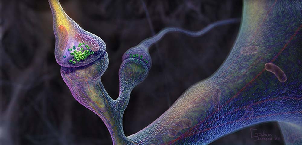
A group of physicists recently built the smallest engine ever created from just a single atom. Like any other engine it converts heat energy into movement — but it does so on a smaller scale than ever seen before. The atom is trapped in a cone of electromagnetic energy and lasers are used to heat it up and cool it down, which causes the atom to move back and forth in the cone like an engine piston.

The scientists from the University of Mainz in Germany who are behind the invention don’t have a particular use in mind for the engine. But it’s a good illustration of how we are increasingly able to replicate the everyday machines we rely on at a tiny scale. This is opening the way for some exciting possibilities in the future, particularly in the use of nanorobots in medicine, that could be sent into the body to release targeted drugs or even fight diseases such as cancer.

Nanotechnology deals with ultra-small objects equivalent to one billionth of a meter in size, which sounds an impossibly tiny scale at which to build machines. But size is relative to how close you are to an object. We can’t see things at the nanoscale with the naked eye, just as we can’t see the outer planets of the solar system. Yet if we zoom in — with a telescope for the planets or a powerful electron microscope for nano-objects — then we change the frame of reference and things look very different.

However, even after getting a closer look, we still can’t build machines at the nanoscale using conventional engineering tools. While regular machines, such as the internal combustion engines in most cars, operate according to the rules of physics laid out by Isaac Newton, things at the nanoscale follow the more complex laws of quantum mechanics. So we need different tools that take into account the quantum world in order to manipulate atoms and molecules in a way that uses them as building blocks for nanomachines. Here are four more tiny machines that could have a big impact.

1- Graphene engine for nanorobots
Researchers from Singapore have recently demonstrated a simple but nano-sized engine made from a highly elastic piece of graphene. Graphene is a two-dimensional sheet of carbon atoms that has exceptional mechanical strength. Inserting some chlorine and fluorine molecules into the graphene lattice and firing a laser at it causes the sheet to expand. Rapidly turning the laser on and off makes the graphene pump back and forth like the piston in an internal combustion engine.

The researchers think the graphene nano-engine could be used to power tiny robots, for example to attack cancer cells in the body. Or it could be used in a so-called “lab-on-a-chip” — a device that shrinks the functions of a chemistry lab into tiny package that can be used for rapid blood tests, among other things.

2- Frictionless nano-rotor
Molecular motor.
Image credit: 
Palma, C.-A.; Kühne, D.; Klappenberger, F.; Barth, J.V.; Technische Universität München


The rotors that produce movement in machines such as aircraft engines and fans all usually suffer from friction, which limits their performance. Nanotechnology can be used to create a motor from a single molecule, which can rotate without any friction. Normal rotors interact with the air according to Newton’s laws as they spin round and so experience friction. But, at the nanoscale, molecular rotors follow quantum law, meaning they don’t interact with the air in the same way and so friction doesn’t affect their performance.

Nature has actually already shown us that molecular motors are possible. Certain proteins can travel along a surface using a rotating mechanism that create movement from chemical energy. These motor proteins are what cause cells to contract and so are responsible for our muscle movements.

Researchers from Germany recently reported creating a molecular rotor by placing moving molecules inside a tiny hexagonal hole known as a nanopore in a thin piece of silver. The position and movement of the molecules meant they began to rotate around the hole like a rotor. Again, this form of nano-engine could be used to power a tiny robot around the body.

3- Controllable nano-rockets


A rocket is the fastest man-made vehicle that can freely travel across the universe. Several groups of researchers have recently constructed a high-speed, remote-controlled nanoscale version of a rocket by combining nanoparticles with biological molecules.

In one case, the body of the rocket was made from a polystyrene bead covered in gold and chromium. This was attached to multiple “catalytic engine” molecules using strands of DNA. When placed in a solution of hydrogen peroxide, the engine molecules caused a chemical reaction that produced oxygen bubbles, forcing the rocket to move in the opposite direction. Shining a beam of ultra-violet light on one side of the rocket causes the DNA to break apart, detaching the engines and changing the rocket’s direction of travel. The researchers hope to develop the rocket so it can be used in any environment, for example to deliver drugs to a target area of the body.

4- Magnetic nano-vehicles for carrying drugs

Magnetic nanoparticles. Image credit: Tapas Sen, author provided
My own research group is among those working on a simpler way to carry drugs through the body that is already being explored with magnetic nanoparticles. Drugs are injected into a magnetic shell structure that can expand in the presence of heat or light. This means that, once inserted into the body, they can be guided to the target area using magnets and then activated to expand and release their drug.

The technology is also being studied for medical imaging. Creating the nanoparticles to gather in certain tissues and then scanning the body with a magnetic resonance imaging (MRI) could help highlight problems such as diabetes.

Tapas Sen, Reader in Nanomaterials Chemistry, University of Central Lancashire

This article was originally published on The Conversation. Read the original article.

ORIGINAL: Singularity Hub

martes, 20 de noviembre de 2012

More complex circuits for synthetic biology lead toward engineered cells


The protein–protein and protein–DNA interactions that can lead to crosstalk between gates are shown as red rectangles. (Credit: Tae Seok Moon et al./Nature)

One possible pathway from current technology to advanced nanotechnology that will comprise atomically precise manufacturing implemented by atomically precise machinery is through adaptation and extension of the complex molecular machine systems evolved by biology. Synthetic biology, which engineers new biological systems and function not evolved in nature, is an intermediate stage along this path. An article on KurzweilAI-net describes a recent achievement by MIT scientists in constructing a synthetic genetic circuit that responds to control signals from four molecules without any one molecule interfering with the responses to any other molecules. From “The most complex synthetic biology circuit yet“:

Christopher Voigt, an associate professor of biological engineering at MIT, and his students have developed circuit components that don’t interfere with one another, allowing them to produce the most complex synthetic circuit ever built.

The circuit integrates four sensors for different molecules. Such circuits could be used in cells to precisely monitor their environments and respond appropriately.

Using genes as interchangeable parts, synthetic biologists design cellular circuits that can perform new functions, such as sensing environmental conditions. However, the complexity that can be achieved in such circuits has been limited by a critical bottleneck: the difficulty in assembling genetic components that don’t interfere with each other.

Unlike electronic circuits on a silicon chip, biological circuits inside a cell cannot be physically isolated from one another. “The cell is sort of a burrito. It has everything mixed together,” says Voigt.

Because all the cellular machinery for reading genes and synthesizing proteins is jumbled together, researchers have to be careful that proteins that control one part of their synthetic circuit don’t hinder other parts of the circuit.

The pathway consists of three components: an activator, a promoter and a chaperone
A promoter is a region of DNA where proteins bind to initiate transcription of a gene. 
An activator is one such protein. 
Some activators also require a chaperone protein before they can bind to DNA to initiate transcription. …

To design synthetic circuits so they can be layered together, their inputs and outputs must mesh. With an electrical circuit, the inputs and outputs are always electricity. With these biological circuits, the inputs and outputs are proteins that control the next circuit (either activators or chaperones).

The research was published in Nature [abstract]. The authors conclude “This work demonstrates the successful layering of orthogonal logic gates, a design strategy that could enable the construction of large, integrated circuits in single cells.Certainly there are many steps between engineering cells to optimize their normal outputs to achieve engineered purposes, and engineering cells for entirely novel nanofabrication of materials not normally found in biology, but the more complex the tools that are available, the more opportunities there will be to advance along this path. Whether or not we can go all the way along this path from engineering biology to molecular manufacturing remains to be seen.
James Lewis, PhD

lunes, 12 de noviembre de 2012

A Moving Kinesin Motor Protein


A Moving Kinesin Motor Protein 
The two heads of the kinesin dimer work in a coordinated manner to move processively along the track. The coiled coil (gray) extends towards the top and leads up to the kinesin cargo.
Each catalytic core (blue) is bound to a tubulin heterodimer (green, beta subunit; white, alpha subunit) along a microtubule protofilament (the cylindrical microtubule is composed of 13 protofilament tracks). To adopt this position, the neck linker points forward on the trailing head (orange; neck linker next to but not tightly docked to the core) and rearward on the leading head (red).

ATP binding to the leading head will initiate neck linker docking. Neck linker docking is completed by the leading head (yellow), which throws the partner head forward by 160 angstroms (arrow) toward the next tubulin binding site. After a random diffusional search, the new leading head docks tightly onto the binding site, which completes the 80 angstrom motion of the attached cargo.

Polymer binding also accelerates ADP release, and during this time, the trailing head hydrolyzes ATP to ADP-Pi.

After ADP dissociates, an ATP binds to the leading head and the neck linker begins to zipper onto the core (partially docked neck indicated by the orange color). The trailing head, which has released its phosphate (Pi) and detached its neck linker (red) from the core, is in the process of being thrown forward.

The surface features of the motors and filaments were rendered by G Johnson (fiVth media: http://www.fiVth.com) using the programs MolView, Strata Studio Pro, and Cinema 4D. Protein Data Bank files used throughout the figures are as follows: human conventional kinesin [prestroke, red: 1BG2], and rat conventional kinesin [poststroke, yellow: 2KIN].

Width of tubulin beta subunit (in green) is approximately 40 angstroms. animation; ATP; kinesin; microtubule; motor protein Movie 2 in: Vale RD, Milligan RA.

The way things move: looking under the hood of molecular motors. Science [serial online]. 2000;288:88-95. Available [subscription required] at:
 http://www.sciencemag.org/cgi/reprint/288/5463/88.pdf

Original resource provided by Ronald D Vale.
Work conducted at Howard Hughes Medical Institute and University of California, San Francisco, CA (RV), Scripps Research Institute, La Jolla, CA (RM).

Biological Sources Cellular Component kinesin complexmicrotubule
Biological Context Biological Process ATP catabolic process
Molecular Function kinesin binding
Attribution Names
Ronald D Vale
Ronald A Milligan
Graham Johnson
Pubmed 10753125

Imaging
Image Type animationcomputer graphic

Processing History
surface rendering

Dimensions
Spatial AxisImage SizePixel Size
X
320px
0.118nm
Y
240px
0.118nm
Time
39 sec
12 frames/s